

Search Count: 14
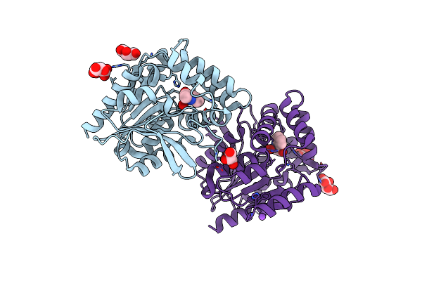 |
Crystal Structure Of The Cofactor-Devoid 1-H-3-Hydroxy-4- Oxoquinaldine 2,4-Dioxygenase (Hod) S101A Variant Complexed With 2-Methyl-Quinolin-4(1H)-One Under Hyperoxyc Conditions
Organism: Paenarthrobacter nitroguajacolicus
Method: X-RAY DIFFRACTION Resolution:2.00 Å Release Date: 2024-01-17 Classification: OXIDOREDUCTASE Ligands: VFH, OXY, TAR, GOL, NA |
Organism: Paenarthrobacter nitroguajacolicus
Method: X-RAY DIFFRACTION
Release Date: 2024-01-17
Ligands: VFH, OXY, TAR, GOL, NA
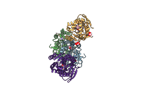 |
Crystal Structure Of The Cofactor-Devoid 1-H-3-Hydroxy-4- Oxoquinaldine 2,4-Dioxygenase (Hod) S101A Variant Complexed With 2-Methyl-Quinolin-4(1H)-One Under Normoxyc Conditions
Organism: Paenarthrobacter nitroguajacolicus
Method: X-RAY DIFFRACTION Resolution:2.00 Å Release Date: 2024-01-17 Classification: OXIDOREDUCTASE Ligands: VFH, GOL, SRT |
Organism: Paenarthrobacter nitroguajacolicus
Method: X-RAY DIFFRACTION
Release Date: 2024-01-17
Ligands: VFH, GOL, SRT
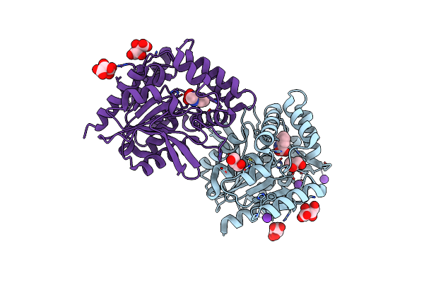 |
Crystal Structure Of The Cofactor-Devoid 1-H-3-Hydroxy-4- Oxoquinaldine 2,4-Dioxygenase (Hod) H251A Variant Complexed With N-Acetylanthranilate As Result Of In Crystallo Turnover Of Its Natural Substrate 1-H-3-Hydroxy-4- Oxoquinaldine Under Hyperoxic Conditions
Organism: Paenarthrobacter nitroguajacolicus
Method: X-RAY DIFFRACTION Resolution:2.00 Å Release Date: 2024-01-17 Classification: OXIDOREDUCTASE Ligands: ZZ8, SRT, GOL, K |
Organism: Paenarthrobacter nitroguajacolicus
Method: X-RAY DIFFRACTION
Release Date: 2024-01-17
Ligands: ZZ8, SRT, GOL, K
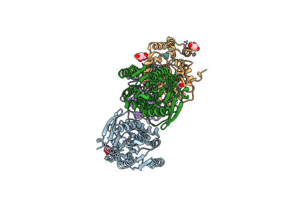 |
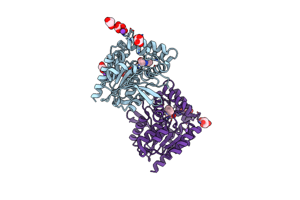
Room Temperature Crystal Structure Of The Cofactor-Devoid 1-H-3-Hydroxy-4- Oxoquinaldine 2,4-Dioxygenase (Hod) Under Xenon Pressure (30 Bar)
Organism: Paenarthrobacter nitroguajacolicus
Method: X-RAY DIFFRACTION Resolution:2.90 Å Release Date: 2023-07-05 Classification: OXIDOREDUCTASE Ligands: XE, TAR |
Organism: Paenarthrobacter nitroguajacolicus
Method: X-RAY DIFFRACTION
Release Date: 2023-07-05
Ligands: XE, TAR
 |
Crystal Structure Of The Cofactor-Devoid 1-H-3-Hydroxy-4- Oxoquinaldine 2,4-Dioxygenase (Hod) Catalytically Inactive H251A Variant Complexed With 2-Methyl-Quinolin-4(1H)-One Under Normoxic Conditions
Organism: Paenarthrobacter nitroguajacolicus
Method: X-RAY DIFFRACTION Resolution:2.00 Å Release Date: 2022-06-01 Classification: OXIDOREDUCTASE Ligands: VFH, K, GOL, SRT, NA |
Organism: Paenarthrobacter nitroguajacolicus
Method: X-RAY DIFFRACTION
Release Date: 2022-06-01
Ligands: VFH, K, GOL, SRT, NA
 |
Crystal Structure Of The Cofactor-Devoid 1-H-3-Hydroxy-4- Oxoquinaldine 2,4-Dioxygenase (Hod) Catalytically Inactive H251A Variant Complexed With 2-Methyl- Quinolin-4(1H)-One Under Hyperoxic Conditions
Organism: Paenarthrobacter nitroguajacolicus
Method: X-RAY DIFFRACTION Resolution:2.10 Å Release Date: 2022-06-01 Classification: OXIDOREDUCTASE Ligands: VFH, K, TAR, GOL |
Organism: Paenarthrobacter nitroguajacolicus
Method: X-RAY DIFFRACTION
Release Date: 2022-06-01
Ligands: VFH, K, TAR, GOL
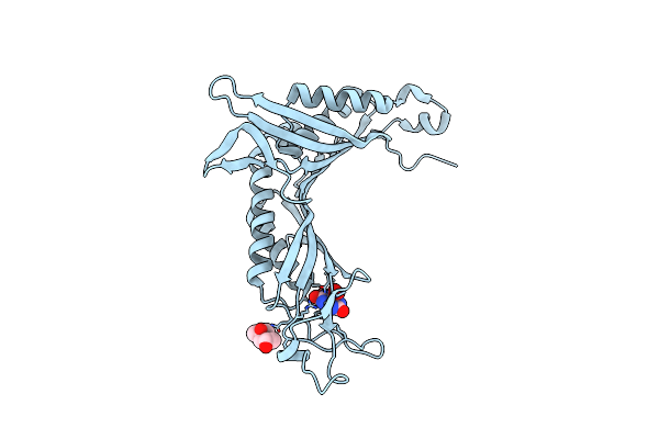 |
Crystal Structure Of Cofactor-Free Urate Oxidase In Complex With Its 5-Peroxoisourate Intermediate (X-Ray Dose, 106 Kgy)
Organism: Aspergillus flavus
Method: X-RAY DIFFRACTION Resolution:1.30 Å Release Date: 2014-11-05 Classification: OXIDOREDUCTASE Ligands: IUP, MPD, OXY, URC |
Organism: Aspergillus flavus
Method: X-RAY DIFFRACTION
Release Date: 2014-11-05
Ligands: IUP, MPD, OXY, URC
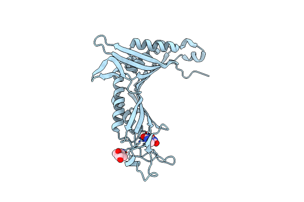 |
Crystal Structure Of Cofactor-Free Urate Oxidase Anaerobically Complexed With 9-Methyl Uric Acid
Organism: Aspergillus flavus
Method: X-RAY DIFFRACTION Resolution:1.50 Å Release Date: 2014-10-29 Classification: OXIDOREDUCTASE Ligands: MPD, MUA |
Organism: Aspergillus flavus
Method: X-RAY DIFFRACTION
Release Date: 2014-10-29
Ligands: MPD, MUA
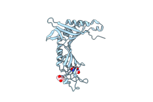 |
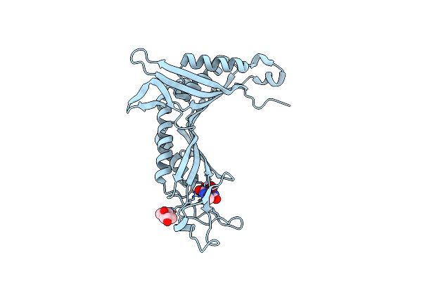
Crystal Structure Of Cofactor-Free Urate Oxidase In Complex With The 5-Peroxo Derivative Of 9-Metyl Uric Acid (X-Ray Dose, 2.5 Kgy)
Organism: Aspergillus flavus
Method: X-RAY DIFFRACTION Resolution:1.32 Å Release Date: 2014-10-29 Classification: OXIDOREDUCTASE Ligands: XDS, MPD |
Organism: Aspergillus flavus
Method: X-RAY DIFFRACTION
Release Date: 2014-10-29
Ligands: XDS, MPD
 |
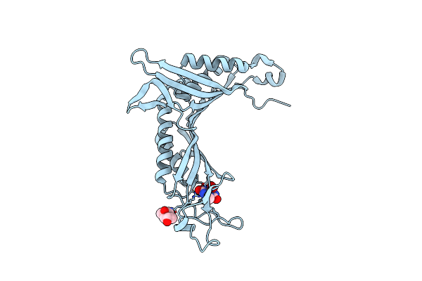
Crystal Structure Of Cofactor-Free Urate Oxidase In Complex With The 5-Peroxo Derivative Of 9-Metyl Uric Acid (X-Ray Dose, 665 Kgy)
Organism: Aspergillus flavus
Method: X-RAY DIFFRACTION Resolution:1.34 Å Release Date: 2014-10-29 Classification: OXIDOREDUCTASE Ligands: XDS, MPD, OXY, MUA |
Organism: Aspergillus flavus
Method: X-RAY DIFFRACTION
Release Date: 2014-10-29
Ligands: XDS, MPD, OXY, MUA
 |
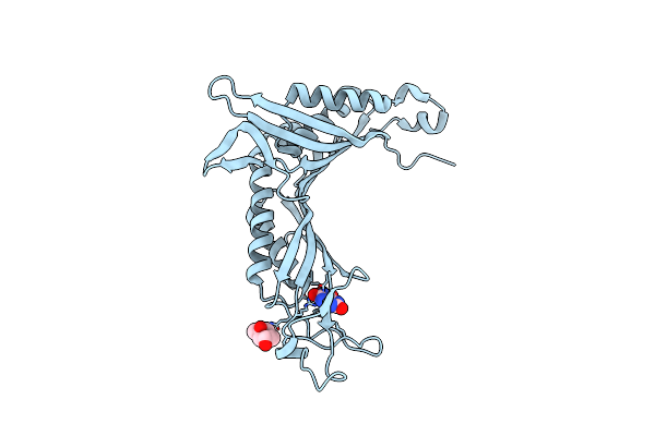
Crystal Structure Of Cofactor-Free Urate Oxidase In Complex With The 5-Peroxo Derivative Of 9-Metyl Uric Acid (X-Ray Dose, 92 Kgy)
Organism: Aspergillus flavus
Method: X-RAY DIFFRACTION Resolution:1.28 Å Release Date: 2014-10-29 Classification: OXIDOREDUCTASE Ligands: XDS, MPD, OXY, MUA |
Organism: Aspergillus flavus
Method: X-RAY DIFFRACTION
Release Date: 2014-10-29
Ligands: XDS, MPD, OXY, MUA
 |
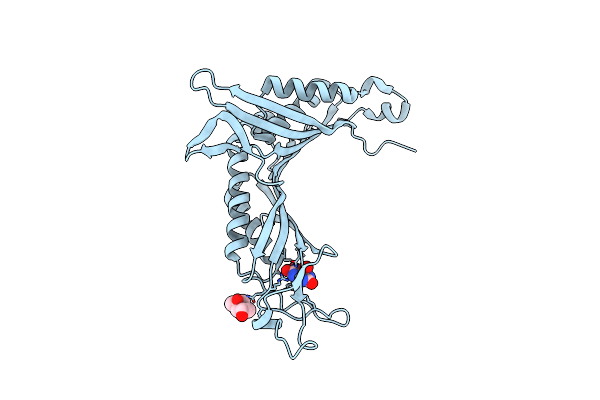
Crystal Structure Of Cofactor-Free Urate Oxidase Anaerobically Complexed With Uric Acid
Organism: Aspergillus flavus
Method: X-RAY DIFFRACTION Resolution:1.40 Å Release Date: 2014-10-29 Classification: OXIDOREDUCTASE Ligands: MPD, URC |
Organism: Aspergillus flavus
Method: X-RAY DIFFRACTION
Release Date: 2014-10-29
Ligands: MPD, URC
 |
Crystal Structure Of Cofactor-Free Urate Oxidase In Complex With Its 5-Peroxoisourate Intermediate (X-Ray Dose, 2.2 Kgy)
Organism: Aspergillus flavus
Method: X-RAY DIFFRACTION Resolution:1.30 Å Release Date: 2014-10-29 Classification: OXIDOREDUCTASE Ligands: IUP, MPD |
Organism: Aspergillus flavus
Method: X-RAY DIFFRACTION
Release Date: 2014-10-29
Ligands: IUP, MPD
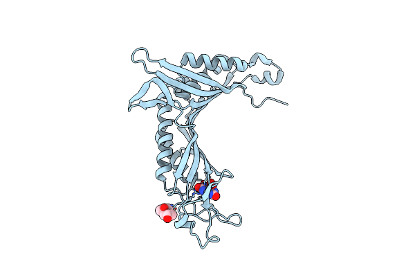 |
Crystal Structure Of Cofactor-Free Urate Oxidase In Complex With Its 5-Peroxoisourate Intermediate (X-Ray Dose, 1.75 Mgy)
Organism: Aspergillus flavus
Method: X-RAY DIFFRACTION Resolution:1.35 Å Release Date: 2014-10-29 Classification: OXIDOREDUCTASE Ligands: IUP, MPD, OXY, URC |
Organism: Aspergillus flavus
Method: X-RAY DIFFRACTION
Release Date: 2014-10-29
Ligands: IUP, MPD, OXY, URC