

Search Count: 148
 |
Metabotropic Glutamate Receptor 7 In Complex With Ecto-Domain Of Extracellular Leucine Rich Repeat And Fibronectin Type Iii Domain Containing 2
Organism: Homo sapiens
Method: ELECTRON MICROSCOPY Resolution:4.60 Å Release Date: 2026-08-05 Classification: MEMBRANE PROTEIN |
Organism: Homo sapiens
Method: ELECTRON MICROSCOPY
Release Date: 2026-08-05
 |
Cryo Em Structure Of The Closed Tetramer Of Rv2531C From Mycobacterium Tuberculosis
Organism: Mycobacterium tuberculosis h37rv
Method: ELECTRON MICROSCOPY Resolution:3.30 Å Release Date: 2025-11-26 Classification: LYASE |
Organism: Mycobacterium tuberculosis h37rv
Method: ELECTRON MICROSCOPY
Release Date: 2025-11-26
 |
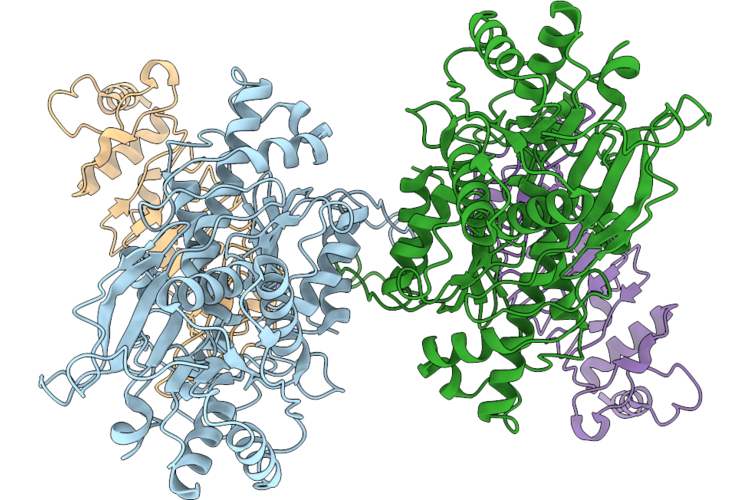
Cryoem Structure Of Rv2531C Dimer From Mycobacterium Tuberculosis
Organism: Mycobacterium tuberculosis h37rv
Method: ELECTRON MICROSCOPY Resolution:2.80 Å Release Date: 2025-11-26 Classification: LYASE Ligands: PLP |
Organism: Mycobacterium tuberculosis h37rv
Method: ELECTRON MICROSCOPY
Release Date: 2025-11-26
Ligands: PLP
 |
Cryo Em Structure Of The Open Tetramer Of Rv2531C From Mycobacterium Tuberculosis.
Organism: Mycobacterium tuberculosis h37rv
Method: ELECTRON MICROSCOPY Resolution:3.40 Å Release Date: 2025-11-26 Classification: LYASE |
Organism: Mycobacterium tuberculosis h37rv
Method: ELECTRON MICROSCOPY
Release Date: 2025-11-26
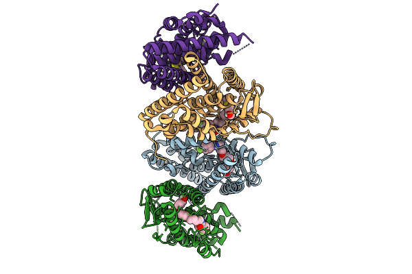 |
Crystal Structure Of The Er-Alpha Ligand-Binding Domain (L372S, L536S) In Complex With Na98
Organism: Homo sapiens
Method: X-RAY DIFFRACTION Resolution:1.45 Å Release Date: 2025-08-27 Classification: NUCLEAR PROTEIN Ligands: A1AIZ |
Organism: Homo sapiens
Method: X-RAY DIFFRACTION
Release Date: 2025-08-27
Ligands: A1AIZ
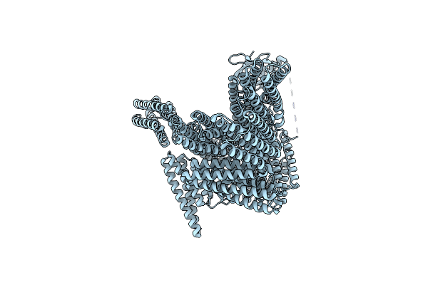 |
Cryogenic Electron Microscopy Model Of Full-Length Talin Lacking F2, R12 And Fabd.
Organism: Mus musculus
Method: ELECTRON MICROSCOPY Release Date: 2024-10-02 Classification: CELL ADHESION |
Organism: Mus musculus
Method: ELECTRON MICROSCOPY
Release Date: 2024-10-02
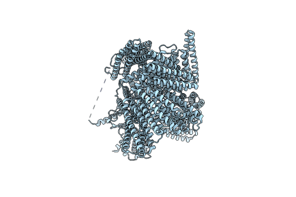 |
Cryogenic Electron Microscopy Model Of Full-Length Talin Without Fabd
Organism: Mus musculus
Method: ELECTRON MICROSCOPY Release Date: 2024-10-02 Classification: CELL ADHESION |
Organism: Mus musculus
Method: ELECTRON MICROSCOPY
Release Date: 2024-10-02
 |
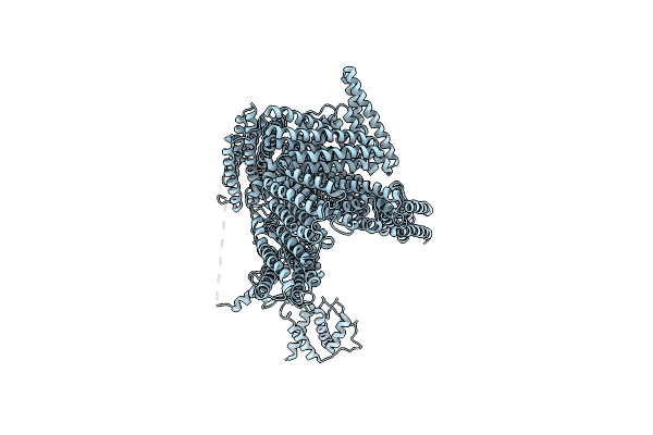
Cryogenic Electron Microscopy Model Of Full-Length Talin
Organism: Mus musculus
Method: ELECTRON MICROSCOPY Release Date: 2024-10-02 Classification: CELL ADHESION |
Organism: Mus musculus
Method: ELECTRON MICROSCOPY
Release Date: 2024-10-02
 |
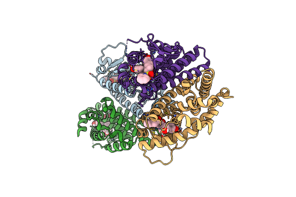
Cryogenic Electron Microscopy Model Of Full-Length Talin Without R12 And Fabd
Organism: Mus musculus
Method: ELECTRON MICROSCOPY Release Date: 2024-10-02 Classification: CELL ADHESION |
Organism: Mus musculus
Method: ELECTRON MICROSCOPY
Release Date: 2024-10-02
 |
Crystal Structure Of The Er-Alpha Ligand-Binding Domain (L372S, L536S) In Complex With K-411
Organism: Homo sapiens
Method: X-RAY DIFFRACTION Resolution:1.61 Å Release Date: 2024-06-12 Classification: NUCLEAR PROTEIN Ligands: A1AHV |
Organism: Homo sapiens
Method: X-RAY DIFFRACTION
Release Date: 2024-06-12
Ligands: A1AHV
 |
Crystal Structure Of The Er-Alpha Ligand-Binding Domain (L372S, L536S) In Complex With K-410
Organism: Homo sapiens
Method: X-RAY DIFFRACTION Resolution:1.69 Å Release Date: 2024-06-12 Classification: NUCLEAR PROTEIN Ligands: A1AHU |
Organism: Homo sapiens
Method: X-RAY DIFFRACTION
Release Date: 2024-06-12
Ligands: A1AHU
 |
Crystal Structure Of The Er-Alpha Ligand-Binding Domain (L372S, L536S) In Complex With K-400
Organism: Homo sapiens
Method: X-RAY DIFFRACTION Resolution:1.86 Å Release Date: 2024-06-12 Classification: NUCLEAR PROTEIN Ligands: A1AHO |
Organism: Homo sapiens
Method: X-RAY DIFFRACTION
Release Date: 2024-06-12
Ligands: A1AHO
 |
Crystal Structure Of The Er-Alpha Ligand-Binding Domain (L372S, L536S) In Complex With K-409
Organism: Homo sapiens
Method: X-RAY DIFFRACTION Resolution:1.82 Å Release Date: 2024-06-12 Classification: NUCLEAR PROTEIN Ligands: A1AHS |
Organism: Homo sapiens
Method: X-RAY DIFFRACTION
Release Date: 2024-06-12
Ligands: A1AHS
 |
Crystal Structure Of The Er-Alpha Ligand-Binding Domain (L372S, L536S) In Complex With K-403
Organism: Homo sapiens
Method: X-RAY DIFFRACTION Resolution:1.72 Å Release Date: 2024-06-12 Classification: NUCLEAR PROTEIN Ligands: A1AHW |
Organism: Homo sapiens
Method: X-RAY DIFFRACTION
Release Date: 2024-06-12
Ligands: A1AHW
 |
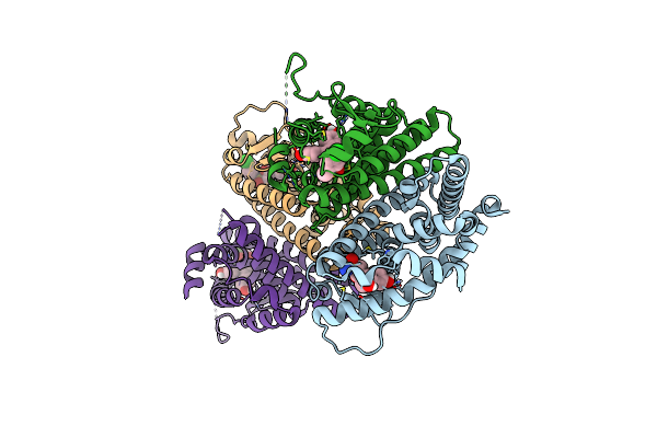
Crystal Structure Of The Er-Alpha Ligand-Binding Domain (L372S, L536S) In Complex With K-406
Organism: Homo sapiens
Method: X-RAY DIFFRACTION Resolution:1.75 Å Release Date: 2024-06-12 Classification: NUCLEAR PROTEIN Ligands: A1AHX, NI |
Organism: Homo sapiens
Method: X-RAY DIFFRACTION
Release Date: 2024-06-12
Ligands: A1AHX, NI
 |
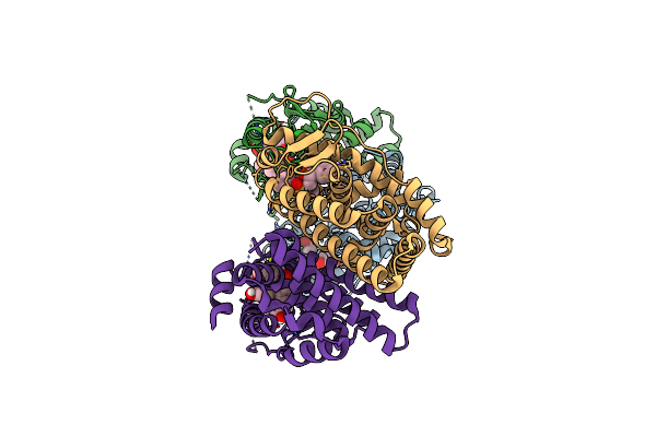
Crystal Structure Of The Er-Alpha Ligand-Binding Domain (L372S, L536S) In Complex With K-1154
Organism: Homo sapiens
Method: X-RAY DIFFRACTION Resolution:1.68 Å Release Date: 2024-06-12 Classification: NUCLEAR PROTEIN Ligands: OBT |
Organism: Homo sapiens
Method: X-RAY DIFFRACTION
Release Date: 2024-06-12
Ligands: OBT
 |
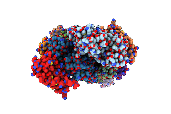
Crystal Structure Of The Er-Alpha Ligand-Binding Domain (L372S, L536S) In Complex With K-402
Organism: Homo sapiens
Method: X-RAY DIFFRACTION Resolution:1.83 Å Release Date: 2024-06-12 Classification: NUCLEAR PROTEIN Ligands: A1AHY, A1AHZ |
Organism: Homo sapiens
Method: X-RAY DIFFRACTION
Release Date: 2024-06-12
Ligands: A1AHY, A1AHZ
 |
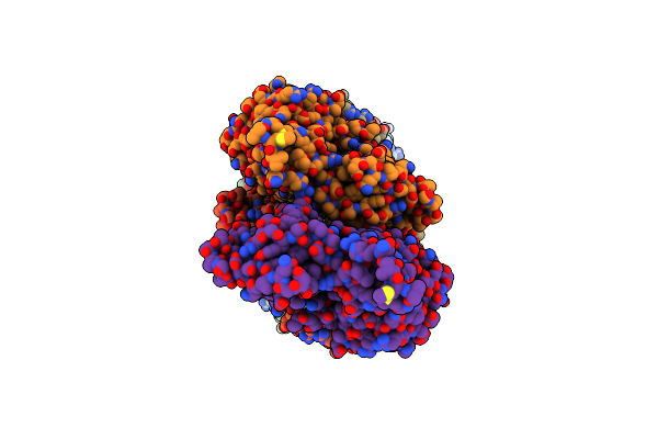
Cryogenic Electron Microscopy 3D Map Of F-Actin Bound By Human Dimeric Alpha-Catenin
Organism: Oryctolagus cuniculus, Homo sapiens
Method: ELECTRON MICROSCOPY Release Date: 2023-03-08 Classification: CELL ADHESION Ligands: ADP, MG |
Organism: Oryctolagus cuniculus, Homo sapiens
Method: ELECTRON MICROSCOPY
Release Date: 2023-03-08
Ligands: ADP, MG
 |
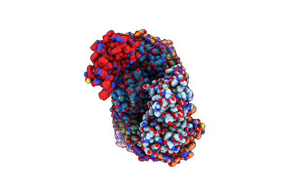
Cryogenic Electron Microscopy 3D Map Of F-Actin
Organism: Oryctolagus cuniculus
Method: ELECTRON MICROSCOPY Release Date: 2023-03-08 Classification: CELL ADHESION Ligands: ADP, MG |
Organism: Oryctolagus cuniculus
Method: ELECTRON MICROSCOPY
Release Date: 2023-03-08
Ligands: ADP, MG
 |
Cryogenic Electron Microscopy 3D Map Of F-Actin Bound By The Actin Binding Domain Of Alpha-Catenin Ortholog, Hmp1
Organism: Oryctolagus cuniculus, Caenorhabditis elegans
Method: ELECTRON MICROSCOPY Release Date: 2023-01-18 Classification: CELL ADHESION Ligands: ADP, MG |
Organism: Oryctolagus cuniculus, Caenorhabditis elegans
Method: ELECTRON MICROSCOPY
Release Date: 2023-01-18
Ligands: ADP, MG