

Search Count: 26
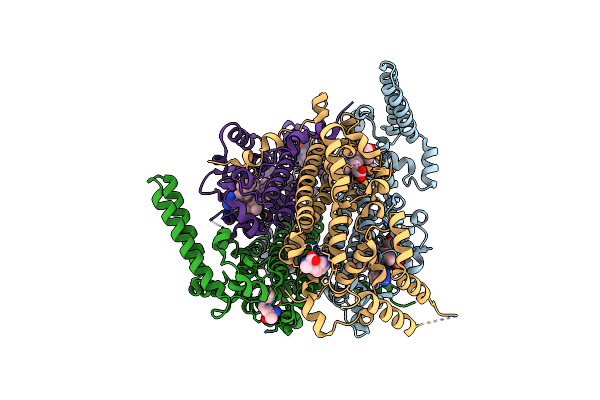 |
Human Tdo (Htdo) In Complex With Lm10
Organism: Homo sapiens
Method: X-RAY DIFFRACTION Resolution:2.29 Å Release Date: 2024-08-28 Classification: CYTOSOLIC PROTEIN Ligands: HEM, ZIQ, A1AER |
Organism: Homo sapiens
Method: X-RAY DIFFRACTION
Release Date: 2024-08-28
Ligands: HEM, ZIQ, A1AER
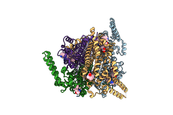 |
Crystal Structure Of Human Tryptophan 2,3-Dioxygenase In Complex With Pan3 Inhibitor
Organism: Homo sapiens
Method: X-RAY DIFFRACTION Resolution:2.25 Å Release Date: 2024-08-28 Classification: OXIDOREDUCTASE/INHIBITOR Ligands: HEM, ZIQ, A1AF5 |
Organism: Homo sapiens
Method: X-RAY DIFFRACTION
Release Date: 2024-08-28
Ligands: HEM, ZIQ, A1AF5
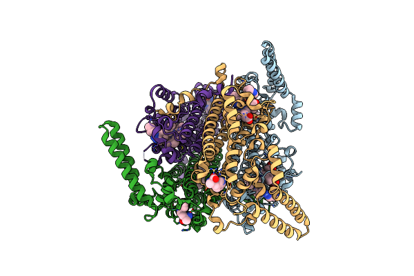 |
Crystal Structure Of Human Tryptophan 2,3-Dioxygenase In Complex With Ppn3 Inhibitor
Organism: Homo sapiens
Method: X-RAY DIFFRACTION Resolution:2.05 Å Release Date: 2024-08-21 Classification: OXIDOREDUCTASE/INHIBITOR Ligands: HEM, ZIQ, A1ADX |
Organism: Homo sapiens
Method: X-RAY DIFFRACTION
Release Date: 2024-08-21
Ligands: HEM, ZIQ, A1ADX
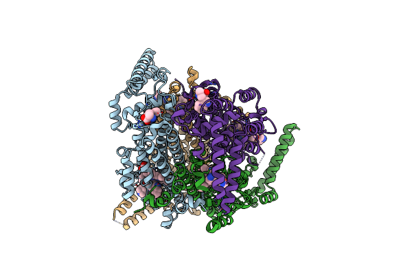 |
Crystal Structure Of Human Tryptophan 2,3-Dioxygenase In Complex With Ppn1 Inhibitor
Organism: Homo sapiens
Method: X-RAY DIFFRACTION Resolution:2.05 Å Release Date: 2024-08-21 Classification: OXIDOREDUCTASE/INHIBITOR Ligands: HEM, ZIQ, A1AD1 |
Organism: Homo sapiens
Method: X-RAY DIFFRACTION
Release Date: 2024-08-21
Ligands: HEM, ZIQ, A1AD1
 |
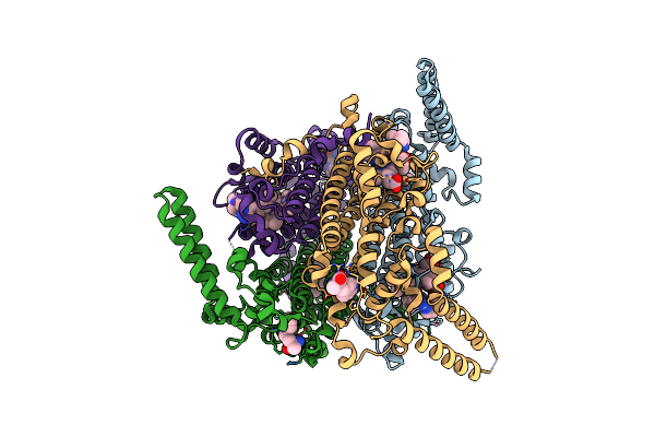
Crystal Structure Of Human Tryptophan 2,3-Dioxygenase In Complex With Pyn3 Inhibitor
Organism: Homo sapiens
Method: X-RAY DIFFRACTION Resolution:2.10 Å Release Date: 2024-08-21 Classification: OXIDOREDUCTASE/INHIBITOR Ligands: HEM, ZIQ, A1AEY |
Organism: Homo sapiens
Method: X-RAY DIFFRACTION
Release Date: 2024-08-21
Ligands: HEM, ZIQ, A1AEY
 |
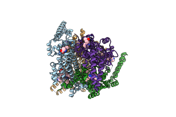
Crystal Structure Of Human Tryptophan 2,3-Dioxygenase In Complex With Pan3F Inhibitor
Organism: Homo sapiens
Method: X-RAY DIFFRACTION Resolution:2.45 Å Release Date: 2024-08-21 Classification: OXIDOREDUCTASE/INHIBITOR Ligands: HEM, ZIQ, A1AE7 |
Organism: Homo sapiens
Method: X-RAY DIFFRACTION
Release Date: 2024-08-21
Ligands: HEM, ZIQ, A1AE7
 |
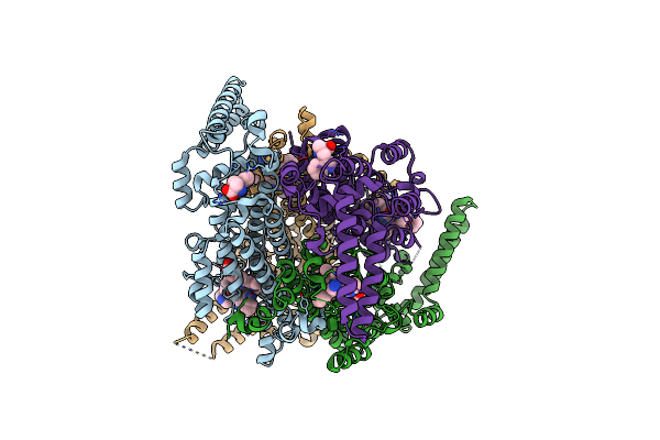
Crystal Structure Of Human Tryptophan 2,3-Dioxygenase In Complex With Pan1 Inhibitor
Organism: Homo sapiens
Method: X-RAY DIFFRACTION Resolution:2.65 Å Release Date: 2024-08-21 Classification: OXIDOREDUCTASE/INHIBITOR Ligands: HEM, A1AH9, ZIQ |
Organism: Homo sapiens
Method: X-RAY DIFFRACTION
Release Date: 2024-08-21
Ligands: HEM, A1AH9, ZIQ
 |
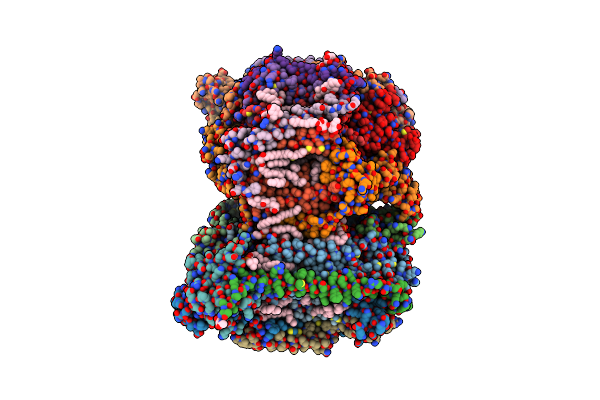
Crystal Structure Of Human Tryptophan 2,3-Dioxygenase In Complex With Pyn1 Inhibitor
Organism: Homo sapiens
Method: X-RAY DIFFRACTION Resolution:2.62 Å Release Date: 2024-08-21 Classification: OXIDOREDUCTASE/INHIBITOR Ligands: HEM, A1AH8, ZIQ |
Organism: Homo sapiens
Method: X-RAY DIFFRACTION
Release Date: 2024-08-21
Ligands: HEM, A1AH8, ZIQ
 |
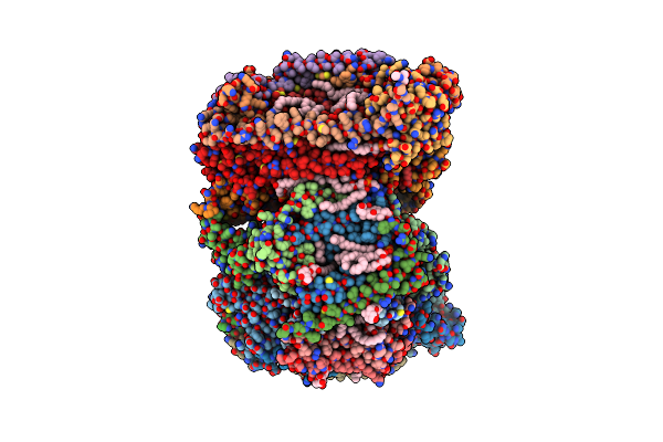
Sfx Structure Of Oxidized Cytochrome C Oxidase At 2.38 Angstrom Resolution
Organism: Bos taurus
Method: X-RAY DIFFRACTION Resolution:2.38 Å Release Date: 2023-09-27 Classification: MEMBRANE PROTEIN Ligands: CU, MG, NA, PGV, HEA, OH, CUA, TGL, PSC, CHD, PEK, CDL, DMU, ZN, SAC |
 |
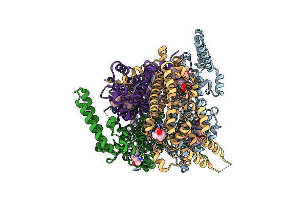
Time-Resolve Sfx Structure Of A Photoproduct Of Carbon Monoxide Complex Of Bovine Cytochrome C Oxidase
Organism: Bos taurus
Method: X-RAY DIFFRACTION Resolution:2.80 Å Release Date: 2023-09-20 Classification: MEMBRANE PROTEIN Ligands: HEA, CU, MG, NA, TGL, PGV, CMO, CUA, CHD, OH, CDL, PSC, ZN, PEK, DMU, SAC |
 |
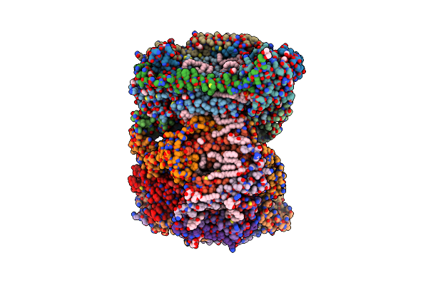
Human Tdo (Htdo) In Complex With Nlg919 Analog
Organism: Homo sapiens
Method: X-RAY DIFFRACTION Resolution:2.30 Å Release Date: 2022-08-24 Classification: OXIDOREDUCTASE Ligands: HEM, ZIQ, 5PK |
Organism: Homo sapiens
Method: X-RAY DIFFRACTION
Release Date: 2022-08-24
Ligands: HEM, ZIQ, 5PK
 |
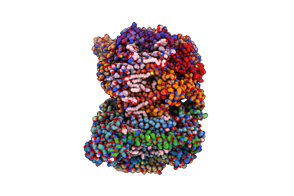
Structure Of Reduced Bovine Cytochrome C Oxidase At 1.93 Angstrom Resolution Obtained By Synchrotron X-Rays
Organism: Bos taurus
Method: X-RAY DIFFRACTION Resolution:1.93 Å Release Date: 2022-03-30 Classification: OXIDOREDUCTASE/MEMBRANE PROTEIN Ligands: CU, MG, NA, PGV, HEA, EDO, TGL, CUA, CHD, DMU, PEK, CDL, PSC, ZN, SAC |
 |
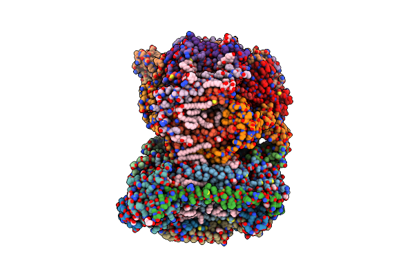
Structure Of Oxidized Bovine Cytochrome C Oxidase At 1.90 Angstrom Resolution Obtained By Synchrotron X-Rays
Organism: Bos taurus
Method: X-RAY DIFFRACTION Resolution:1.90 Å Release Date: 2022-03-30 Classification: OXIDOREDUCTASE/MEMBRANE PROTEIN Ligands: CU, MG, NA, PGV, HEA, EDO, TGL, OH, CUA, PSC, CHD, PEK, CDL, DMU, ZN, SAC |
 |
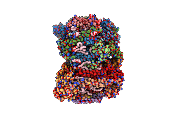
Structure Of Oxidized Bovine Cytochrome C Oxidase With Reduced Metal Centers Induced By Synchrotron X-Ray Exposure
Organism: Bos taurus
Method: X-RAY DIFFRACTION Resolution:2.35 Å Release Date: 2022-03-30 Classification: MEMBRANE PROTEIN Ligands: CU, MG, NA, PGV, HEA, EDO, OH, CUA, TGL, PSC, CHD, CDL, DMU, PEK, ZN, SAC |
 |
Annealed Structure Of Oxidized Bovine Cytochrome C Oxidase With Reduced Metal Centers Induced By Synchrotron X-Ray Exposure
Organism: Bos taurus
Method: X-RAY DIFFRACTION Resolution:2.45 Å Release Date: 2022-03-30 Classification: MEMBRANE PROTEIN Ligands: HEA, CU, MG, NA, PGV, EDO, OH, TGL, CUA, DMU, PEK, CDL, CHD, PSC, ZN, SAC |
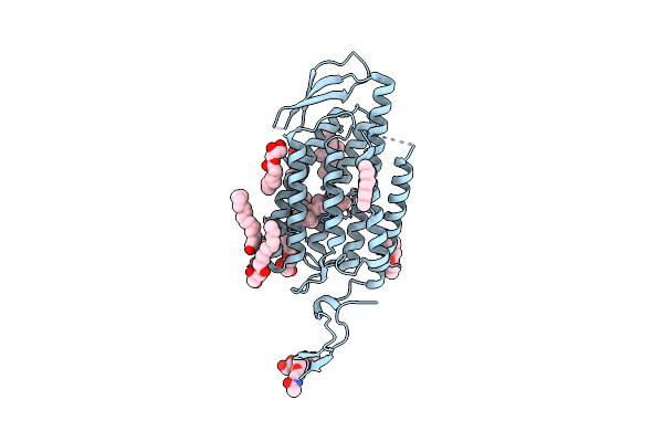 |
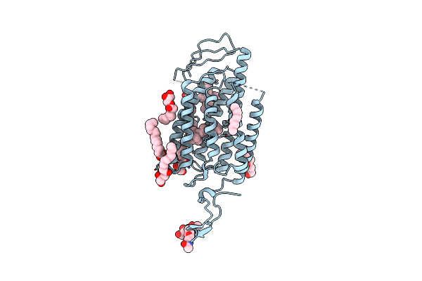
Time-Resolved Serial Femtosecond Crystallography Reveals Early Structural Changes In Channelrhodopsin: Dark State Structure
Organism: Chlamydomonas reinhardtii
Method: X-RAY DIFFRACTION Resolution:2.30 Å Release Date: 2021-04-07 Classification: MEMBRANE PROTEIN Ligands: RET, OLC |
Organism: Chlamydomonas reinhardtii
Method: X-RAY DIFFRACTION
Release Date: 2021-04-07
Ligands: RET, OLC
 |
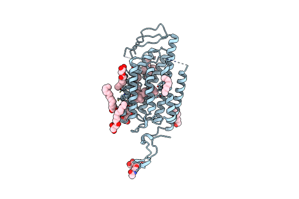
Time-Resolved Serial Femtosecond Crystallography Reveals Early Structural Changes In Channelrhodopsin: 4 Ms Structure
Organism: Chlamydomonas reinhardtii
Method: X-RAY DIFFRACTION Resolution:2.50 Å Release Date: 2021-04-07 Classification: MEMBRANE PROTEIN Ligands: RET, OLC |
Organism: Chlamydomonas reinhardtii
Method: X-RAY DIFFRACTION
Release Date: 2021-04-07
Ligands: RET, OLC
 |
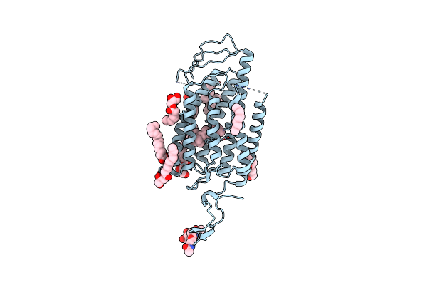
Time-Resolved Serial Femtosecond Crystallography Reveals Early Structural Changes In Channelrhodopsin: 1 Microsecond Structure
Organism: Chlamydomonas reinhardtii
Method: X-RAY DIFFRACTION Resolution:2.50 Å Release Date: 2021-04-07 Classification: MEMBRANE PROTEIN Ligands: RET, OLC |
Organism: Chlamydomonas reinhardtii
Method: X-RAY DIFFRACTION
Release Date: 2021-04-07
Ligands: RET, OLC
 |
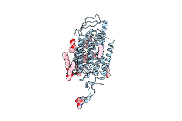
Time-Resolved Serial Femtosecond Crystallography Reveals Early Structural Changes In Channelrhodopsin: 50 Microsecond Structure
Organism: Chlamydomonas reinhardtii
Method: X-RAY DIFFRACTION Resolution:2.50 Å Release Date: 2021-04-07 Classification: MEMBRANE PROTEIN Ligands: RET, OLC |
Organism: Chlamydomonas reinhardtii
Method: X-RAY DIFFRACTION
Release Date: 2021-04-07
Ligands: RET, OLC
 |
Time-Resolved Serial Femtosecond Crystallography Reveals Early Structural Changes In Channelrhodopsin: 250 Microsecond Structure
Organism: Chlamydomonas reinhardtii
Method: X-RAY DIFFRACTION Resolution:2.50 Å Release Date: 2021-04-07 Classification: MEMBRANE PROTEIN Ligands: RET, OLC |
Organism: Chlamydomonas reinhardtii
Method: X-RAY DIFFRACTION
Release Date: 2021-04-07
Ligands: RET, OLC