

Search Count: 21
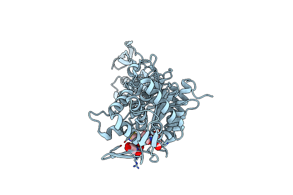 |
Crystal Structure Of Pbp3 In Complex With Compound 14 ((2E)-2-({[(2S)-2-{[(2Z)-2-(2-Amino-1,3-Thiazol-4-Yl)-2-{[(1,5-Dihydroxy-4-Oxo-1,4-Dihydropyridin-2-Yl)Methoxy]Imino}Acetyl]Amino}-3-Oxopropyl]Oxy}Imino)Pentanedioic Acid)
Organism: Pseudomonas aeruginosa pa1r
Method: X-RAY DIFFRACTION Resolution:2.30 Å Release Date: 2014-05-07 Classification: transferase/transferase inhibitor Ligands: 2U2 |
Organism: Pseudomonas aeruginosa pa1r
Method: X-RAY DIFFRACTION
Release Date: 2014-05-07
Ligands: 2U2
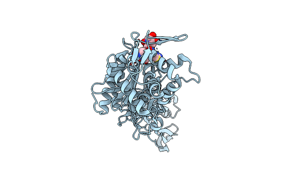 |
Crystal Structure Of Pbp3 In Complex With Bal30072 ((2Z)-2-(2-Amino-1,3-Thiazol-4-Yl)-2-{[(1,5-Dihydroxy-4-Oxo-1,4-Dihydropyridin-2-Yl)Methoxy]Imino}-N-{(2S)-1-Hydroxy-3-Methyl-3-[(Sulfooxy)Amino]Butan-2-Yl}Ethanamide)
Organism: Pseudomonas aeruginosa pa1r
Method: X-RAY DIFFRACTION Resolution:2.00 Å Release Date: 2014-05-07 Classification: transferase/transferase inhibitor Ligands: 2U3 |
Organism: Pseudomonas aeruginosa pa1r
Method: X-RAY DIFFRACTION
Release Date: 2014-05-07
Ligands: 2U3
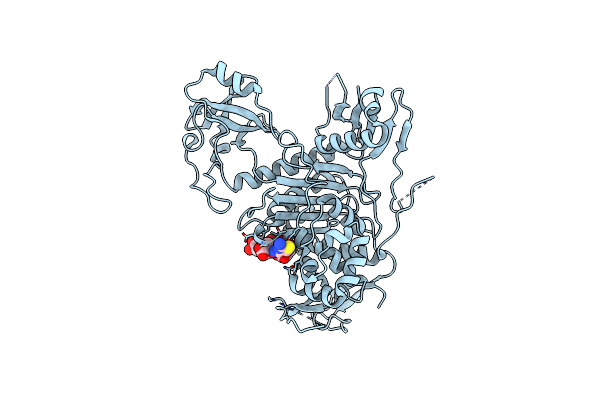 |
Crystal Structure Of Pbp1A In Complex With Compound 17 ((4Z,8S,11E,14S)-5-(2-Amino-1,3-Thiazol-4-Yl)-14-(5,6-Dihydroxy-1,3-Dioxo-1,3-Dihydro-2H-Isoindol-2-Yl)-8-Formyl-2-Methyl-6-Oxo-3,10-Dioxa-4,7,11-Triazatetradeca-4,11-Diene-2,12,14-Tricarboxylic Acid)
Organism: Pseudomonas aeruginosa pao1
Method: X-RAY DIFFRACTION Resolution:3.20 Å Release Date: 2014-05-07 Classification: transferase/transferase inhibitor Ligands: 2U4 |
Organism: Pseudomonas aeruginosa pao1
Method: X-RAY DIFFRACTION
Release Date: 2014-05-07
Ligands: 2U4
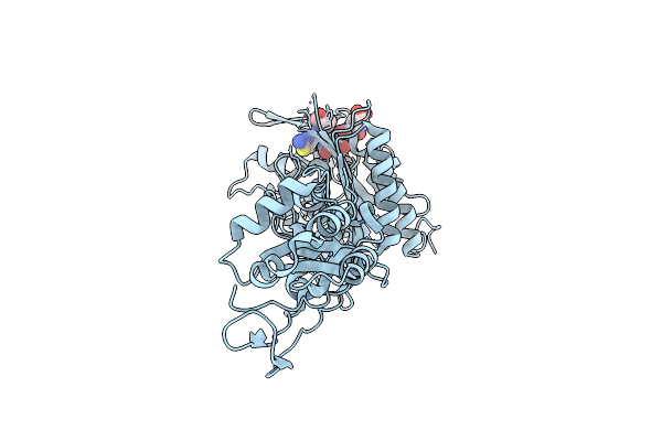 |
Crystal Structure Of P.Aeruginosa Pbp3 In Complex With Compound 4
Organism: Pseudomonas aeruginosa
Method: X-RAY DIFFRACTION Resolution:2.10 Å Release Date: 2013-08-21 Classification: Penicillin Binding Protein/Antibiotic Ligands: PFV |
Organism: Pseudomonas aeruginosa
Method: X-RAY DIFFRACTION
Release Date: 2013-08-21
Ligands: PFV
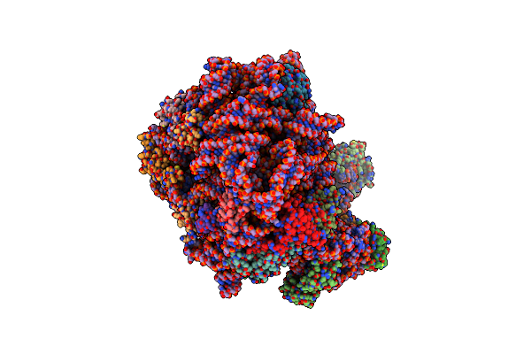 |
Crystal Structure Of Compound 4D Bound To Large Ribosomal Subunit (50S) From Deinococcus Radiodurans
Organism: Deinococcus radiodurans
Method: X-RAY DIFFRACTION Resolution:3.20 Å Release Date: 2013-03-06 Classification: RIBOSOME/RIBOSOME INHIBITOR Ligands: MG, 1F2 |
Organism: Deinococcus radiodurans
Method: X-RAY DIFFRACTION
Release Date: 2013-03-06
Ligands: MG, 1F2
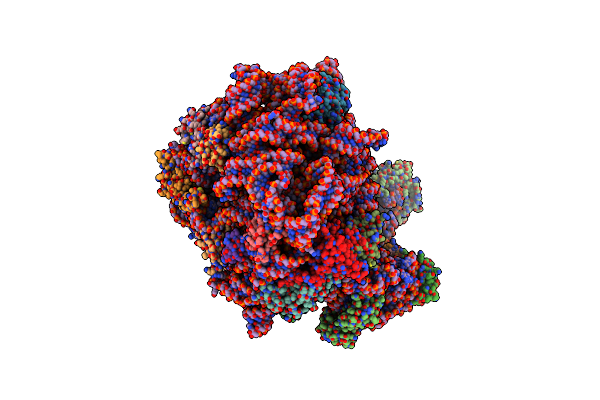 |
Crystal Structure Of Compound 4E Bound To Large Ribosomal Subunit (50S) From Deinococcus Radiodurans
Organism: Deinococcus radiodurans
Method: X-RAY DIFFRACTION Resolution:3.20 Å Release Date: 2013-03-06 Classification: RIBOSOME/RIBOSOME INHIBITOR Ligands: MG, 1F3 |
Organism: Deinococcus radiodurans
Method: X-RAY DIFFRACTION
Release Date: 2013-03-06
Ligands: MG, 1F3
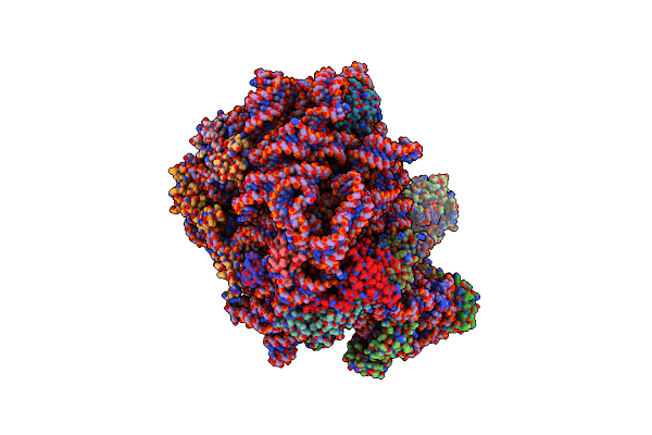 |
Crystal Structure Of Compound 4F Bound To Large Ribosomal Subunit (50S) From Deinococcus Radiodurans
Organism: Deinococcus radiodurans
Method: X-RAY DIFFRACTION Resolution:3.60 Å Release Date: 2013-03-06 Classification: RIBOSOME/RIBOSOME INHIBITOR Ligands: MG, 1F4 |
Organism: Deinococcus radiodurans
Method: X-RAY DIFFRACTION
Release Date: 2013-03-06
Ligands: MG, 1F4
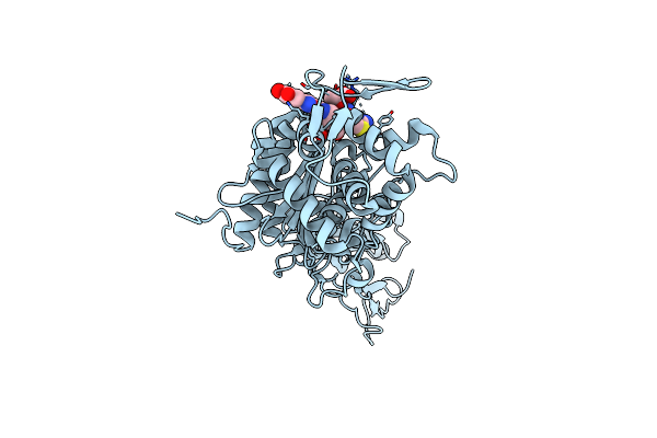 |
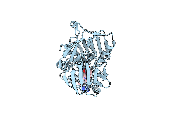
Crystal Structure Of Pseudomonas Aeruginosa Pbp3 Complexed With Compound 14
Organism: Pseudomonas aeruginosa
Method: X-RAY DIFFRACTION Resolution:2.20 Å Release Date: 2012-10-17 Classification: PENICILLIN-BINDING PROTEIN/ANTIBIOTIC Ligands: 0W0 |
Organism: Pseudomonas aeruginosa
Method: X-RAY DIFFRACTION
Release Date: 2012-10-17
Ligands: 0W0
 |
Udp-3-O-[3-Hydroxymyristoyl] N-Acetylglucosamine Deacetylase In Complex With Inhibitor
Organism: Pseudomonas aeruginosa
Method: X-RAY DIFFRACTION Resolution:1.26 Å Release Date: 2012-02-01 Classification: Hydrolase/Hydrolase inhibitor Ligands: ZN, RFN, GAI, EDO |
Organism: Pseudomonas aeruginosa
Method: X-RAY DIFFRACTION
Release Date: 2012-02-01
Ligands: ZN, RFN, GAI, EDO
 |
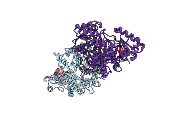
Potent Inhibitors Of Lpxc For The Treatment Of Gram-Negative Infections
Organism: Pseudomonas aeruginosa
Method: X-RAY DIFFRACTION Resolution:2.00 Å Release Date: 2012-01-04 Classification: Hydrolase/Hydrolase inhibitor Ligands: ZN, 03I |
Organism: Pseudomonas aeruginosa
Method: X-RAY DIFFRACTION
Release Date: 2012-01-04
Ligands: ZN, 03I
 |
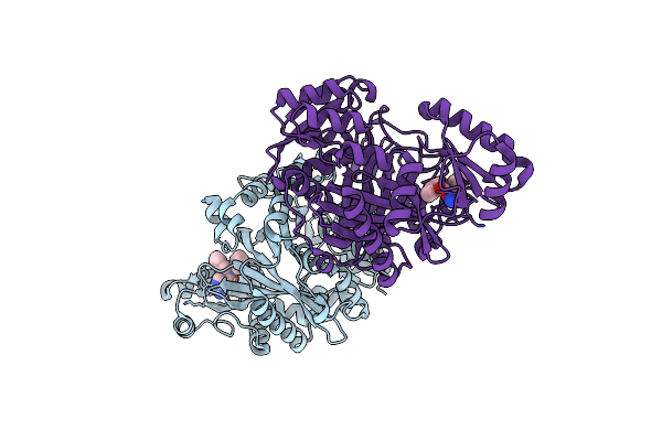
Crystal Structure Of Biotin Carboxylase From E. Coli In Complex With Amino-Oxazole Fragment Series
Organism: Escherichia coli
Method: X-RAY DIFFRACTION Resolution:2.00 Å Release Date: 2009-05-19 Classification: LIGASE Ligands: OA1, CL |
Organism: Escherichia coli
Method: X-RAY DIFFRACTION
Release Date: 2009-05-19
Ligands: OA1, CL
 |
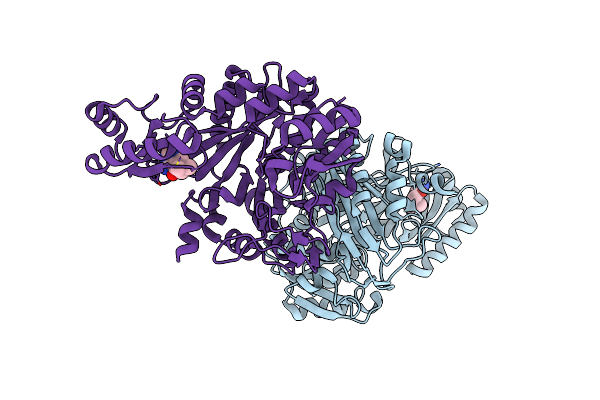
Crystal Structure Of Biotin Carboxylase From E. Coli In Complex With Amino-Oxazole Fragment Series
Organism: Escherichia coli
Method: X-RAY DIFFRACTION Resolution:1.87 Å Release Date: 2009-05-19 Classification: LIGASE Ligands: OA2, CL |
Organism: Escherichia coli
Method: X-RAY DIFFRACTION
Release Date: 2009-05-19
Ligands: OA2, CL
 |
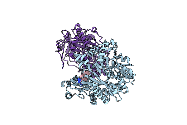
Crystal Structure Of Biotin Carboxylase From E. Coli In Complex With 4-Amino-7,7-Dimethyl-7,8-Dihydro-Quinazolinone Fragment
Organism: Escherichia coli
Method: X-RAY DIFFRACTION Resolution:2.50 Å Release Date: 2009-05-19 Classification: LIGASE Ligands: OA3, CL |
Organism: Escherichia coli
Method: X-RAY DIFFRACTION
Release Date: 2009-05-19
Ligands: OA3, CL
 |
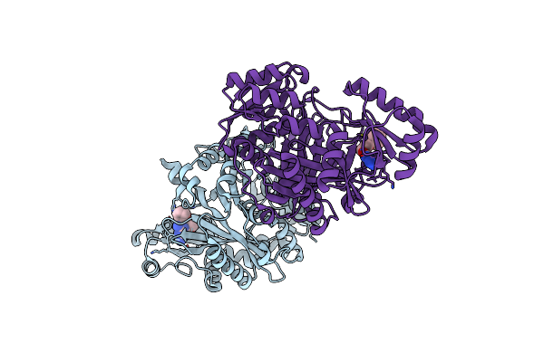
Crystal Structure Of Biotin Carboxylase From E. Coli In Complex With 5-Methyl-6-Phenyl-Quinazoline-2,4-Diamine
Organism: Escherichia coli
Method: X-RAY DIFFRACTION Resolution:1.85 Å Release Date: 2009-05-19 Classification: LIGASE Ligands: OA4 |
Organism: Escherichia coli
Method: X-RAY DIFFRACTION
Release Date: 2009-05-19
Ligands: OA4
 |
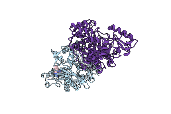
Crystal Structure Of Biotin Carboxylase From E. Coli In Complex With The Triazine-2,4-Diamine Fragment
Organism: Escherichia coli
Method: X-RAY DIFFRACTION Resolution:2.05 Å Release Date: 2009-05-19 Classification: LIGASE Ligands: OA5, CL |
Organism: Escherichia coli
Method: X-RAY DIFFRACTION
Release Date: 2009-05-19
Ligands: OA5, CL
 |
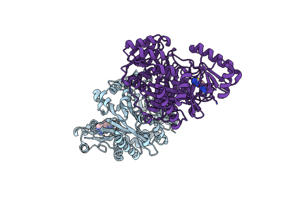
Crystal Structure Of Biotin Carboxylase From E. Coli In Complex With The 3-(3-Methyl-But-2-Enyl)-3H-Purin-6-Ylamine Fragment
Organism: Escherichia coli
Method: X-RAY DIFFRACTION Resolution:1.90 Å Release Date: 2009-05-19 Classification: LIGASE Ligands: L21, CL |
Organism: Escherichia coli
Method: X-RAY DIFFRACTION
Release Date: 2009-05-19
Ligands: L21, CL
 |
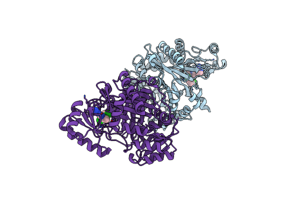
Crystal Structure Of Biotin Carboxylase From E. Coli In Complex With The Amino-Thiazole-Pyrimidine Fragment
Organism: Escherichia coli
Method: X-RAY DIFFRACTION Resolution:1.77 Å Release Date: 2009-05-19 Classification: LIGASE Ligands: L22, CL |
Organism: Escherichia coli
Method: X-RAY DIFFRACTION
Release Date: 2009-05-19
Ligands: L22, CL
 |
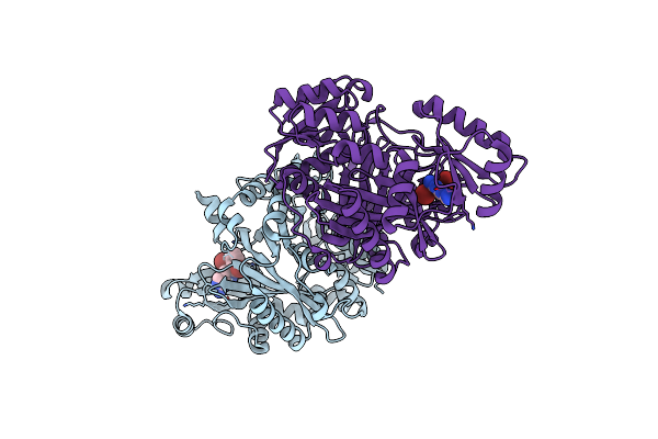
Crystal Structure Of Biotin Carboxylase From E. Coli In Complex With The Imidazole-Pyrimidine Inhibitor
Organism: Escherichia coli
Method: X-RAY DIFFRACTION Resolution:1.99 Å Release Date: 2009-05-19 Classification: LIGASE Ligands: CL, L23 |
Organism: Escherichia coli
Method: X-RAY DIFFRACTION
Release Date: 2009-05-19
Ligands: CL, L23
 |
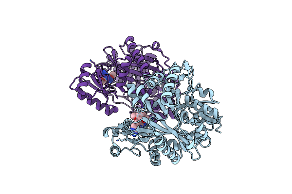
Crystal Structure Of Biotin Carboxylase From E.Coli In Complex With Potent Inhibitor 1
Organism: Escherichia coli
Method: X-RAY DIFFRACTION Resolution:2.10 Å Release Date: 2009-01-13 Classification: LIGASE Ligands: LZJ, CL |
Organism: Escherichia coli
Method: X-RAY DIFFRACTION
Release Date: 2009-01-13
Ligands: LZJ, CL
 |
Crystal Structure Of Biotin Carboxylase From E.Coli In Complex With Potent Inhibitor 2
Organism: Escherichia coli
Method: X-RAY DIFFRACTION Resolution:2.40 Å Release Date: 2009-01-13 Classification: LIGASE Ligands: LZK |
Organism: Escherichia coli
Method: X-RAY DIFFRACTION
Release Date: 2009-01-13
Ligands: LZK