

Search Count: 10
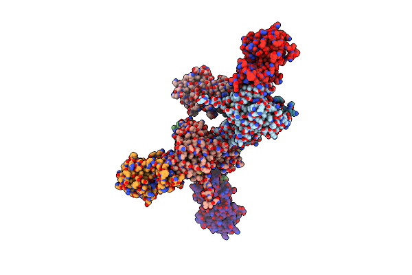 |
Exoenzyme S From Pseudomonas Aeruginosa In Complex With Human 14-3-3 Protein Beta, Tetrameric Crystal Form
Organism: Homo sapiens, Pseudomonas aeruginosa
Method: X-RAY DIFFRACTION Resolution:3.24 Å Release Date: 2018-09-26 Classification: TOXIN |
Organism: Homo sapiens, Pseudomonas aeruginosa
Method: X-RAY DIFFRACTION
Release Date: 2018-09-26
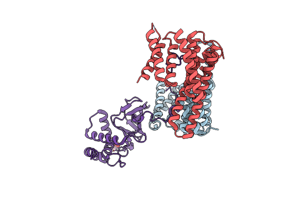 |
Exoenzyme S From Pseudomonas Aeruginosa In Complex With Human 14-3-3 Protein Beta, Trimeric Crystal Form
Organism: Homo sapiens, Pseudomonas aeruginosa
Method: X-RAY DIFFRACTION Resolution:2.34 Å Release Date: 2018-09-26 Classification: TOXIN Ligands: MPD |
Organism: Homo sapiens, Pseudomonas aeruginosa
Method: X-RAY DIFFRACTION
Release Date: 2018-09-26
Ligands: MPD
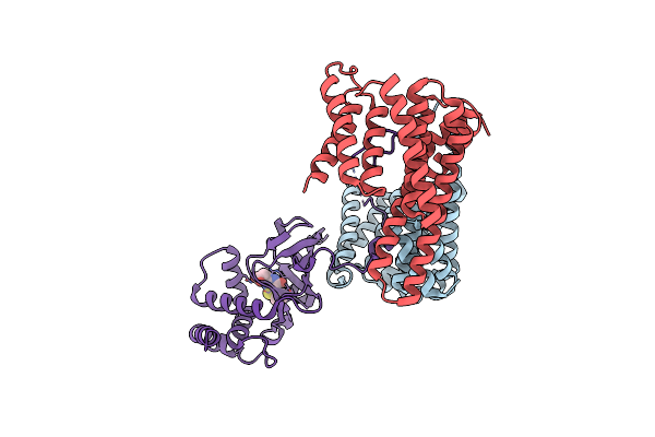 |
Exoenzyme S From Pseudomonas Aeruginosa In Complex With Human 14-3-3 Protein Beta, Trimeric Crystal Form In Complex With Sto1101
Organism: Homo sapiens, Pseudomonas aeruginosa
Method: X-RAY DIFFRACTION Resolution:3.24 Å Release Date: 2018-09-26 Classification: TOXIN Ligands: F4W |
Organism: Homo sapiens, Pseudomonas aeruginosa
Method: X-RAY DIFFRACTION
Release Date: 2018-09-26
Ligands: F4W
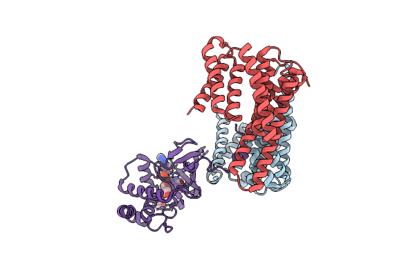 |
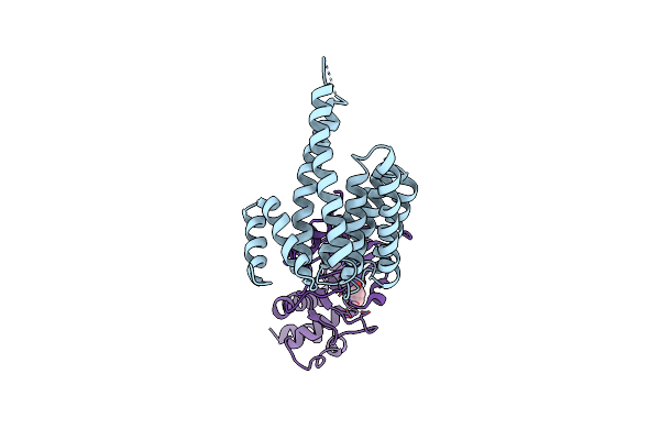
Exoenzyme S From Pseudomonas Aeruginosa In Complex With Human 14-3-3 Protein Beta, Trimeric Crystal Form Bound To Carba-Nad
Organism: Homo sapiens, Pseudomonas aeruginosa
Method: X-RAY DIFFRACTION Resolution:2.55 Å Release Date: 2018-09-26 Classification: TOXIN Ligands: CNA, MPD |
Organism: Homo sapiens, Pseudomonas aeruginosa
Method: X-RAY DIFFRACTION
Release Date: 2018-09-26
Ligands: CNA, MPD
 |
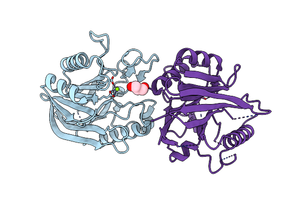
Exoenzyme T From Pseudomonas Aeruginosa In Complex With Human 14-3-3 Protein Beta, Tetrameric Crystal Form Bound To Sto1101
Organism: Homo sapiens, pseudomonas aeruginosa pao1
Method: X-RAY DIFFRACTION Resolution:3.79 Å Release Date: 2018-09-26 Classification: TOXIN Ligands: F4W |
Organism: Homo sapiens, pseudomonas aeruginosa pao1
Method: X-RAY DIFFRACTION
Release Date: 2018-09-26
Ligands: F4W
 |
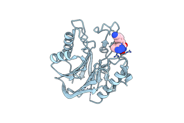
Crystal Structure Of The Catalytic Domain Of Human Tyrosyl Dna Phosphodiesterase 2
Organism: Homo sapiens
Method: X-RAY DIFFRACTION Resolution:3.10 Å Release Date: 2016-05-04 Classification: HYDROLASE Ligands: MG, GOL |
Organism: Homo sapiens
Method: X-RAY DIFFRACTION
Release Date: 2016-05-04
Ligands: MG, GOL
 |
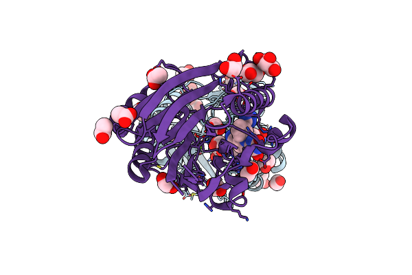
Crystal Structure Of The Catalytic Domain Of Human Tyrosyl Dna Phosphodiesterase 2 In Complex With A Small Molecule Inhibitor
Organism: Homo sapiens
Method: X-RAY DIFFRACTION Resolution:3.40 Å Release Date: 2016-05-04 Classification: HYDROLASE Ligands: 6FQ |
Organism: Homo sapiens
Method: X-RAY DIFFRACTION
Release Date: 2016-05-04
Ligands: 6FQ
 |
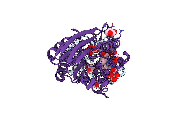
Crystal Structure Of M2Htdp2-Cat In Complex With A Small Molecule Inhibitor
Organism: Mus musculus
Method: X-RAY DIFFRACTION Resolution:1.80 Å Release Date: 2016-05-04 Classification: HYDROLASE Ligands: 6FQ, MG, GOL, EDO, ACT |
Organism: Mus musculus
Method: X-RAY DIFFRACTION
Release Date: 2016-05-04
Ligands: 6FQ, MG, GOL, EDO, ACT
 |
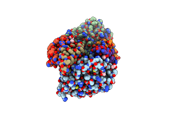
Crystal Structure Of M2Htdp2-Cat In Complex With A Small Molecule Inhibitor
Organism: Mus musculus
Method: X-RAY DIFFRACTION Resolution:1.70 Å Release Date: 2016-05-04 Classification: HYDROLASE Ligands: 6FV, TLA, GOL, EDO, MN |
Organism: Mus musculus
Method: X-RAY DIFFRACTION
Release Date: 2016-05-04
Ligands: 6FV, TLA, GOL, EDO, MN
 |
Crystallographic Structure Of Hhv-1 Uracil-Dna Glycosylase Complexed With The Bacillus Phage Pza Inhibitor Protein P56
Organism: Herpes simplex virus type 1, Bacillus phage pza
Method: X-RAY DIFFRACTION Resolution:2.16 Å Release Date: 2013-08-07 Classification: HYDROLASE/HYDROLASE INHIBITOR Ligands: ACT |
Organism: Herpes simplex virus type 1, Bacillus phage pza
Method: X-RAY DIFFRACTION
Release Date: 2013-08-07
Ligands: ACT