

Search Count: 16
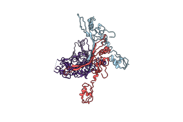 |
Cryo-Em Structure Of The Anti-Feeding Prophage (Afp) Baseplate In Extended State, 3-Fold Symmetrised
Organism: Serratia entomophila
Method: ELECTRON MICROSCOPY Release Date: 2019-04-24 Classification: VIRUS LIKE PARTICLE |
Organism: Serratia entomophila
Method: ELECTRON MICROSCOPY
Release Date: 2019-04-24
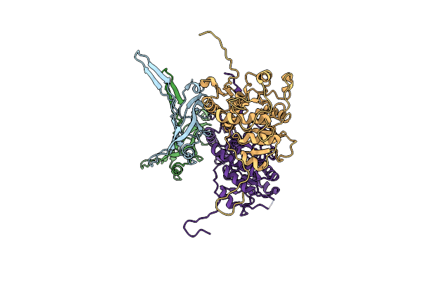 |
Cryo-Em Structure Of The Anti-Feeding Prophage (Afp) Helical Sheath-Tube Complex In Extended State
Organism: Serratia entomophila
Method: ELECTRON MICROSCOPY Release Date: 2019-04-24 Classification: VIRUS LIKE PARTICLE |
Organism: Serratia entomophila
Method: ELECTRON MICROSCOPY
Release Date: 2019-04-24
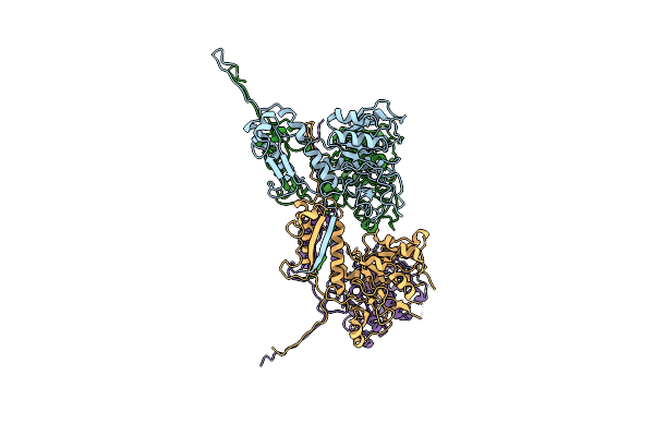 |
Cryo-Em Structure Of The Anti-Feeding Prophage (Afp) Baseplate In Contracted State
Organism: Serratia entomophila
Method: ELECTRON MICROSCOPY Release Date: 2019-04-24 Classification: VIRUS LIKE PARTICLE |
Organism: Serratia entomophila
Method: ELECTRON MICROSCOPY
Release Date: 2019-04-24
 |
Cryo-Em Structure Of The Anti-Feeding Prophage (Afp) Baseplate, 6-Fold Symmetrised
Organism: Serratia entomophila
Method: ELECTRON MICROSCOPY Release Date: 2019-04-17 Classification: VIRUS LIKE PARTICLE |
Organism: Serratia entomophila
Method: ELECTRON MICROSCOPY
Release Date: 2019-04-17
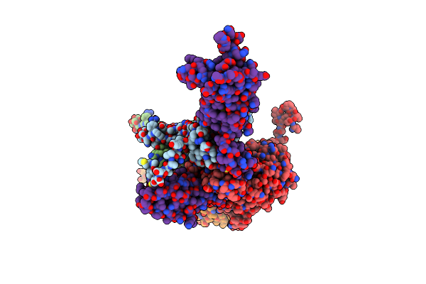 |
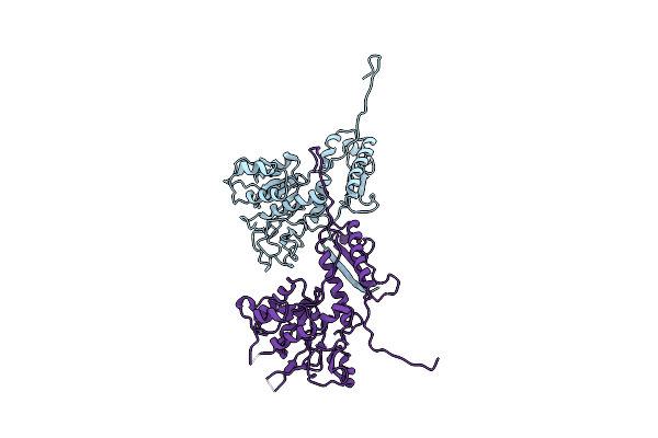
Cryo-Em Structure Of The Anti-Feeding Prophage Cap (Afp Tube Terminating Cap)
Organism: Serratia entomophila
Method: ELECTRON MICROSCOPY Release Date: 2019-04-17 Classification: VIRUS LIKE PARTICLE |
Organism: Serratia entomophila
Method: ELECTRON MICROSCOPY
Release Date: 2019-04-17
 |
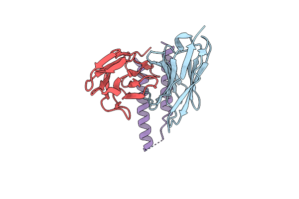
Cryo-Em Structure Of The Anti-Feeding Prophage (Afp) Helical Sheath In Contracted State
Organism: Serratia entomophila
Method: ELECTRON MICROSCOPY Release Date: 2019-04-17 Classification: VIRUS LIKE PARTICLE |
Organism: Serratia entomophila
Method: ELECTRON MICROSCOPY
Release Date: 2019-04-17
 |
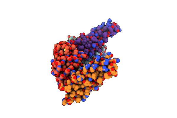
Hiv-1 Rev Ntd Dimers With Variable Crossing Angles
Organism: Oryctolagus cuniculus, Human immunodeficiency virus 1
Method: X-RAY DIFFRACTION Resolution:4.30 Å Release Date: 2016-06-29 Classification: IMMUNE SYSTEM |
Organism: Oryctolagus cuniculus, Human immunodeficiency virus 1
Method: X-RAY DIFFRACTION
Release Date: 2016-06-29
 |
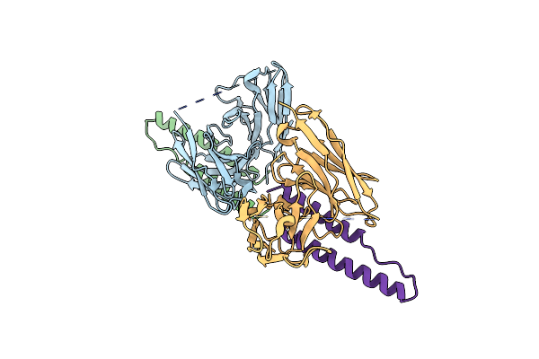
Hiv-1 Rev Ntd Dimers With Variable Crossing Angles
Organism: Oryctolagus cuniculus, Human immunodeficiency virus 1
Method: X-RAY DIFFRACTION Resolution:2.30 Å Release Date: 2016-06-22 Classification: IMMUNE SYSTEM Ligands: CL |
Organism: Oryctolagus cuniculus, Human immunodeficiency virus 1
Method: X-RAY DIFFRACTION
Release Date: 2016-06-22
Ligands: CL
 |
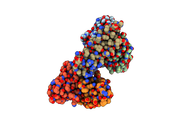
Hiv-1 Rev Ntd Dimers With Variable Crossing Angles
Organism: Oryctolagus cuniculus, Human immunodeficiency virus 1
Method: X-RAY DIFFRACTION Resolution:2.90 Å Release Date: 2016-06-22 Classification: IMMUNE SYSTEM |
Organism: Oryctolagus cuniculus, Human immunodeficiency virus 1
Method: X-RAY DIFFRACTION
Release Date: 2016-06-22
 |
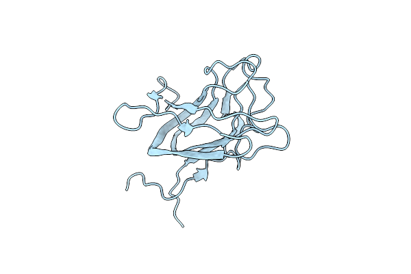
Hiv-1 Rev Ntd Dimers With Variable Crossing Angles
Organism: Oryctolagus cuniculus, Human immunodeficiency virus 1
Method: X-RAY DIFFRACTION Resolution:3.10 Å Release Date: 2016-06-22 Classification: IMMUNE SYSTEM |
Organism: Oryctolagus cuniculus, Human immunodeficiency virus 1
Method: X-RAY DIFFRACTION
Release Date: 2016-06-22
 |
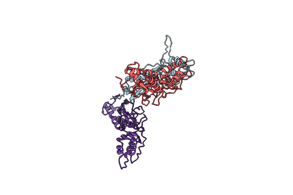
Double Octamer Structure Of Retinoschisin, A Cell-Cell Adhesion Protein Of The Retina
Organism: Homo sapiens
Method: ELECTRON MICROSCOPY Release Date: 2016-05-11 Classification: CELL ADHESION |
Organism: Homo sapiens
Method: ELECTRON MICROSCOPY
Release Date: 2016-05-11
 |
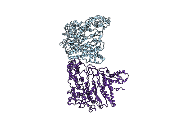
Myxococcus Xanthus Encapsulin Protein (Enca)
Organism: Myxococcus xanthus
Method: ELECTRON MICROSCOPY Resolution:4.60 Å Release Date: 2014-07-30 Classification: VIRUS LIKE PARTICLE |
Organism: Myxococcus xanthus
Method: ELECTRON MICROSCOPY
Release Date: 2014-07-30
 |
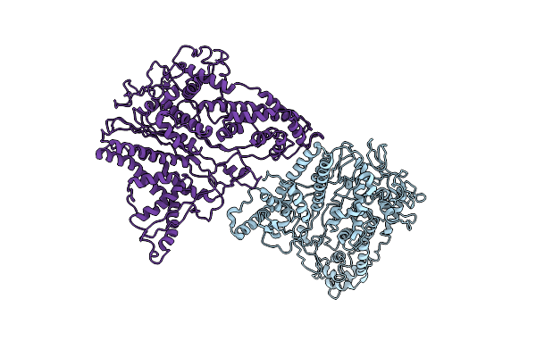
Coordinates Of The Bacteriophage Phi6 Capsid Subunits Fitted Into The Cryoem Map Emd-1206
Organism: Pseudomonas phage phi6
Method: ELECTRON MICROSCOPY Resolution:7.50 Å Release Date: 2013-12-11 Classification: VIRAL PROTEIN |
Organism: Pseudomonas phage phi6
Method: ELECTRON MICROSCOPY
Release Date: 2013-12-11
 |
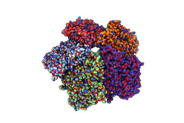
Coordinates Of The Bacteriophage Phi6 Capsid Subunits (P1A And P1B) Fitted Into The Cryoem Reconstruction Of The Procapsid At 4.4 A Resolution
Organism: Pseudomonas phage phi6
Method: ELECTRON MICROSCOPY Resolution:4.40 Å Release Date: 2013-08-14 Classification: VIRUS |
Organism: Pseudomonas phage phi6
Method: ELECTRON MICROSCOPY
Release Date: 2013-08-14
 |
Major Capsid Protein P1 Of The Pseudomonas Phage Phi6
Organism: Pseudomonas phage phi6
Method: X-RAY DIFFRACTION Resolution:3.60 Å Release Date: 2013-08-14 Classification: VIRAL PROTEIN |
Organism: Pseudomonas phage phi6
Method: X-RAY DIFFRACTION
Release Date: 2013-08-14
 |
Structure Of Aquaporin-1 At 3.8 A Resolution By Electron Crystallography
Organism: Homo sapiens
Method: ELECTRON CRYSTALLOGRAPHY Resolution:3.80 Å Release Date: 2000-10-18 Classification: MEMBRANE PROTEIN |
Organism: Homo sapiens
Method: ELECTRON CRYSTALLOGRAPHY
Release Date: 2000-10-18