

Search Count: 65
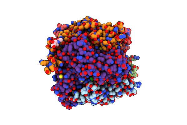 |
Crystal Structure Of Achbp From Aplysia Californica Complex With 2-Aminopyrimidine At Ph 8.0 Spacegroup P21
Organism: Aplysia californica
Method: X-RAY DIFFRACTION Resolution:2.40 Å Release Date: 2017-11-15 Classification: METAL BINDING PROTEIN Ligands: 7KO, DMS, NAG |
Organism: Aplysia californica
Method: X-RAY DIFFRACTION
Release Date: 2017-11-15
Ligands: 7KO, DMS, NAG
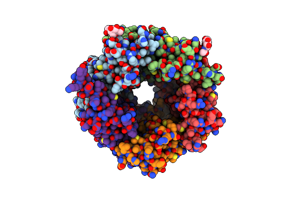 |
Crystal Structure Of Achbp From Aplysia Californica Complex With 2-Aminopyrimidine At Ph 7.0 Spacegroup P212121
Organism: Aplysia californica
Method: X-RAY DIFFRACTION Resolution:2.29 Å Release Date: 2017-10-25 Classification: METAL BINDING PROTEIN Ligands: 7KO, NAG |
Organism: Aplysia californica
Method: X-RAY DIFFRACTION
Release Date: 2017-10-25
Ligands: 7KO, NAG
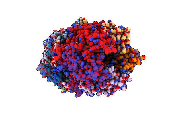 |
X-Ray Crystal Structure Of Acetylcholine Binding Protein (Achbp) In Complex With N4,N4-Bis[(Pyridin-2-Yl)Methyl]-6-(Thiophen-3-Yl)Pyrimidine-2,4-Diamine
Organism: Lymnaea stagnalis
Method: X-RAY DIFFRACTION Resolution:2.04 Å Release Date: 2017-03-08 Classification: ACETYLCHOLINE-BINDING PROTEIN Ligands: PO4, NAG, 6GH, GOL, DMS |
Organism: Lymnaea stagnalis
Method: X-RAY DIFFRACTION
Release Date: 2017-03-08
Ligands: PO4, NAG, 6GH, GOL, DMS
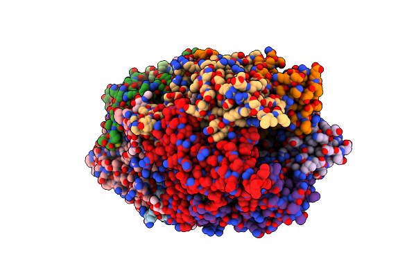 |
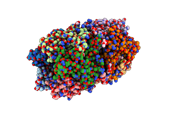
X-Ray Crystal Structure Of Acetylcholine Binding Protein (Achbp) In Complex With 6-(4-Methoxyphenyl)-N4,N4-Bis[(Pyridin-2-Yl)Methyl]Pyrimidine-2,4-Diamine
Organism: Lymnaea stagnalis
Method: X-RAY DIFFRACTION Resolution:2.04 Å Release Date: 2017-03-08 Classification: ACETYLCHOLINE-BINDING PROTEIN Ligands: PO4, NAG, 6GF |
Organism: Lymnaea stagnalis
Method: X-RAY DIFFRACTION
Release Date: 2017-03-08
Ligands: PO4, NAG, 6GF
 |
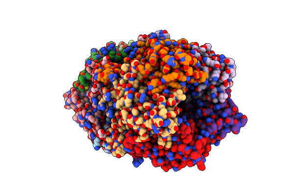
X-Ray Structure Of Acetylcholine Binding Protein (Achbp) In Complex With 6-(2-Methoxyphenyl)-N4,N4-Bis[(Pyridin-2-Yl)Methyl]Pyrimidine-2,4-Diamine
Organism: Lymnaea stagnalis
Method: X-RAY DIFFRACTION Resolution:2.70 Å Release Date: 2017-03-08 Classification: ACETYLCHOLINE-BINDING PROTEIN Ligands: 6GK, NAG, PO4 |
Organism: Lymnaea stagnalis
Method: X-RAY DIFFRACTION
Release Date: 2017-03-08
Ligands: 6GK, NAG, PO4
 |
X-Ray Crystal Structure Of Acetylcholine Binding Protein (Achbp) In Complex With 4-(2-Amino-6-{Bis[(Pyridin-2-Yl)Methyl]Amino}Pyrimidin-4-Yl)Phenol
Organism: Lymnaea stagnalis
Method: X-RAY DIFFRACTION Resolution:2.33 Å Release Date: 2017-03-08 Classification: ACETYLCHOLINE-BINDING PROTEIN, AChBP Ligands: NAG, 6GM, PO4 |
Organism: Lymnaea stagnalis
Method: X-RAY DIFFRACTION
Release Date: 2017-03-08
Ligands: NAG, 6GM, PO4
 |
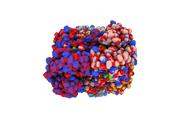
Crystal Structure Of Aminotransferase Atms13 From Actinomadura Melliaura
Organism: Actinomadura melliaura
Method: X-RAY DIFFRACTION Resolution:1.50 Å Release Date: 2015-06-03 Classification: TRANSFERASE Ligands: GOL |
Organism: Actinomadura melliaura
Method: X-RAY DIFFRACTION
Release Date: 2015-06-03
Ligands: GOL
 |
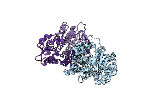
Ensemble Refinement Of Cystathione Gamma Lyase (Cale6) D7G From Micromonospora Echinospora
Organism: Micromonospora echinospora
Method: X-RAY DIFFRACTION Resolution:2.10 Å Release Date: 2015-04-15 Classification: LYASE Ligands: MES, GOL, CL, FMT |
Organism: Micromonospora echinospora
Method: X-RAY DIFFRACTION
Release Date: 2015-04-15
Ligands: MES, GOL, CL, FMT
 |
Crystal Structure Of Cals8 From Micromonospora Echinospora Cocrystallized With Nad And Tdp-Glucose
Organism: Micromonospora echinospora
Method: X-RAY DIFFRACTION Resolution:1.95 Å Release Date: 2015-02-11 Classification: OXIDOREDUCTASE Ligands: NAD, NA, TMP, GOL |
Organism: Micromonospora echinospora
Method: X-RAY DIFFRACTION
Release Date: 2015-02-11
Ligands: NAD, NA, TMP, GOL
 |
Crystal Structure Of Cals8 From Micromonospora Echinospora (P294S Mutant)
Organism: Micromonospora echinospora
Method: X-RAY DIFFRACTION Resolution:2.55 Å Release Date: 2015-02-11 Classification: OXIDOREDUCTASE Ligands: GOL |
Organism: Micromonospora echinospora
Method: X-RAY DIFFRACTION
Release Date: 2015-02-11
Ligands: GOL
 |
Crystal Structure Of Ats13 From Actinomadura Melliaura
Organism: Actinomadura melliaura
Method: X-RAY DIFFRACTION Resolution:3.00 Å Release Date: 2014-12-24 Classification: TRANSFERASE Ligands: PLP |
Organism: Actinomadura melliaura
Method: X-RAY DIFFRACTION
Release Date: 2014-12-24
Ligands: PLP
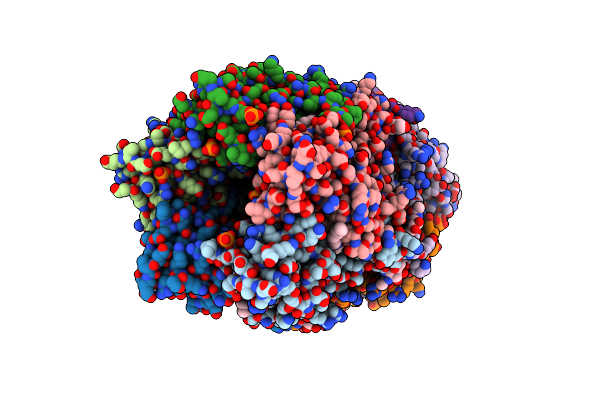 |
X-Ray Structure Of Acetylcholine Binding Protein (Achbp) In Complex With 6-(4-Methoxyphenyl)-N4-Octylpyrimidine-2,4-Diamine
Organism: Lymnaea stagnalis
Method: X-RAY DIFFRACTION Resolution:2.70 Å Release Date: 2014-07-16 Classification: Acetylcholine-Binding Protein Ligands: KK1, NAG, PO4 |
Organism: Lymnaea stagnalis
Method: X-RAY DIFFRACTION
Release Date: 2014-07-16
Ligands: KK1, NAG, PO4
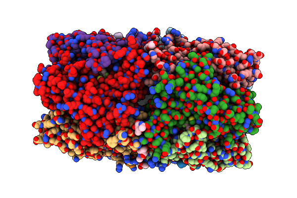 |
X-Ray Structure Of Acetylcholine Binding Protein (Achbp) In Complex With 4-(Morpholin-4-Yl)-6-[4-(Trifluoromethyl)Phenyl]Pyrimidin-2-Amine
Organism: Lymnaea stagnalis
Method: X-RAY DIFFRACTION Resolution:2.98 Å Release Date: 2014-07-16 Classification: Acetylcholine-Binding Protein Ligands: KK2, NAG, PO4 |
Organism: Lymnaea stagnalis
Method: X-RAY DIFFRACTION
Release Date: 2014-07-16
Ligands: KK2, NAG, PO4
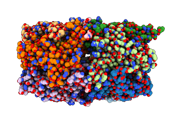 |
X-Ray Structure Of Acetylcholine Binding Protein (Achbp) In Complex With 4-(4-Methylpiperidin-1-Yl)-6-(4-(Trifluoromethyl)Phenyl)Pyrimidin-2-Amine
Organism: Lymnaea stagnalis
Method: X-RAY DIFFRACTION Resolution:2.10 Å Release Date: 2014-07-16 Classification: Acetylcholine-Binding Protein Ligands: PO4, NAG, KK3 |
Organism: Lymnaea stagnalis
Method: X-RAY DIFFRACTION
Release Date: 2014-07-16
Ligands: PO4, NAG, KK3
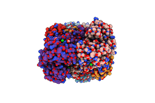 |
The Crystal Structure Of Cystathione Gamma Lyase (Cale6) From Micromonospora Echinospora
Organism: Micromonospora echinospora
Method: X-RAY DIFFRACTION Resolution:2.10 Å Release Date: 2014-05-07 Classification: LYASE Ligands: MES, CL, GOL, FMT |
Organism: Micromonospora echinospora
Method: X-RAY DIFFRACTION
Release Date: 2014-05-07
Ligands: MES, CL, GOL, FMT
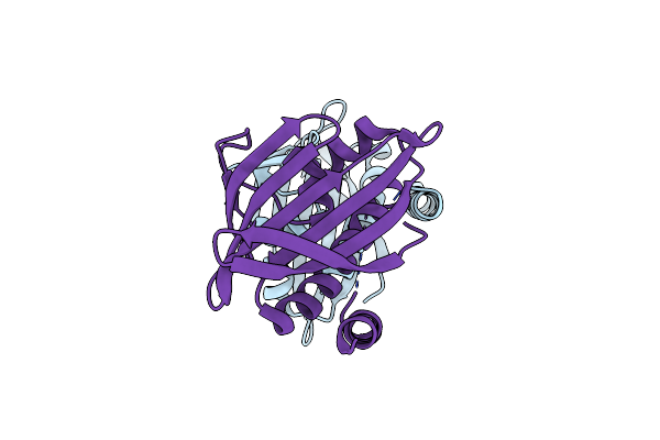 |
Crystal Structure Of Calu16 From Micromonospora Echinospora. Northeast Structural Genomics Consortium Target Mir12.
Organism: Micromonospora echinospora
Method: X-RAY DIFFRACTION Resolution:2.50 Å Release Date: 2012-12-12 Classification: Structural Genomics, Unknown Function |
Organism: Micromonospora echinospora
Method: X-RAY DIFFRACTION
Release Date: 2012-12-12
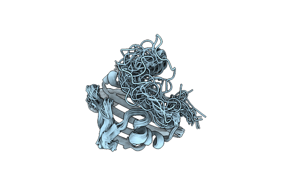 |
Solution Nmr Structure Of Calu16 From Micromonospora Echinospora, Northeast Structural Genomics Consortium (Nesg) Target Mir12
Organism: Micromonospora echinospora
Method: SOLUTION NMR Release Date: 2012-10-03 Classification: STRUCTURAL GENOMICS, UNKNOWN FUNCTION |
Organism: Micromonospora echinospora
Method: SOLUTION NMR
Release Date: 2012-10-03
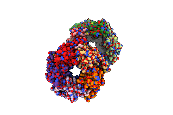 |
Crystal Structure Of Acetylcholine Binding Protein Complexed With Metocurine
Organism: Aplysia californica
Method: X-RAY DIFFRACTION Resolution:2.10 Å Release Date: 2011-10-12 Classification: CHOLINE-BINDING PROTEIN Ligands: CU9 |
Organism: Aplysia californica
Method: X-RAY DIFFRACTION
Release Date: 2011-10-12
Ligands: CU9
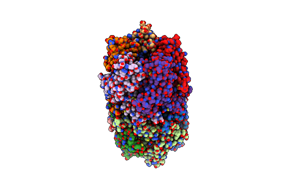 |
Crystal Structure Of The Complex Of Acetylcholine Binding Protein And D-Tubocurarine
Organism: Aplysia californica
Method: X-RAY DIFFRACTION Resolution:2.44 Å Release Date: 2011-10-05 Classification: CHOLINE-BINDING PROTEIN Ligands: MG, TUB |
Organism: Aplysia californica
Method: X-RAY DIFFRACTION
Release Date: 2011-10-05
Ligands: MG, TUB
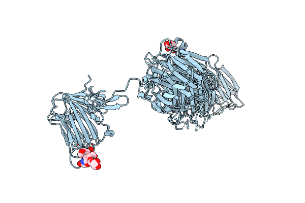 |
Crystal Structure Of The Alpha-Neurexin-1 Ectodomain, Lns 2-6
Organism: Bos taurus
Method: X-RAY DIFFRACTION Resolution:3.02 Å Release Date: 2011-06-08 Classification: CELL ADHESION Ligands: BGC |
Organism: Bos taurus
Method: X-RAY DIFFRACTION
Release Date: 2011-06-08
Ligands: BGC