

Search Count: 18
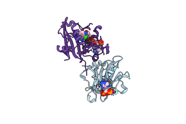 |
Candida Albicans Dihydrofolate Reductase Complexed With Dihydro-Nicotinamide-Adenine-Dinucleotide Phosphate (Nadph) And 5-Chloryl-2,4,6-Quinazolinetriamine (Gw1225)
Organism: Candida albicans
Method: X-RAY DIFFRACTION Resolution:1.71 Å Release Date: 2003-03-04 Classification: OXIDOREDUCTASE Ligands: NDP, CLZ |
Organism: Candida albicans
Method: X-RAY DIFFRACTION
Release Date: 2003-03-04
Ligands: NDP, CLZ
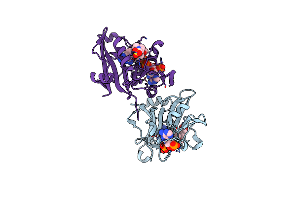 |
Candida Albicans Dihydrofolate Reductase Complexed With Dihydro-Nicotinamide-Adenine-Dinucleotide Phosphate (Nadph) And 5-(4-Methoxyphenoxy)-2,4-Quinazolinediamine (Gw1466)
Organism: Candida albicans
Method: X-RAY DIFFRACTION Resolution:1.70 Å Release Date: 2003-03-04 Classification: OXIDOREDUCTASE Ligands: NDP, MQ1, MES |
Organism: Candida albicans
Method: X-RAY DIFFRACTION
Release Date: 2003-03-04
Ligands: NDP, MQ1, MES
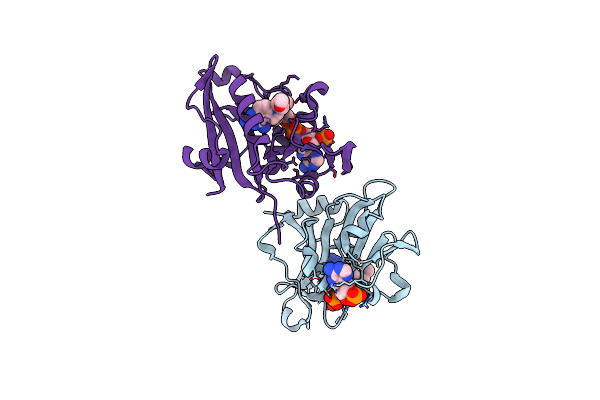 |
Candida Albicans Dihydrofolate Reductase Complexed With Dihydro-Nicotinamide-Adenine-Dinucleotide Phosphate (Nadph) And 7-[2-Methoxy-1-(Methoxymethyl)Ethyl]-7H-Pyrrolo[3,2-F] Quinazoline-1,3-Diamine (Gw557)
Organism: Candida albicans
Method: X-RAY DIFFRACTION Resolution:1.76 Å Release Date: 2003-03-04 Classification: OXIDOREDUCTASE Ligands: NDP, MQU |
Organism: Candida albicans
Method: X-RAY DIFFRACTION
Release Date: 2003-03-04
Ligands: NDP, MQU
 |
Candida Albicans Dihydrofolate Reductase Complexed With Dihydro-Nicotinamide-Adenine-Dinucleotide Phosphate (Nadph) And 5-(Phenylsulfanyl)-2,4-Quinazolinediamine (Gw997)
Organism: Candida albicans
Method: X-RAY DIFFRACTION Resolution:1.70 Å Release Date: 2001-04-11 Classification: OXIDOREDUCTASE Ligands: PO4, NDP, TQ3 |
Organism: Candida albicans
Method: X-RAY DIFFRACTION
Release Date: 2001-04-11
Ligands: PO4, NDP, TQ3
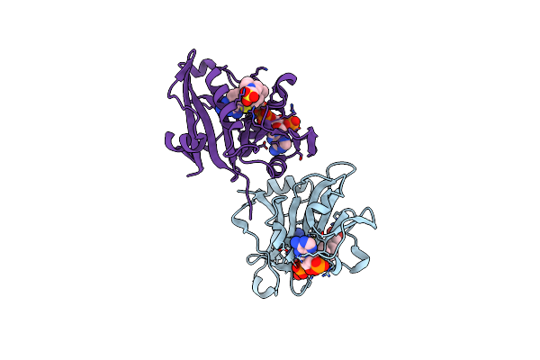 |
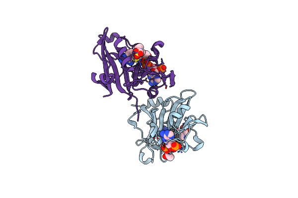
Candida Albicans Dihydrofolate Reductase Complexed With Dihydro-Nicotinamide-Adenine-Dinucleotide Phosphate (Nadph) And 5-[(4-Methylphenyl)Sulfanyl]-2,4-Quinazolinediamine (Gw578)
Organism: Candida albicans
Method: X-RAY DIFFRACTION Resolution:1.82 Å Release Date: 2001-04-11 Classification: OXIDOREDUCTASE Ligands: NDP, TQ4, MES |
Organism: Candida albicans
Method: X-RAY DIFFRACTION
Release Date: 2001-04-11
Ligands: NDP, TQ4, MES
 |
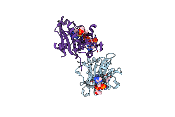
Candida Albicans Dihydrofolate Reductase Complex In Which The Dihydronicotinamide Moiety Of Dihydro-Nicotinamide-Adenine-Dinucleotide Phosphate (Nadph) Is Displaced By 5-[(4-Tert-Butylphenyl)Sulfanyl]-2,4-Quinazolinediamine (Gw995)
Organism: Candida albicans
Method: X-RAY DIFFRACTION Resolution:1.78 Å Release Date: 2001-04-11 Classification: OXIDOREDUCTASE Ligands: NDP, TQ5, MES |
Organism: Candida albicans
Method: X-RAY DIFFRACTION
Release Date: 2001-04-11
Ligands: NDP, TQ5, MES
 |
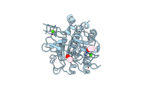
Candida Albicans Dihydrofolate Reductase Complex In Which The Dihydronicotinamide Moiety Of Dihydro-Nicotinamide-Adenine-Dinucleotide Phosphate (Nadph) Is Displaced By 5-{[4-(4-Morpholinyl)Phenyl]Sulfanyl}-2,4-Quinazolinediamin (Gw2021)
Organism: Candida albicans
Method: X-RAY DIFFRACTION Resolution:1.85 Å Release Date: 2001-04-11 Classification: OXIDOREDUCTASE Ligands: NDP, TQ6, MES |
Organism: Candida albicans
Method: X-RAY DIFFRACTION
Release Date: 2001-04-11
Ligands: NDP, TQ6, MES
 |
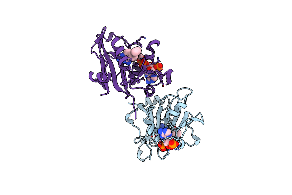
Subtilisin Bpn' Mutant 7186
Organism: Bacillus amyloliquefaciens
Method: X-RAY DIFFRACTION Resolution:1.80 Å Release Date: 1998-04-29 Classification: HYDROLASE Ligands: CA, ACN |
Organism: Bacillus amyloliquefaciens
Method: X-RAY DIFFRACTION
Release Date: 1998-04-29
Ligands: CA, ACN
 |
Candida Albicans Dihydrofolate Reductase Complexed With Dihydro-Nicotinamide-Adenine-Dinucleotide Phosphate (Nadph) And 1,3-Diamino-7-(1-Ethyepropye)-7H-Pyrralo-[3,2-F]Quinazoline (Gw345)
Organism: Candida albicans
Method: X-RAY DIFFRACTION Resolution:1.60 Å Release Date: 1998-01-07 Classification: OXIDOREDUCTASE Ligands: NDP, GW3 |
Organism: Candida albicans
Method: X-RAY DIFFRACTION
Release Date: 1998-01-07
Ligands: NDP, GW3
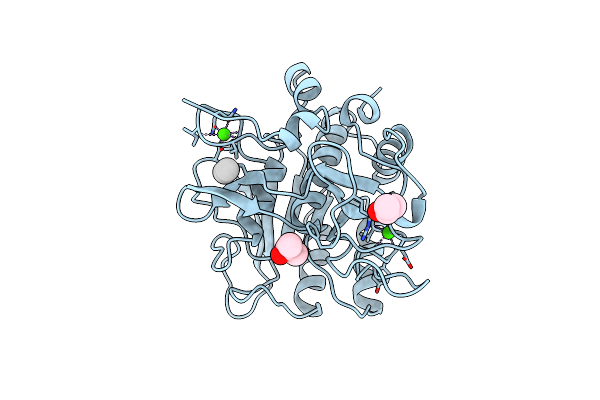 |
Subtilisin Bpn' Mutant 8324 In Citrate
Organism: Bacillus amyloliquefaciens
Method: X-RAY DIFFRACTION Resolution:1.80 Å Release Date: 1997-12-31 Classification: SERINE PROTEASE Ligands: CA, UNX, IPA |
Organism: Bacillus amyloliquefaciens
Method: X-RAY DIFFRACTION
Release Date: 1997-12-31
Ligands: CA, UNX, IPA
 |
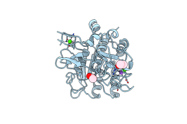
Candida Albicans Dihydrofolate Reductase
Organism: Candida albicans
Method: X-RAY DIFFRACTION Resolution:1.85 Å Release Date: 1997-11-12 Classification: OXIDOREDUCTASE Ligands: NDP |
Organism: Candida albicans
Method: X-RAY DIFFRACTION
Release Date: 1997-11-12
Ligands: NDP
 |
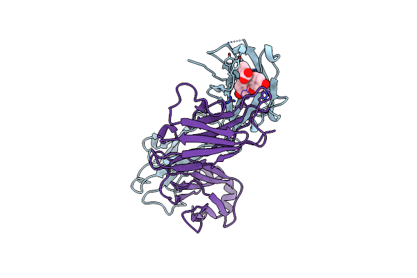
Subtilisin Mutant 8321
Organism: Bacillus amyloliquefaciens
Method: X-RAY DIFFRACTION Resolution:1.80 Å Release Date: 1997-11-12 Classification: SERINE PROTEASE Ligands: CA, NA, IPA |
Organism: Bacillus amyloliquefaciens
Method: X-RAY DIFFRACTION
Release Date: 1997-11-12
Ligands: CA, NA, IPA
 |
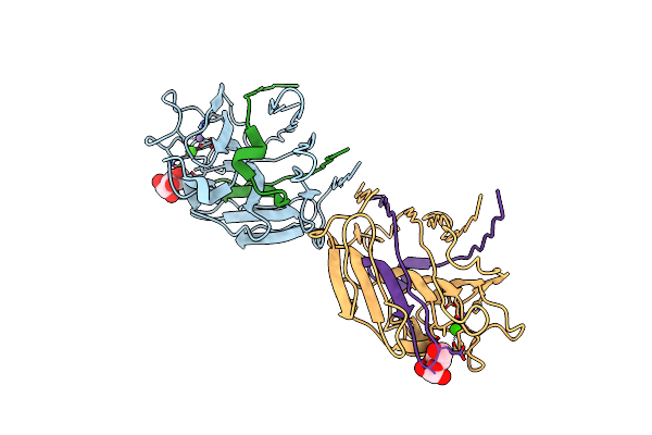
4-4-20 Fab Fragment
Organism: Mus musculus
Method: X-RAY DIFFRACTION Resolution:1.85 Å Release Date: 1995-09-15 Classification: IMMUNOGLOBULIN Ligands: FLU |
Organism: Mus musculus
Method: X-RAY DIFFRACTION
Release Date: 1995-09-15
Ligands: FLU
 |
X-Ray Crystal Structure Of A Pea Lectin-Trimannoside Complex At 2.6 Angstroms Resolution
Organism: Pisum sativum
Method: X-RAY DIFFRACTION Resolution:2.60 Å Release Date: 1993-10-31 Classification: LECTIN Ligands: MAN, MN, CA |
Organism: Pisum sativum
Method: X-RAY DIFFRACTION
Release Date: 1993-10-31
Ligands: MAN, MN, CA
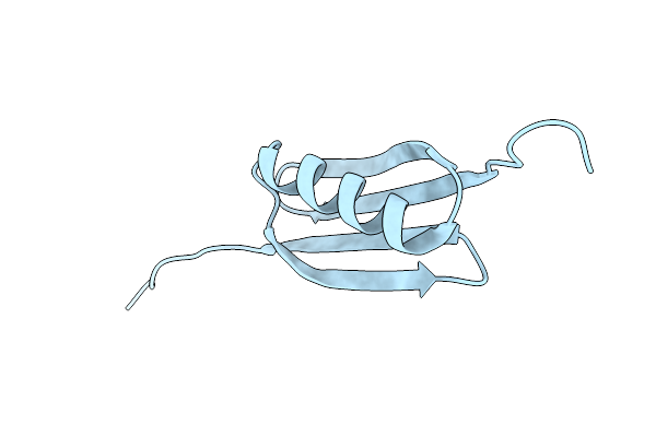 |
The 1.66 Angstroms X-Ray Structure Of The B2 Immunoglobulin-Binding Domain Of Streptococcal Protein G And Comparison To The Nmr Structure Of The B1 Domain
Organism: Streptococcus
Method: X-RAY DIFFRACTION Resolution:1.66 Å Release Date: 1992-07-15 Classification: IMMUNOGLOBULIN BINDING PROTEIN |
Organism: Streptococcus
Method: X-RAY DIFFRACTION
Release Date: 1992-07-15
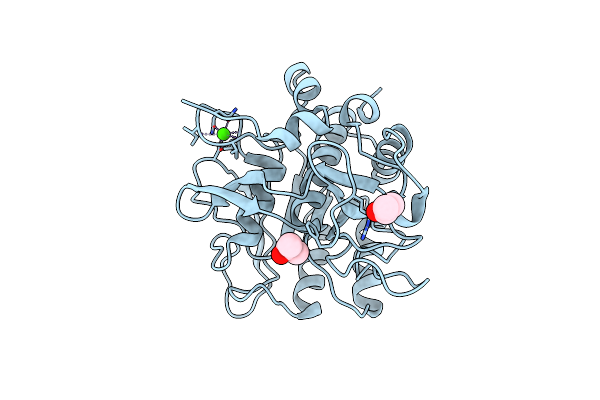 |
Large Increases In General Stability For Subtilisin Bpn(Prime) Through Incremental Changes In The Free Energy Of Unfolding
Organism: Bacillus amyloliquefaciens
Method: X-RAY DIFFRACTION Resolution:1.70 Å Release Date: 1990-10-15 Classification: HYDROLASE Ligands: CA, IPA |
Organism: Bacillus amyloliquefaciens
Method: X-RAY DIFFRACTION
Release Date: 1990-10-15
Ligands: CA, IPA
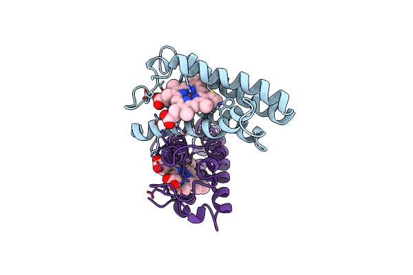 |
Structure Of Ferricytochrome C(Prime) From Rhodospirillum Molischianum At 1.67 Angstroms Resolution
Organism: Phaeospirillum molischianum
Method: X-RAY DIFFRACTION Resolution:1.67 Å Release Date: 1986-01-21 Classification: ELECTRON TRANSPORT (HEME PROTEIN) Ligands: HEC |
Organism: Phaeospirillum molischianum
Method: X-RAY DIFFRACTION
Release Date: 1986-01-21
Ligands: HEC
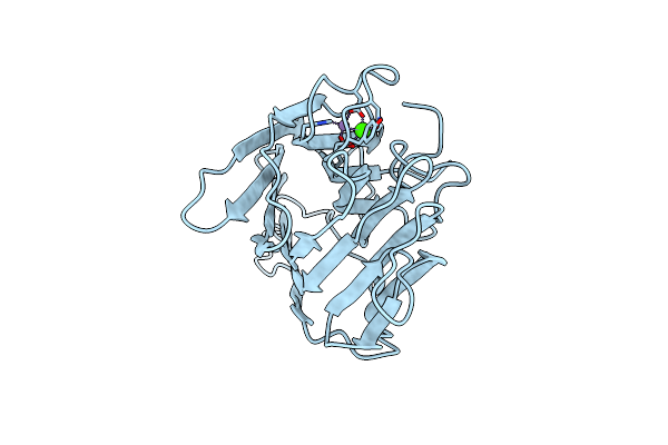 |
Structure Of Concanavalin A At 2.4 Angstroms Resolution
Organism: Canavalia ensiformis
Method: X-RAY DIFFRACTION Resolution:2.40 Å Release Date: 1976-10-11 Classification: LECTIN (AGGLUTININ) Ligands: MN, CA |
Organism: Canavalia ensiformis
Method: X-RAY DIFFRACTION
Release Date: 1976-10-11
Ligands: MN, CA