

Search Count: 10
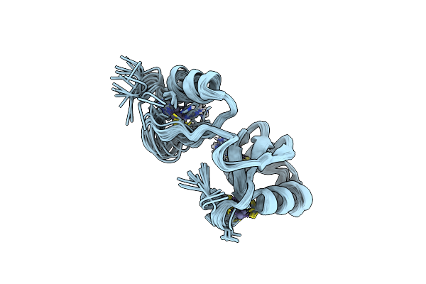 |
Phosphotyrosine Binding Domain
Organism: Mus musculus
Method: SOLUTION NMR Release Date: 2014-08-06 Classification: LIGASE Ligands: ZN |
Organism: Mus musculus
Method: SOLUTION NMR
Release Date: 2014-08-06
Ligands: ZN
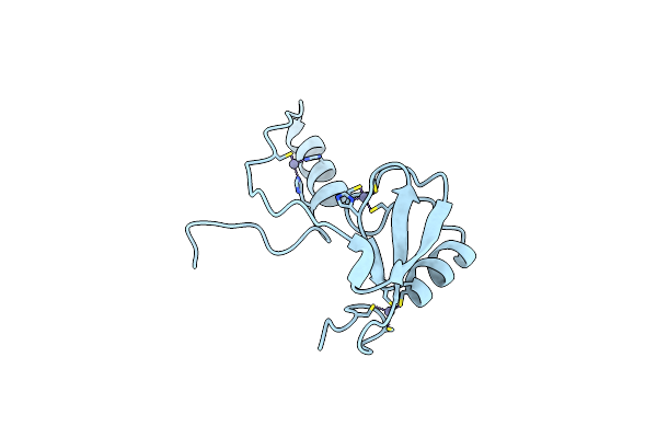 |
Crystal Structure Of A Phosphotyrosine Binding Domain
Organism: Mus musculus
Method: X-RAY DIFFRACTION Resolution:1.90 Å Release Date: 2012-01-25 Classification: LIGASE Ligands: ZN |
Organism: Mus musculus
Method: X-RAY DIFFRACTION
Release Date: 2012-01-25
Ligands: ZN
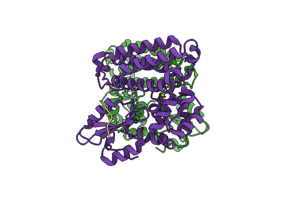 |
Reverse Binding Mode Of Metrd Peptide Complexed With C-Cbl Tkb Domain
Organism: Homo sapiens
Method: X-RAY DIFFRACTION Resolution:1.92 Å Release Date: 2010-12-01 Classification: PROTEIN BINDING/LIGASE Ligands: CA, CL |
Organism: Homo sapiens
Method: X-RAY DIFFRACTION
Release Date: 2010-12-01
Ligands: CA, CL
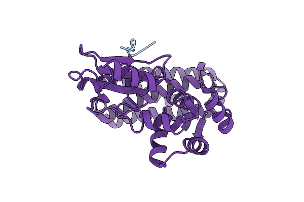 |
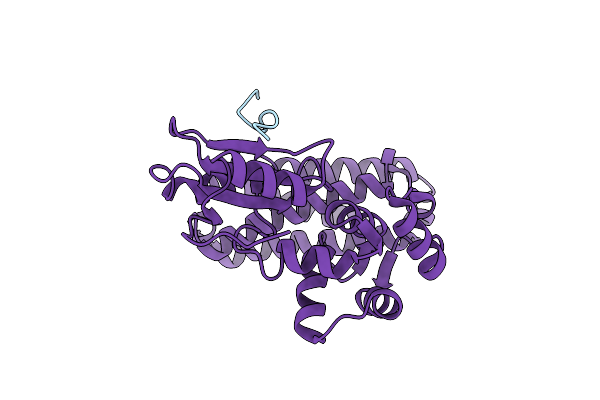
Crystal Structure Of C-Cbl Tkb Domain In Complex With Double Phosphorylated Spry2 Peptide
Organism: Homo sapiens
Method: X-RAY DIFFRACTION Resolution:2.20 Å Release Date: 2010-08-18 Classification: Ligase/signaling Protein |
Organism: Homo sapiens
Method: X-RAY DIFFRACTION
Release Date: 2010-08-18
 |
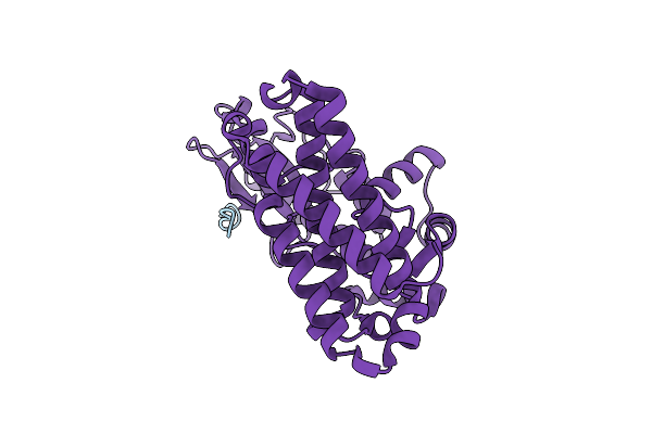
Crystal Structure Of C-Cbl Tkb Domain In Complex With Double Phosphorylated Egfr Peptide
Organism: Homo sapiens
Method: X-RAY DIFFRACTION Resolution:2.10 Å Release Date: 2010-08-18 Classification: Ligase/signaling Protein |
Organism: Homo sapiens
Method: X-RAY DIFFRACTION
Release Date: 2010-08-18
 |
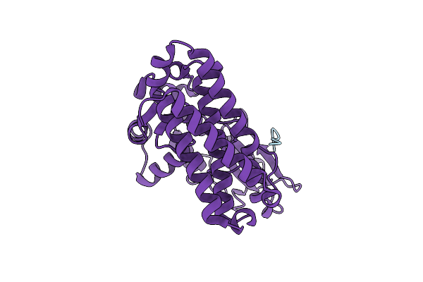
Crystal Structure Of C-Cbl-Tkb Domain Complexed With Its Binding Motif In Sprouty2
Organism: Homo sapiens
Method: X-RAY DIFFRACTION Resolution:2.00 Å Release Date: 2008-02-26 Classification: LIGASE |
Organism: Homo sapiens
Method: X-RAY DIFFRACTION
Release Date: 2008-02-26
 |
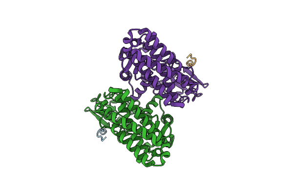
Crystal Structure Of C-Cbl-Tkb Domain Complexed With Its Binding Motif In Sprouty4
Organism: Homo sapiens
Method: X-RAY DIFFRACTION Resolution:2.00 Å Release Date: 2008-02-26 Classification: LIGASE/SIGNALING PROTEIN |
Organism: Homo sapiens
Method: X-RAY DIFFRACTION
Release Date: 2008-02-26
 |
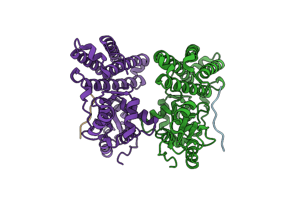
Crystal Structure Of C-Cbl-Tkb Domain Complexed With Its Binding Motif In Egf Receptor'
Organism: Homo sapiens
Method: X-RAY DIFFRACTION Resolution:2.60 Å Release Date: 2008-02-26 Classification: LIGASE/SIGNALING PROTEIN |
Organism: Homo sapiens
Method: X-RAY DIFFRACTION
Release Date: 2008-02-26
 |
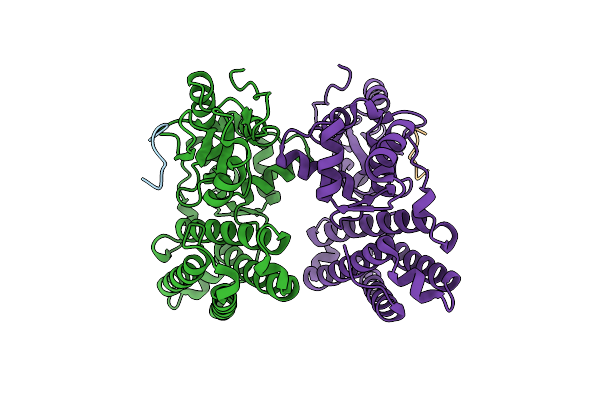
Crystal Structure Of C-Cbl-Tkb Domain Complexed With Its Binding Motif In Syk
Organism: Homo sapiens
Method: X-RAY DIFFRACTION Resolution:1.45 Å Release Date: 2008-02-26 Classification: LIGASE/SIGNALING PROTEIN |
Organism: Homo sapiens
Method: X-RAY DIFFRACTION
Release Date: 2008-02-26
 |
Crystal Structure Of C-Cbl-Tkb Domain Complexed With Its Binding Motif In C-Met
Organism: Homo sapiens
Method: X-RAY DIFFRACTION Resolution:1.35 Å Release Date: 2008-02-26 Classification: LIGASE/SIGNALING PROTEIN |
Organism: Homo sapiens
Method: X-RAY DIFFRACTION
Release Date: 2008-02-26