

Search Count: 31
 |
Rbd In Complex With Fab14
Organism: Homo sapiens, Severe acute respiratory syndrome coronavirus 2
Method: ELECTRON MICROSCOPY Release Date: 2022-09-14 Classification: IMMUNE SYSTEM/VIRAL PROTEIN Ligands: NAG |
Organism: Homo sapiens, Severe acute respiratory syndrome coronavirus 2
Method: ELECTRON MICROSCOPY
Release Date: 2022-09-14
Ligands: NAG
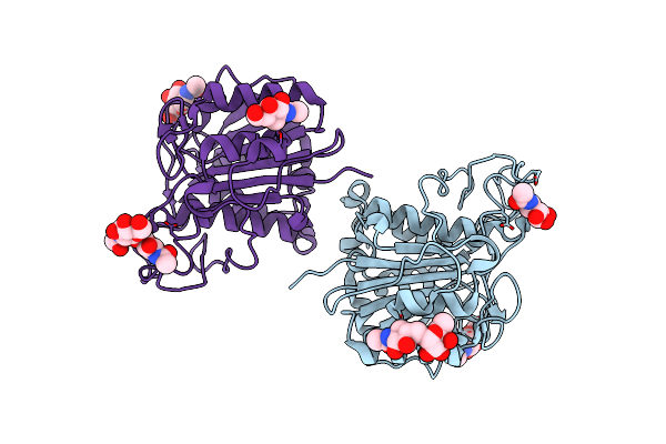 |
The Crystal Structure Of Vypal2-C214A, A Dead Mutant Of Vypal2 Peptide Asparaginyl Ligase In Form Ii
Organism: Viola philippica
Method: X-RAY DIFFRACTION Resolution:1.80 Å Release Date: 2022-07-13 Classification: PLANT PROTEIN Ligands: NAG |
Organism: Viola philippica
Method: X-RAY DIFFRACTION
Release Date: 2022-07-13
Ligands: NAG
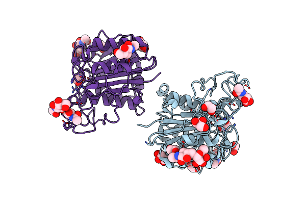 |
The Crystal Structure Of Vypal2-I244V, A More Efficient Mutant Of Vypal2 Peptide Asparaginyl Ligase In Its Active Enzyme Form
Organism: Viola philippica
Method: X-RAY DIFFRACTION Resolution:1.59 Å Release Date: 2022-06-29 Classification: PLANT PROTEIN Ligands: NAG, EDO, GOL |
Organism: Viola philippica
Method: X-RAY DIFFRACTION
Release Date: 2022-06-29
Ligands: NAG, EDO, GOL
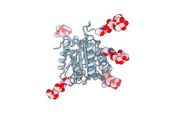 |
The Crystal Structure Of Vypal2-C214A, A Dead Mutant Of Vypal2 Peptide Asparaginyl Ligase In Form I
Organism: Viola philippica
Method: X-RAY DIFFRACTION Resolution:1.90 Å Release Date: 2022-06-29 Classification: PLANT PROTEIN |
Organism: Viola philippica
Method: X-RAY DIFFRACTION
Release Date: 2022-06-29
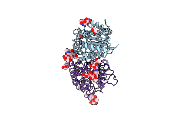 |
The Crystal Structure Of Vypal2 Peptide Asparaginyl Ligase In Its Active Enzyme Form
Organism: Viola philippica
Method: X-RAY DIFFRACTION Resolution:2.30 Å Release Date: 2022-06-29 Classification: PLANT PROTEIN Ligands: NAG, EDO |
Organism: Viola philippica
Method: X-RAY DIFFRACTION
Release Date: 2022-06-29
Ligands: NAG, EDO
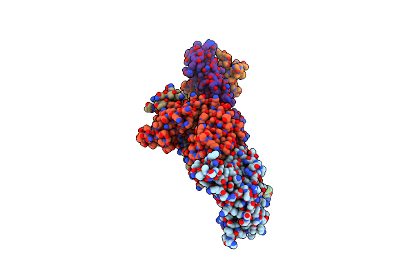 |
Sars-Cov2 Rbd Bound To Fab06
Organism: Severe acute respiratory syndrome coronavirus 2, Homo sapiens
Method: X-RAY DIFFRACTION Resolution:2.89 Å Release Date: 2022-03-30 Classification: VIRAL PROTEIN/IMMUNE SYSTEM Ligands: NAG |
Organism: Severe acute respiratory syndrome coronavirus 2, Homo sapiens
Method: X-RAY DIFFRACTION
Release Date: 2022-03-30
Ligands: NAG
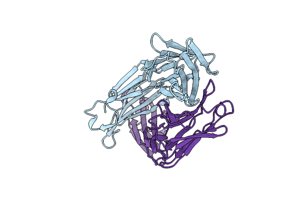 |
Fab14 - A Sars-Cov2 Rbd Neutralising Antibody
Organism: Homo sapiens
Method: X-RAY DIFFRACTION Resolution:2.46 Å Release Date: 2022-03-30 Classification: IMMUNE SYSTEM |
Organism: Homo sapiens
Method: X-RAY DIFFRACTION
Release Date: 2022-03-30
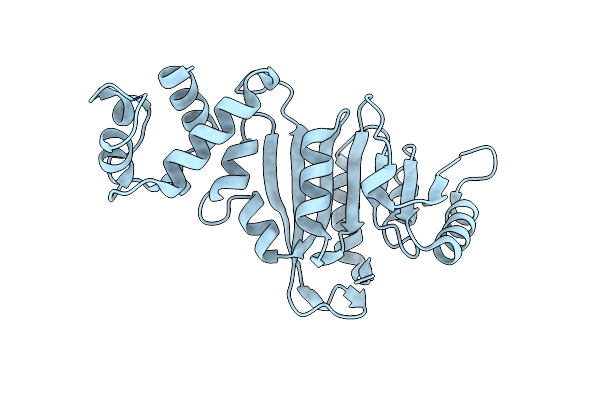 |
Crystal Structure Of Rrna Methyltransferase Erm38
Organism: Mycolicibacterium smegmatis
Method: X-RAY DIFFRACTION Resolution:1.90 Å Release Date: 2022-01-12 Classification: TRANSFERASE |
Organism: Mycolicibacterium smegmatis
Method: X-RAY DIFFRACTION
Release Date: 2022-01-12
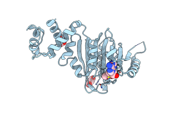 |
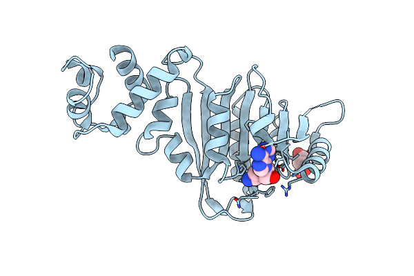
Crystal Structure Of Rrna Methyltransferase Erm38 In Complex With Sam
Organism: Mycolicibacterium smegmatis
Method: X-RAY DIFFRACTION Resolution:2.25 Å Release Date: 2022-01-12 Classification: TRANSFERASE Ligands: SAM, SIN |
Organism: Mycolicibacterium smegmatis
Method: X-RAY DIFFRACTION
Release Date: 2022-01-12
Ligands: SAM, SIN
 |
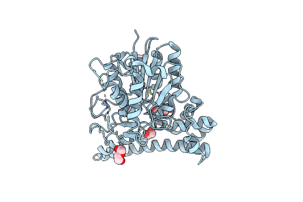
Crystal Structure Of Rrna Methyltransferase Erm38 In Complex With Sinefungin
Organism: Mycolicibacterium smegmatis
Method: X-RAY DIFFRACTION Resolution:2.25 Å Release Date: 2022-01-12 Classification: TRANSFERASE Ligands: SFG, SIN |
Organism: Mycolicibacterium smegmatis
Method: X-RAY DIFFRACTION
Release Date: 2022-01-12
Ligands: SFG, SIN
 |
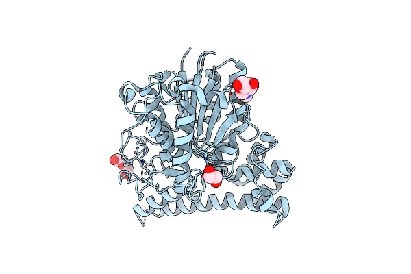
Turning An Asparaginyl Endopeptidase Into A Peptide Ligase
Organism: Clitoria ternatea
Method: X-RAY DIFFRACTION Resolution:1.35 Å Release Date: 2020-10-07 Classification: HYDROLASE Ligands: EDO, PEG |
Organism: Clitoria ternatea
Method: X-RAY DIFFRACTION
Release Date: 2020-10-07
Ligands: EDO, PEG
 |
Turning An Asparaginyl Endopeptidase Into A Peptide Ligase
Organism: Clitoria ternatea
Method: X-RAY DIFFRACTION Resolution:1.66 Å Release Date: 2020-10-07 Classification: HYDROLASE Ligands: NAG, EDO, PEG |
Organism: Clitoria ternatea
Method: X-RAY DIFFRACTION
Release Date: 2020-10-07
Ligands: NAG, EDO, PEG
 |
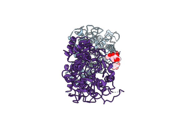
Turning An Asparaginyl Endopeptidase Into A Peptide Ligase
Organism: Clitoria ternatea
Method: X-RAY DIFFRACTION Resolution:2.64 Å Release Date: 2020-10-07 Classification: HYDROLASE |
Organism: Clitoria ternatea
Method: X-RAY DIFFRACTION
Release Date: 2020-10-07
 |
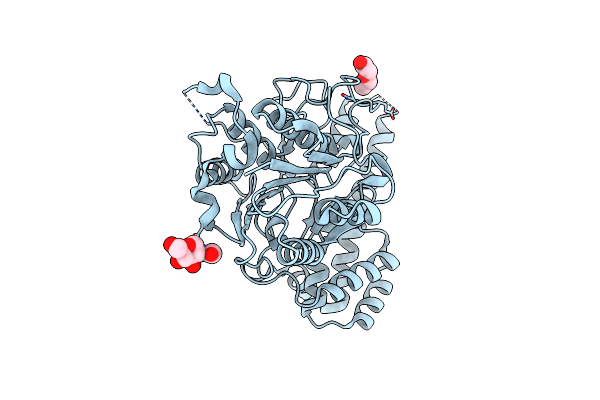
Turning An Asparaginyl Endopeptidase Into A Peptide Ligase
Organism: Clitoria ternatea
Method: X-RAY DIFFRACTION Resolution:1.50 Å Release Date: 2020-10-07 Classification: HYDROLASE Ligands: NAG, EDO, PEG |
Organism: Clitoria ternatea
Method: X-RAY DIFFRACTION
Release Date: 2020-10-07
Ligands: NAG, EDO, PEG
 |
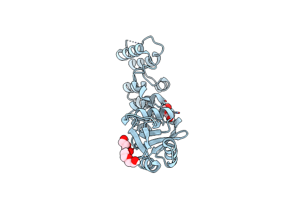
Turning An Asparaginyl Endopeptidase Into A Peptide Ligase
Organism: Clitoria ternatea
Method: X-RAY DIFFRACTION Resolution:2.00 Å Release Date: 2020-10-07 Classification: HYDROLASE Ligands: NAG, PEG |
Organism: Clitoria ternatea
Method: X-RAY DIFFRACTION
Release Date: 2020-10-07
Ligands: NAG, PEG
 |
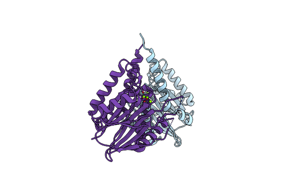
Crystal Structure Of A Periplasmic Sensor Domain Of Histidine Kinase Vbrk
Organism: Vibrio parahaemolyticus
Method: X-RAY DIFFRACTION Resolution:2.28 Å Release Date: 2020-09-16 Classification: SIGNALING PROTEIN Ligands: CL, PEG |
Organism: Vibrio parahaemolyticus
Method: X-RAY DIFFRACTION
Release Date: 2020-09-16
Ligands: CL, PEG
 |
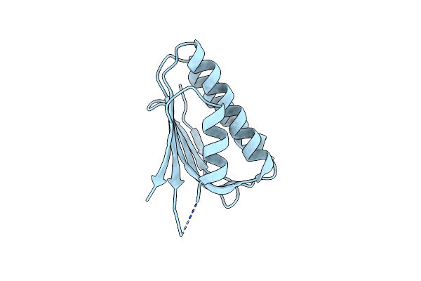
Siaa-Pp2C Domain Of Pseudomonas Aeruginosa
Organism: Pseudomonas aeruginosa
Method: X-RAY DIFFRACTION Resolution:2.09 Å Release Date: 2019-06-26 Classification: SIGNALING PROTEIN Ligands: MG |
Organism: Pseudomonas aeruginosa
Method: X-RAY DIFFRACTION
Release Date: 2019-06-26
Ligands: MG
 |
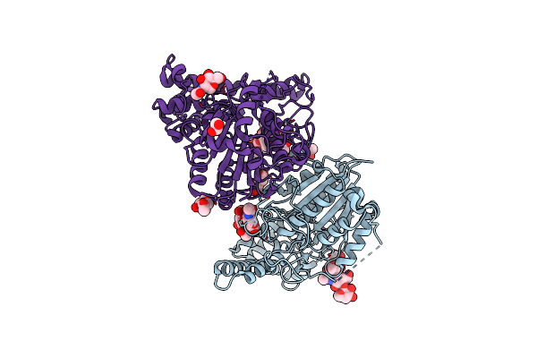
Siac Of Pseudomonas Aeruginosa
Organism: Pseudomonas aeruginosa
Method: X-RAY DIFFRACTION Resolution:1.74 Å Release Date: 2019-06-26 Classification: BIOSYNTHETIC PROTEIN |
Organism: Pseudomonas aeruginosa
Method: X-RAY DIFFRACTION
Release Date: 2019-06-26
 |
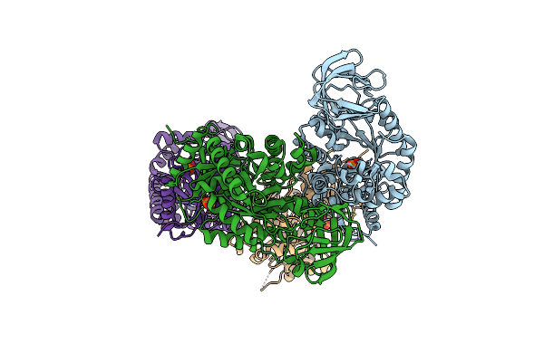
Peptide Asparaginyl Ligases From Viola Yedoensis
Organism: Viola philippica
Method: X-RAY DIFFRACTION Resolution:2.40 Å Release Date: 2019-05-15 Classification: LIGASE Ligands: NAG, EDO, PEG |
Organism: Viola philippica
Method: X-RAY DIFFRACTION
Release Date: 2019-05-15
Ligands: NAG, EDO, PEG
 |
T-State Crystal Structure Of Pyruvate Kinase From Mycobacterium Tuberculosis
Organism: Mycobacterium tuberculosis (strain atcc 25618 / h37rv)
Method: X-RAY DIFFRACTION Resolution:2.85 Å Release Date: 2017-11-15 Classification: TRANSFERASE Ligands: PO4 |
Organism: Mycobacterium tuberculosis (strain atcc 25618 / h37rv)
Method: X-RAY DIFFRACTION
Release Date: 2017-11-15
Ligands: PO4