

Search Count: 34
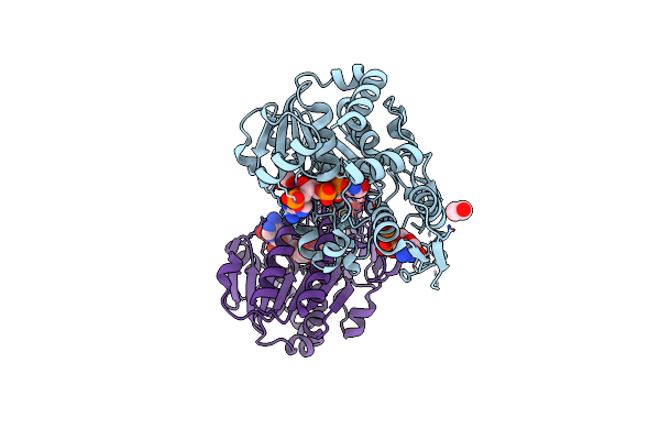 |
X-Ray Structure Of The Nadp-Dependent Reductase From Campylobacter Jejuni Responsible For The Synthesis Of Cdp-Glucitol In The Presence Of Cdp And Nadp
Organism: Campylobacter jejuni
Method: X-RAY DIFFRACTION Resolution:2.00 Å Release Date: 2023-12-20 Classification: OXIDOREDUCTASE Ligands: NAP, CDP, CL, EDO |
Organism: Campylobacter jejuni
Method: X-RAY DIFFRACTION
Release Date: 2023-12-20
Ligands: NAP, CDP, CL, EDO
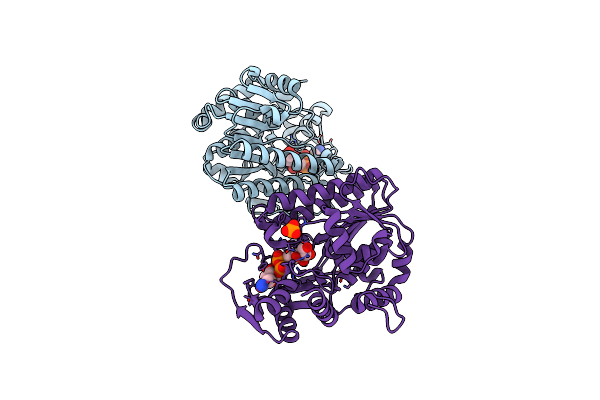 |
X-Ray Structure Of The Nadp-Dependent Reductase From Campylobacter Jejuni Responsible For The Synthesis Of Cdp-Glucitol In The Presence Of Cdp-Glucitol
Organism: Campylobacter jejuni
Method: X-RAY DIFFRACTION Resolution:2.20 Å Release Date: 2023-12-20 Classification: OXIDOREDUCTASE Ligands: YCX, CL, PO4, NA |
Organism: Campylobacter jejuni
Method: X-RAY DIFFRACTION
Release Date: 2023-12-20
Ligands: YCX, CL, PO4, NA
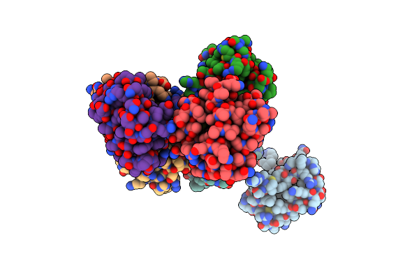 |
Crystal Structure Of Harmonin-Homology Domain 2 (Hhd2) Of Human Rtel1
Organism: Homo sapiens
Method: X-RAY DIFFRACTION Resolution:1.60 Å Release Date: 2023-08-09 Classification: PROTEIN BINDING |
Organism: Homo sapiens
Method: X-RAY DIFFRACTION
Release Date: 2023-08-09
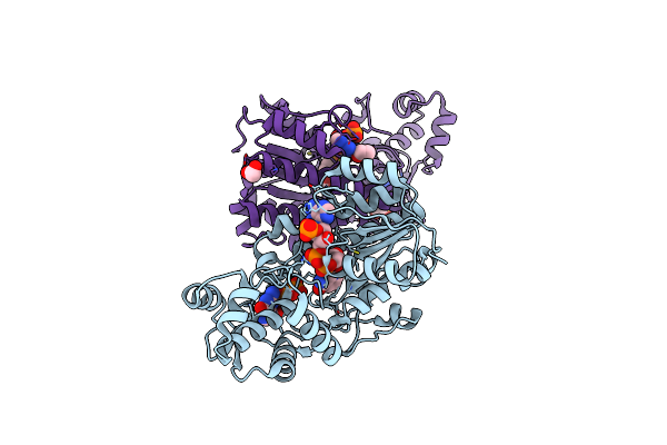 |
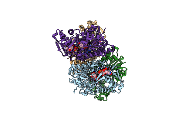
X-Ray Structure Of The Gdp-6-Deoxy-4-Keto-D-Lyxo-Heptose-4-Reductase From Campylobacter Jejuni Hs:15
Organism: Campylobacter jejuni
Method: X-RAY DIFFRACTION Resolution:1.45 Å Release Date: 2022-11-09 Classification: BIOSYNTHETIC PROTEIN, OXIDOREDUCTASE Ligands: GDP, NDP, EDO |
Organism: Campylobacter jejuni
Method: X-RAY DIFFRACTION
Release Date: 2022-11-09
Ligands: GDP, NDP, EDO
 |
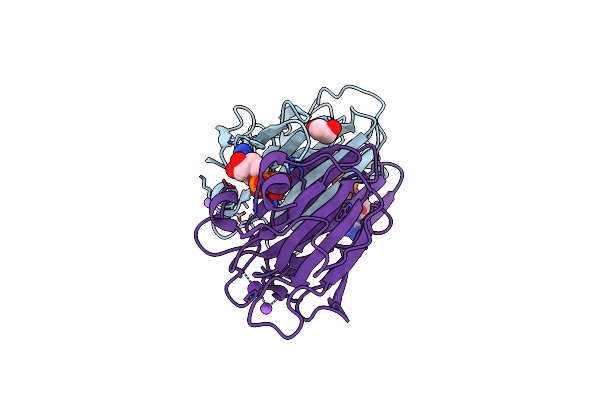
X-Ray Crystal Structure Of Gdp-D-Glycero-D-Manno-Heptose 4,6-Dehydratase From Campylobacter Jejuni
Organism: Campylobacter jejuni
Method: X-RAY DIFFRACTION Resolution:2.10 Å Release Date: 2022-07-06 Classification: OXIDOREDUCTASE Ligands: NAD, GDP, EDO |
Organism: Campylobacter jejuni
Method: X-RAY DIFFRACTION
Release Date: 2022-07-06
Ligands: NAD, GDP, EDO
 |
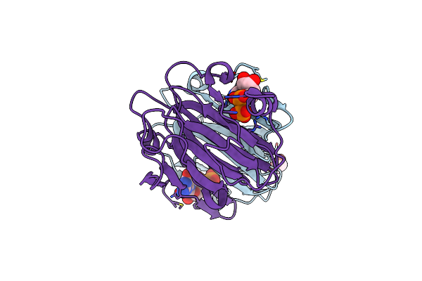
Crystal Structure Of The Gdp-D-Glycero-4-Keto-D-Lyxo-Heptose-3-Epimerase From Campylobacter Jejuni, Serotype Hs:23/36
Organism: Campylobacter jejuni
Method: X-RAY DIFFRACTION Resolution:1.55 Å Release Date: 2022-06-29 Classification: ISOMERASE Ligands: GDP, K, EDO, NA |
Organism: Campylobacter jejuni
Method: X-RAY DIFFRACTION
Release Date: 2022-06-29
Ligands: GDP, K, EDO, NA
 |
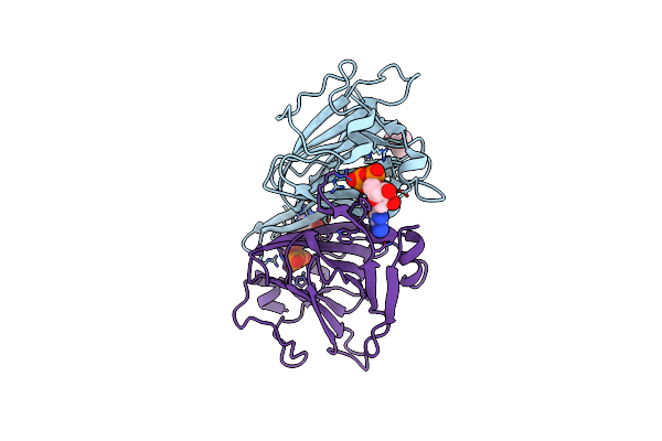
Crystal Structure Of The Gdp-D-Glycero-4-Keto-D-Lyxo-Heptose-3,5-Epimerase From Campylobacter Jejuni, Serotype Hs:42
Organism: Campylobacter jejuni
Method: X-RAY DIFFRACTION Resolution:1.90 Å Release Date: 2022-06-29 Classification: ISOMERASE Ligands: GDP, CL, EDO |
Organism: Campylobacter jejuni
Method: X-RAY DIFFRACTION
Release Date: 2022-06-29
Ligands: GDP, CL, EDO
 |
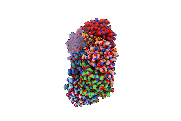
Crystal Structure Of The Gdp-D-Glycero-4-Keto-D-Lyxo-Heptose-3-Epimerase From Campylobacter Jejuni, Serotype Hs:3
Organism: Campylobacter jejuni
Method: X-RAY DIFFRACTION Resolution:1.50 Å Release Date: 2022-06-22 Classification: ISOMERASE Ligands: GDP, CL, TMA |
Organism: Campylobacter jejuni
Method: X-RAY DIFFRACTION
Release Date: 2022-06-22
Ligands: GDP, CL, TMA
 |
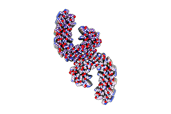
Crystal Structure Of The Gdp-D-Glycero-4-Keto-D-Lyxo-Heptose-3,5-Epimerase From Campylobacter Jejuni, Serotype Hs:15
Organism: Campylobacter jejuni
Method: X-RAY DIFFRACTION Resolution:1.90 Å Release Date: 2022-06-22 Classification: ISOMERASE Ligands: GDP, CL |
Organism: Campylobacter jejuni
Method: X-RAY DIFFRACTION
Release Date: 2022-06-22
Ligands: GDP, CL
 |
Paired Helical Tau Filament Extracted From Prp-Caa Patient Brain Tissue | Tau Filament | Phf Tau
Organism: Homo sapiens
Method: ELECTRON MICROSCOPY Release Date: 2021-07-28 Classification: PROTEIN FIBRIL |
Organism: Homo sapiens
Method: ELECTRON MICROSCOPY
Release Date: 2021-07-28
 |
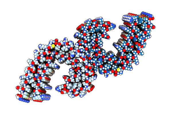
Straight Tau Filament Extracted From Prp-Caa Patient Brain Tissue | Tau Filament | Sf Tau
Organism: Homo sapiens
Method: ELECTRON MICROSCOPY Release Date: 2021-07-28 Classification: PROTEIN FIBRIL |
Organism: Homo sapiens
Method: ELECTRON MICROSCOPY
Release Date: 2021-07-28
 |
Paired Helical Tau Filament Extracted From Gss Patient Brain Tissue | Tau Filament From Gerstmann Straussler Scheinker Disease | Phf Tau
Organism: Homo sapiens
Method: ELECTRON MICROSCOPY Release Date: 2021-07-28 Classification: PROTEIN FIBRIL |
Organism: Homo sapiens
Method: ELECTRON MICROSCOPY
Release Date: 2021-07-28
 |
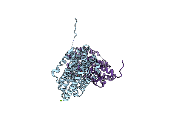
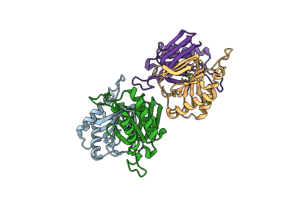
Improved High Resolution Structure Of Mhc Class Ii Complex
Organism: Gallus gallus
Method: X-RAY DIFFRACTION Resolution:1.70 Å Release Date: 2020-11-25 Classification: IMMUNE SYSTEM Ligands: GOL, ACT |
Organism: Gallus gallus
Method: X-RAY DIFFRACTION
Release Date: 2020-11-25
Ligands: GOL, ACT
 |
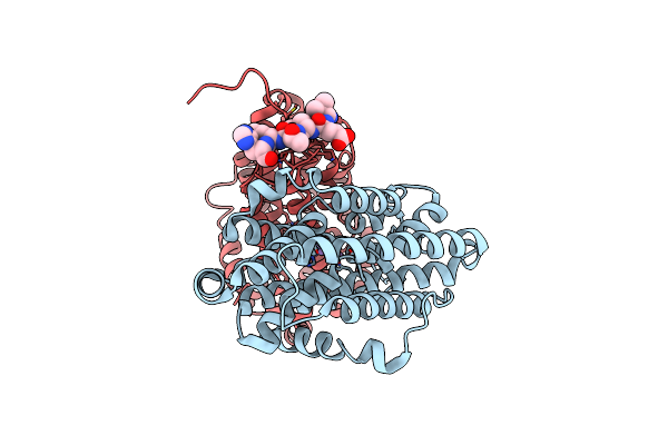
X-Ray Crystal Structure Of Flavobacterium Johnsoniae Dimanganese(Ii) Ribonucleotide Reductase Beta Subunit (Aerobic)
Organism: Flavobacterium johnsoniae (strain atcc 17061 / dsm 2064 / uw101)
Method: X-RAY DIFFRACTION Resolution:1.87 Å Release Date: 2018-06-27 Classification: OXIDOREDUCTASE Ligands: MN, MG |
Organism: Flavobacterium johnsoniae (strain atcc 17061 / dsm 2064 / uw101)
Method: X-RAY DIFFRACTION
Release Date: 2018-06-27
Ligands: MN, MG
 |
X-Ray Crystal Structure Of Flavobacterium Johnsoniae Dimanganese(Ii) Ribonucleotide Reductase Beta Subunit (Anaerobic)
Organism: Flavobacterium johnsoniae (strain atcc 17061 / dsm 2064 / uw101), Flavobacterium johnsoniae
Method: X-RAY DIFFRACTION Resolution:1.92 Å Release Date: 2018-04-18 Classification: OXIDOREDUCTASE Ligands: MN |
Organism: Flavobacterium johnsoniae (strain atcc 17061 / dsm 2064 / uw101), Flavobacterium johnsoniae
Method: X-RAY DIFFRACTION
Release Date: 2018-04-18
Ligands: MN
 |
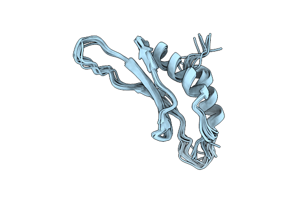
X-Ray Crystal Structure Of Flavobacterium Johnsoniae Dimanganese(Ii) Ribonucleotide Reductase Beta Subunit (As-Isolated)
Organism: Flavobacterium johnsoniae (strain atcc 17061 / dsm 2064 / uw101)
Method: X-RAY DIFFRACTION Resolution:1.90 Å Release Date: 2018-04-18 Classification: OXIDOREDUCTASE Ligands: MN, MG |
Organism: Flavobacterium johnsoniae (strain atcc 17061 / dsm 2064 / uw101)
Method: X-RAY DIFFRACTION
Release Date: 2018-04-18
Ligands: MN, MG
 |
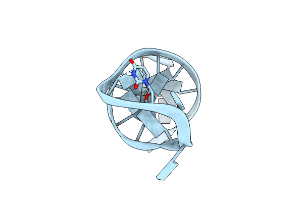
Drosha Double-Stranded Rna Binding Motif
Organism: Homo sapiens
Method: SOLUTION NMR Release Date: 2010-02-23 Classification: GENE REGULATION,NUCLEAR PROTEIN |
Organism: Homo sapiens
Method: SOLUTION NMR
Release Date: 2010-02-23
 |
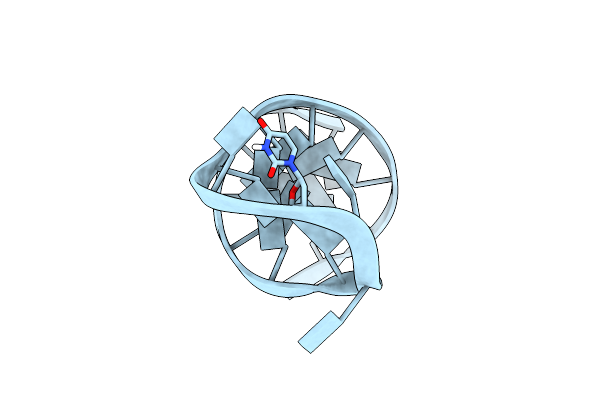
Solution Structure Of A B-Dna Hairpin At Low Pressure.
|
Method: SOLUTION NMR
Release Date: 2007-09-11
 |
Solution Structure Of A B-Dna Hairpin At High Pressure
|
Method: SOLUTION NMR
Release Date: 2007-09-11
 |
C-Myc Dna Unwinding Element Binding Protein
Organism: Homo sapiens
Method: X-RAY DIFFRACTION Resolution:2.00 Å Release Date: 2007-01-30 Classification: HYDROLASE Ligands: MG |
Organism: Homo sapiens
Method: X-RAY DIFFRACTION
Release Date: 2007-01-30
Ligands: MG