

Search Count: 37
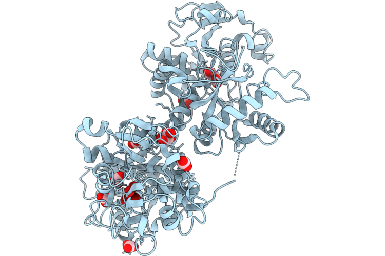 |
Crystal Structure Of The Adduct Formed Upon Reaction Of [V(Iv)O(Acetylacetonate)2] With Human Serum Transferrin With Fe(Iii) Bound At The C-Lobe Only
Organism: Homo sapiens
Method: X-RAY DIFFRACTION Resolution:1.93 Å Release Date: 2026-07-15 Classification: METAL TRANSPORT Ligands: BCT, GOL, FE, MLI, A1JJT |
Organism: Homo sapiens
Method: X-RAY DIFFRACTION
Release Date: 2026-07-15
Ligands: BCT, GOL, FE, MLI, A1JJT
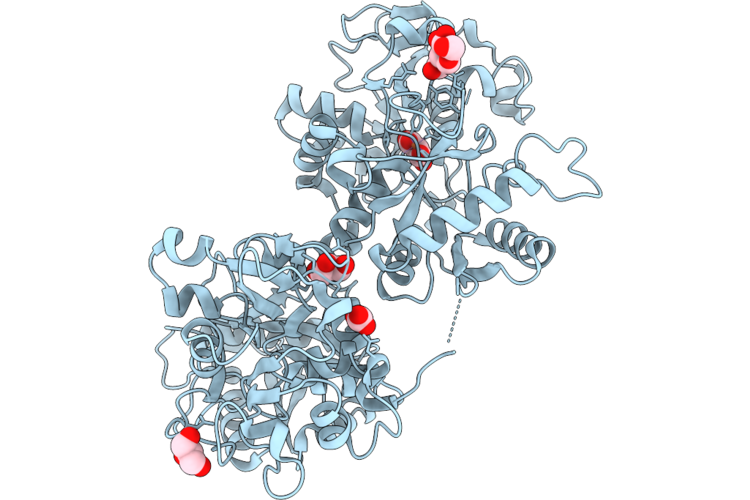 |
Crystal Structure Of The Human Serum Transferrin With Fe(Iii) Bound At The C-Lobe Only
Organism: Homo sapiens
Method: X-RAY DIFFRACTION Resolution:2.16 Å Release Date: 2026-07-15 Classification: METAL TRANSPORT Ligands: FE, MLI, NAG, GOL, BCT |
Organism: Homo sapiens
Method: X-RAY DIFFRACTION
Release Date: 2026-07-15
Ligands: FE, MLI, NAG, GOL, BCT
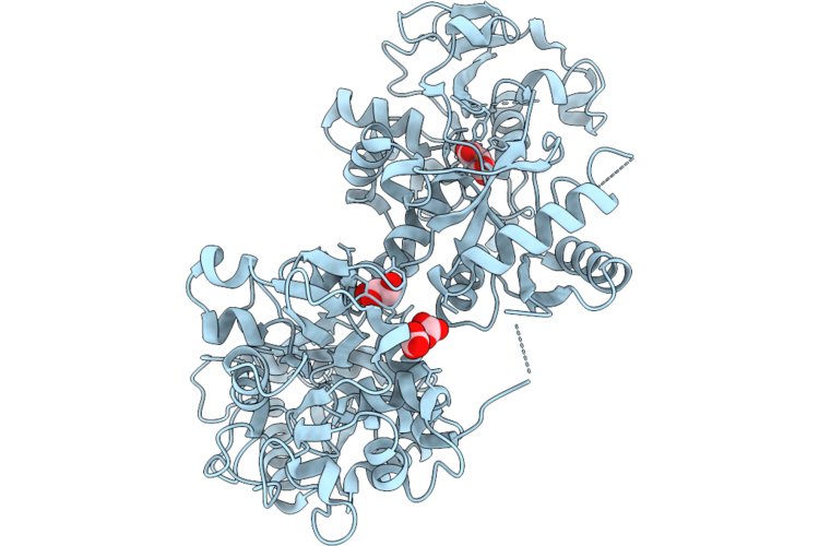 |
Crystal Structure Of The Human Serum Transferrin With Fe(Iii) Bound At The C-Lobe Only (Treated With Dmso)
Organism: Homo sapiens
Method: X-RAY DIFFRACTION Resolution:2.52 Å Release Date: 2026-07-15 Classification: METAL TRANSPORT Ligands: FE, MLI, GOL, BCT |
Organism: Homo sapiens
Method: X-RAY DIFFRACTION
Release Date: 2026-07-15
Ligands: FE, MLI, GOL, BCT
 |
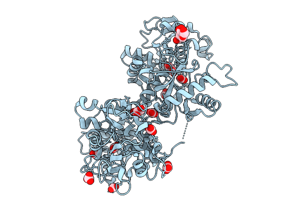
Crystal Structure Of The Adduct Formed Upon Reaction Of [V(Iv)O(Acetylacetonate)2] With Human Serum Transferrin With Fe(Iii) Bound At The C-Lobe Only
Organism: Homo sapiens
Method: X-RAY DIFFRACTION Resolution:2.55 Å Release Date: 2026-01-28 Classification: METAL TRANSPORT Ligands: BCT, GOL, FE, MLA, A1JJT |
Organism: Homo sapiens
Method: X-RAY DIFFRACTION
Release Date: 2026-01-28
Ligands: BCT, GOL, FE, MLA, A1JJT
 |
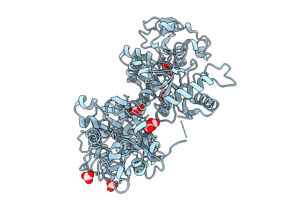
Crystal Structure Of The Human Serum Transferrin With Fe(Iii) Bound At The C-Lobe Only
Organism: Homo sapiens
Method: X-RAY DIFFRACTION Resolution:2.44 Å Release Date: 2026-01-28 Classification: METAL TRANSPORT Ligands: FE, MLA, NAG, GOL, BCT |
Organism: Homo sapiens
Method: X-RAY DIFFRACTION
Release Date: 2026-01-28
Ligands: FE, MLA, NAG, GOL, BCT
 |
Crystal Structure Of The Human Serum Transferrin With Fe(Iii) Bound At The C-Lobe Only (Treated With Dmso)
Organism: Homo sapiens
Method: X-RAY DIFFRACTION Resolution:3.20 Å Release Date: 2026-01-28 Classification: METAL TRANSPORT Ligands: FE, MLA, GOL, BCT |
Organism: Homo sapiens
Method: X-RAY DIFFRACTION
Release Date: 2026-01-28
Ligands: FE, MLA, GOL, BCT
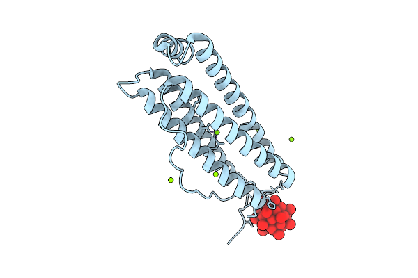 |
X-Ray Structure Of A Polyoxidovanadate/Human H-Ferritin Adduct Obtained When The Protein Is Treated Overnight With [Vivo(Acac)2]
Organism: Homo sapiens
Method: X-RAY DIFFRACTION Resolution:1.38 Å Release Date: 2025-12-03 Classification: METAL TRANSPORT Ligands: CL, MG, A1JGJ |
Organism: Homo sapiens
Method: X-RAY DIFFRACTION
Release Date: 2025-12-03
Ligands: CL, MG, A1JGJ
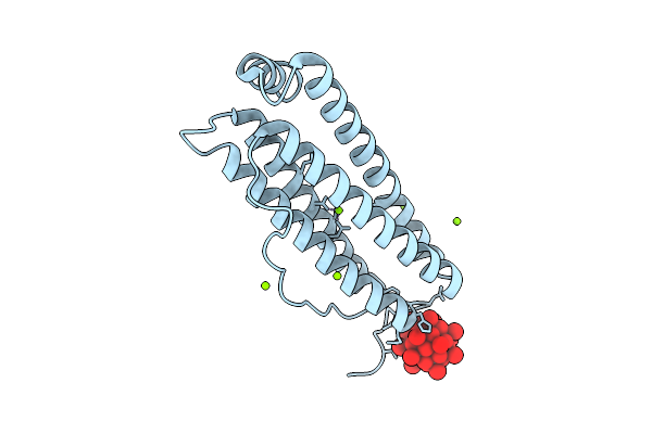 |
X-Ray Structure Of A Polyoxidovanadate/Human H-Ferritin Adduct Obtained When The Protein Is Treated 6 Days With [Vivo(Acac)2]
Organism: Homo sapiens
Method: X-RAY DIFFRACTION Resolution:1.54 Å Release Date: 2025-12-03 Classification: METAL TRANSPORT Ligands: CL, MG, A1JGJ |
Organism: Homo sapiens
Method: X-RAY DIFFRACTION
Release Date: 2025-12-03
Ligands: CL, MG, A1JGJ
 |
X-Ray Structure Of A Polyoxidovanadate/Human H-Ferritin Adduct Obtained When The Protein Is Treated 24 H With [Vivo(Acac)2]
Organism: Homo sapiens
Method: X-RAY DIFFRACTION Resolution:1.50 Å Release Date: 2025-12-03 Classification: METAL TRANSPORT Ligands: CL, MG, A1JGJ |
Organism: Homo sapiens
Method: X-RAY DIFFRACTION
Release Date: 2025-12-03
Ligands: CL, MG, A1JGJ
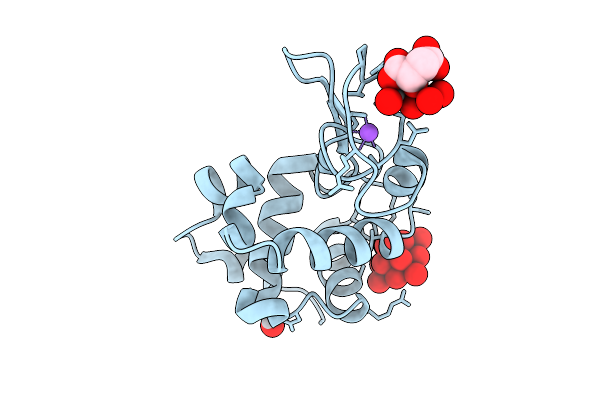 |
X-Ray Structure Of Decavanadate/Lysozyme Adduct Obtained When The Protein Is Treated With Cs2[V(V)2O4(Mal)2]2H2O (Structure A)
Organism: Gallus gallus
Method: X-RAY DIFFRACTION Resolution:1.46 Å Release Date: 2025-08-13 Classification: HYDROLASE Ligands: CL, NA, DVT, A1JFD, VVO |
Organism: Gallus gallus
Method: X-RAY DIFFRACTION
Release Date: 2025-08-13
Ligands: CL, NA, DVT, A1JFD, VVO
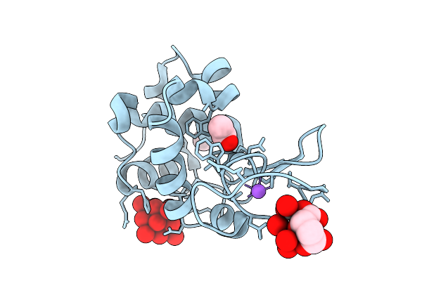 |
X-Ray Structure Of Decavanadate/Lysozyme Adduct Obtained When The Protein Is Treated With Cs2[V(V)2O4(Mal)2]2H2O (Structure B)
Organism: Gallus gallus
Method: X-RAY DIFFRACTION Resolution:1.18 Å Release Date: 2025-08-13 Classification: HYDROLASE Ligands: CL, NA, ACT, DVT, A1JFD |
Organism: Gallus gallus
Method: X-RAY DIFFRACTION
Release Date: 2025-08-13
Ligands: CL, NA, ACT, DVT, A1JFD
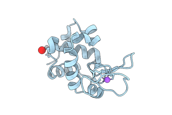 |
X-Ray Structure Of Lysozyme When Is Treated With Cs2[V(V)2O4(Mal)2]2H2O At 310K
Organism: Gallus gallus
Method: X-RAY DIFFRACTION Resolution:2.09 Å Release Date: 2025-08-13 Classification: HYDROLASE Ligands: CL, NA, VVO |
Organism: Gallus gallus
Method: X-RAY DIFFRACTION
Release Date: 2025-08-13
Ligands: CL, NA, VVO
 |
X-Ray Structure Of A Polyoxidovanadate/Lysozyme Adduct Obtained When The Protein Is Treated With [Vivo(Acac)2</Sub>] At 310 K
Organism: Gallus gallus
Method: X-RAY DIFFRACTION Resolution:1.83 Å Release Date: 2025-05-14 Classification: HYDROLASE Ligands: CL, NA, A1ICR, A1H8D |
Organism: Gallus gallus
Method: X-RAY DIFFRACTION
Release Date: 2025-05-14
Ligands: CL, NA, A1ICR, A1H8D
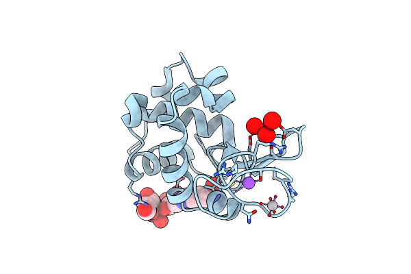 |
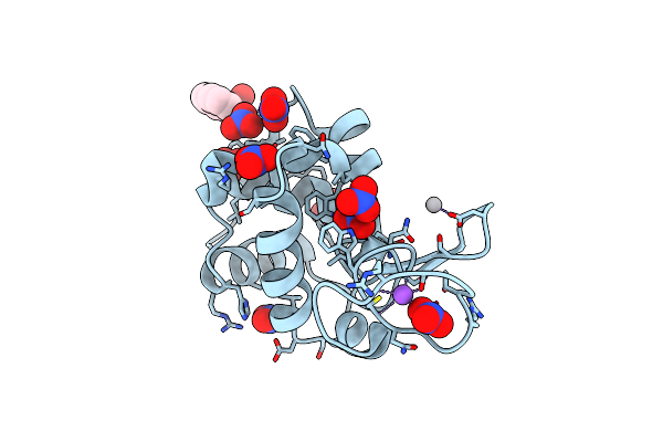
X-Ray Structure Of The Adduct Of Bis(Maltolato)Oxidovanadium(Iv) With Lysozyme Upon The Breakage Of The Maltolate Ring
Organism: Gallus gallus
Method: X-RAY DIFFRACTION Resolution:1.09 Å Release Date: 2025-04-16 Classification: HYDROLASE Ligands: A1IE3, VVB, A1IE4, CL, NA |
Organism: Gallus gallus
Method: X-RAY DIFFRACTION
Release Date: 2025-04-16
Ligands: A1IE3, VVB, A1IE4, CL, NA
 |
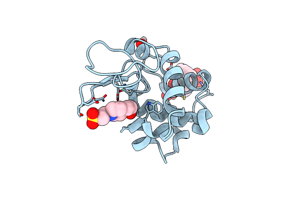
X-Ray Structure Of Lysozyme Obtained Upon Reaction With [Vivo(8-Hq)2] In Ethylene Glycol
Organism: Gallus gallus
Method: X-RAY DIFFRACTION Resolution:1.27 Å Release Date: 2025-02-12 Classification: HYDROLASE |
Organism: Gallus gallus
Method: X-RAY DIFFRACTION
Release Date: 2025-02-12
 |
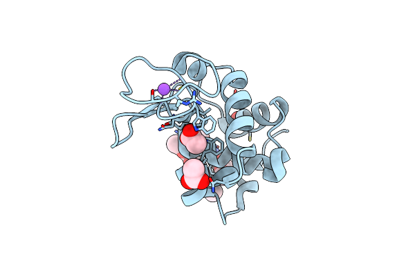
X-Ray Structure Of Lysozyme Obtained Upon Reaction With [Vivo(8-Hq)2] In Sodium Formate
Organism: Gallus gallus
Method: X-RAY DIFFRACTION Resolution:1.21 Å Release Date: 2025-02-12 Classification: HYDROLASE Ligands: EPE, A1H23, VVO, VVB |
Organism: Gallus gallus
Method: X-RAY DIFFRACTION
Release Date: 2025-02-12
Ligands: EPE, A1H23, VVO, VVB
 |
X-Ray Structure Of Lysozyme Obtained Upon Reaction With The Dioxidovanadium(V) Complex With Furan-2-Carboxylic Acid (3-Ethoxy-2-Hydroxybenzylidene)Hydrazide
Organism: Gallus gallus
Method: X-RAY DIFFRACTION Resolution:1.24 Å Release Date: 2024-12-18 Classification: HYDROLASE Ligands: CL, NA, ACT, VVB, A1IOM |
Organism: Gallus gallus
Method: X-RAY DIFFRACTION
Release Date: 2024-12-18
Ligands: CL, NA, ACT, VVB, A1IOM
 |
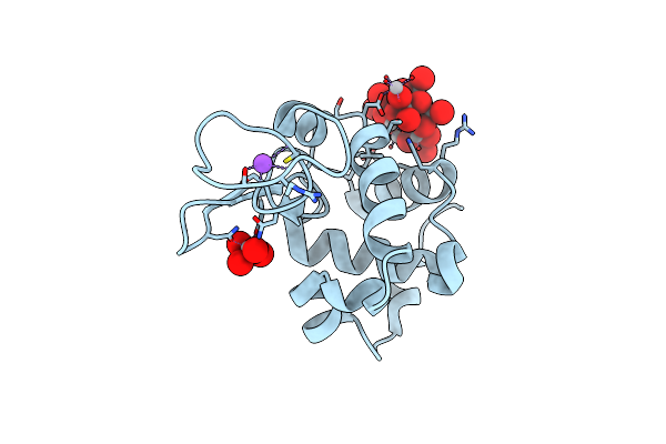
X-Ray Structure Of Lysozyme Obtained Upon Reaction With The Dioxidovanadium(V) Complex With (E)-N'-(1-(2-Hydroxy-5-Methoxyphenyl)Ethylidene)Furan-2-Carbohydrazide)
Organism: Gallus gallus
Method: X-RAY DIFFRACTION Resolution:1.18 Å Release Date: 2024-12-18 Classification: HYDROLASE |
Organism: Gallus gallus
Method: X-RAY DIFFRACTION
Release Date: 2024-12-18
 |
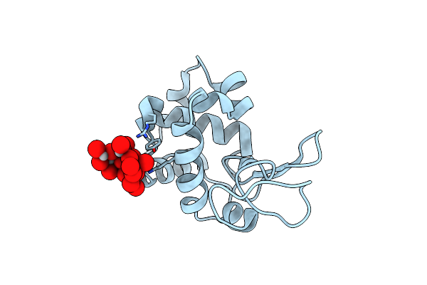
X-Ray Structure Of A Polyoxidovanadate/Lysozyme Adduct Obtained When The Protein Is Treated With [Vivo(Acac)2] In 1.1 M Nacl, 0.1 M Sodium Acetate At Ph 4.0 (Structure A)
Organism: Gallus
Method: X-RAY DIFFRACTION Resolution:1.45 Å Release Date: 2024-06-26 Classification: HYDROLASE Ligands: CL, NA, A1ICR, VO4 |
Organism: Gallus
Method: X-RAY DIFFRACTION
Release Date: 2024-06-26
Ligands: CL, NA, A1ICR, VO4
 |
X-Ray Structure Of A Polyoxidovanadate/Lysozyme Adduct Obtained When The Protein Is Treated With [Vivo(Acac)2] In 1.1 M Nacl, 0.1 M Sodium Acetate At Ph 4.0 (Structure B)
Organism: Gallus gallus
Method: X-RAY DIFFRACTION Resolution:1.79 Å Release Date: 2024-06-26 Classification: HYDROLASE Ligands: CL, A1H8D |
Organism: Gallus gallus
Method: X-RAY DIFFRACTION
Release Date: 2024-06-26
Ligands: CL, A1H8D