

Search Count: 557
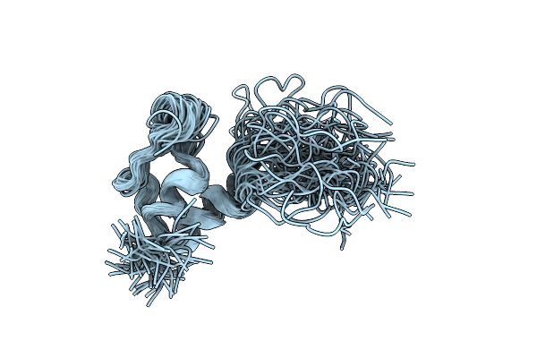 |
Three-Dimensional Solution Structure Of The E3-Binding Domain Of The Dihydrolipoamide Succinyltransferase Core From The 2-Oxoglutarate Dehydrogenase Multienzyme Complex Of (Escherichia Coli)
|
Organism: Escherichia coli
Method: SOLUTION NMR
Release Date: 1994-01-31
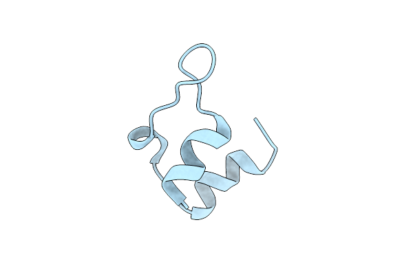 |
Three-Dimensional Solution Structure Of The E3-Binding Domain Of The Dihydrolipoamide Succinyltransferase Core From The 2-Oxoglutarate Dehydrogenase Multienzyme Complex Of Escherichia Coli
|
Organism: Escherichia coli
Method: SOLUTION NMR
Release Date: 1994-01-31
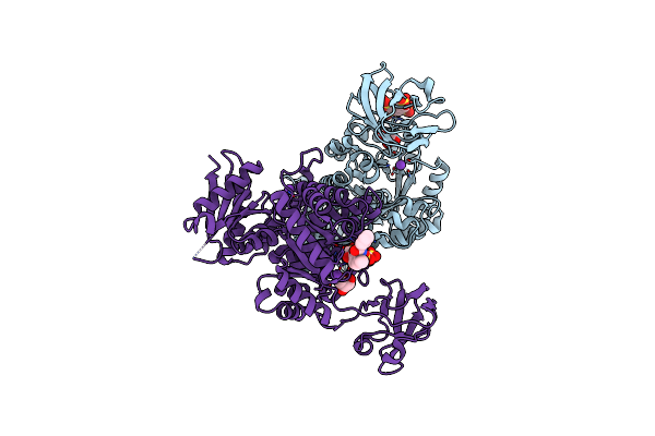 |
Crystal Structure Of Leishmania Mexicana Pyruvate Kinase In Complex With The Drug Suramin, An Inhibitor Of Glycolysis.
Organism: Leishmania mexicana
Method: X-RAY DIFFRACTION Resolution:2.35 Å Release Date: 2010-12-15 Classification: TRANSFERASE/TRANSFERASE INHIBITOR Ligands: PTK, GOL, K, SVR |
Organism: Leishmania mexicana
Method: X-RAY DIFFRACTION
Release Date: 2010-12-15
Ligands: PTK, GOL, K, SVR
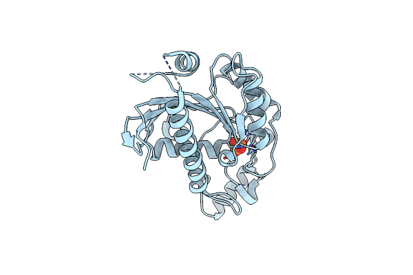 |
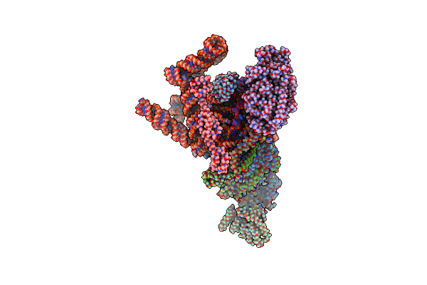
Crystal Structure A Tp53-Induced Glycolysis And Apoptosis Regulator Protein From Homo Sapiens.
Organism: Homo sapiens
Method: X-RAY DIFFRACTION Resolution:1.75 Å Release Date: 2008-07-15 Classification: apoptosis regulator Ligands: PO4 |
Organism: Homo sapiens
Method: X-RAY DIFFRACTION
Release Date: 2008-07-15
Ligands: PO4
 |
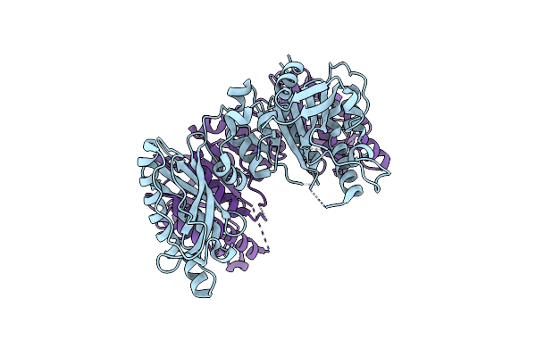
S. Cerevisiae Spliceosomal E Complex (Ubc4)
Organism: Saccharomyces cerevisiae (strain atcc 204508 / s288c), Saccharomyces cerevisiae s288c
Method: ELECTRON MICROSCOPY Release Date: 2019-09-18 Classification: RNA BINDING PROTEIN Ligands: ZN |
Organism: Saccharomyces cerevisiae (strain atcc 204508 / s288c), Saccharomyces cerevisiae s288c
Method: ELECTRON MICROSCOPY
Release Date: 2019-09-18
Ligands: ZN
 |
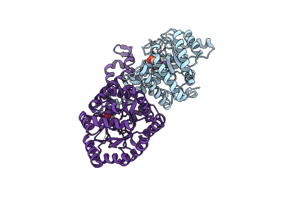
Cofactor-Independent Phosphoglycerate Mutase From Thermoplasma Acidophilum Dsm 1728
Organism: Thermoplasma acidophilum
Method: X-RAY DIFFRACTION Resolution:2.60 Å Release Date: 2009-12-01 Classification: ISOMERASE |
Organism: Thermoplasma acidophilum
Method: X-RAY DIFFRACTION
Release Date: 2009-12-01
 |
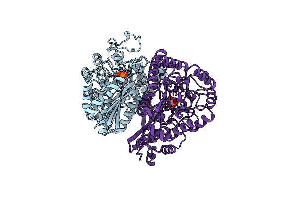
Class Ii Fructose-1,6-Bisphosphate Aldolase Of Escherichia Coli With Dhap
Organism: Escherichia coli (strain k12)
Method: X-RAY DIFFRACTION Resolution:1.70 Å Release Date: 2018-04-11 Classification: LYASE |
Organism: Escherichia coli (strain k12)
Method: X-RAY DIFFRACTION
Release Date: 2018-04-11
 |
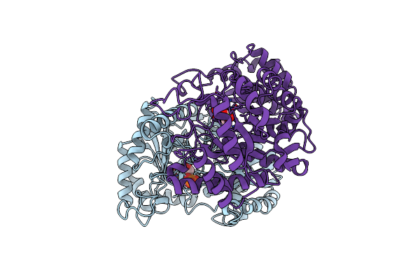
Yeast Enolase Complexed With An Equilibrium Mixture Of 2'-Phosphoglyceate And Phosphoenolpyruvate
Organism: Saccharomyces cerevisiae
Method: X-RAY DIFFRACTION Resolution:1.80 Å Release Date: 1997-01-11 Classification: LYASE Ligands: MG, PEP, 2PG |
Organism: Saccharomyces cerevisiae
Method: X-RAY DIFFRACTION
Release Date: 1997-01-11
Ligands: MG, PEP, 2PG
 |
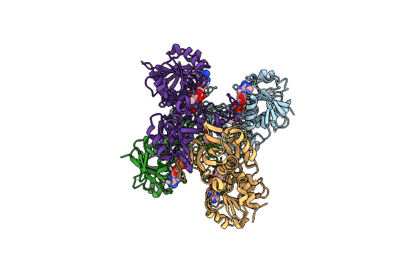
Asymmetric Yeast Enolase Dimer Complexed With Resolved 2'-Phosphoglycerate And Phosphoenolpyruvate
Organism: Saccharomyces cerevisiae
Method: X-RAY DIFFRACTION Resolution:2.00 Å Release Date: 1998-01-14 Classification: LYASE Ligands: MG, LI, 2PG, PEP |
Organism: Saccharomyces cerevisiae
Method: X-RAY DIFFRACTION
Release Date: 1998-01-14
Ligands: MG, LI, 2PG, PEP
 |
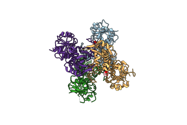
Crystal Structure Of H178N Mutant Of Glyceraldehyde-3-Phosphate-Dehydrogenase 1 (Gapdh 1) From Staphylococcus Aureus Mrsa252 Complexed With Nad At 2.0 Angstrom Resolution.
Organism: Staphylococcus aureus
Method: X-RAY DIFFRACTION Resolution:2.00 Å Release Date: 2010-08-18 Classification: OXIDOREDUCTASE Ligands: NAD, GOL |
Organism: Staphylococcus aureus
Method: X-RAY DIFFRACTION
Release Date: 2010-08-18
Ligands: NAD, GOL
 |
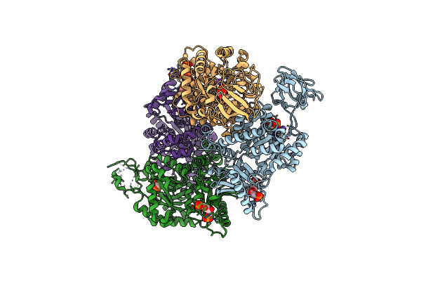
Crystal Structure Of Apo Glyceraldehyde-3-Phosphate Dehydrogenase 1 (Gapdh1) From Methicllin Resistant Staphylococcus Aureus (Mrsa252)
Organism: Staphylococcus aureus
Method: X-RAY DIFFRACTION Resolution:2.50 Å Release Date: 2010-08-18 Classification: OXIDOREDUCTASE Ligands: GOL |
Organism: Staphylococcus aureus
Method: X-RAY DIFFRACTION
Release Date: 2010-08-18
Ligands: GOL
 |
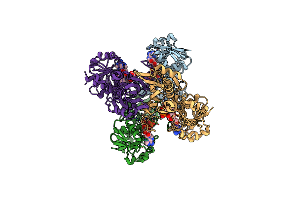
Human Erythrocyte Pyruvate Kinase: R486W Mutant
Organism: Homo sapiens
Method: X-RAY DIFFRACTION Resolution:2.87 Å Release Date: 2007-11-20 Classification: TRANSFERASE Ligands: FBP, PGA, K, MN |
Organism: Homo sapiens
Method: X-RAY DIFFRACTION
Release Date: 2007-11-20
Ligands: FBP, PGA, K, MN
 |
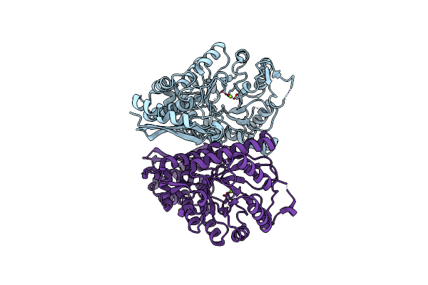
Crystal Structure Of C151S+H178N Mutant Of Glyceraldehyde-3-Phosphate-Dehydrogenase 1 (Gapdh 1) From Staphylococcus Aureus Mrsa252 Complexed With Nad And G3P
Organism: Staphylococcus aureus subsp. aureus
Method: X-RAY DIFFRACTION Resolution:2.60 Å Release Date: 2010-08-18 Classification: OXIDOREDUCTASE Ligands: NAD, 3PG |
Organism: Staphylococcus aureus subsp. aureus
Method: X-RAY DIFFRACTION
Release Date: 2010-08-18
Ligands: NAD, 3PG
 |
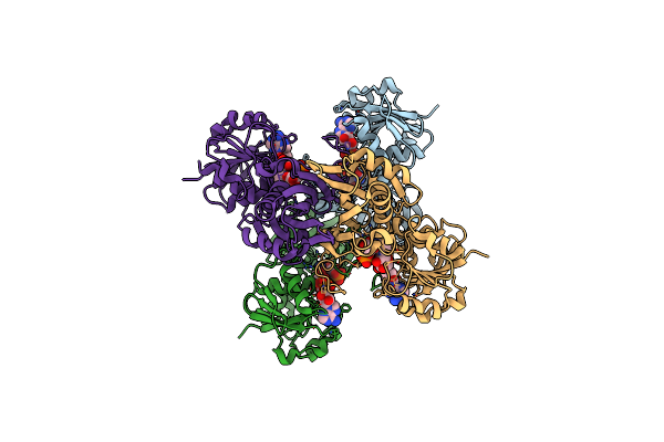
Aspergillus Fumigatus Enolase
Organism: Neosartorya fumigata (strain atcc mya-4609 / af293 / cbs 101355 / fgsc a1100)
Method: X-RAY DIFFRACTION Resolution:2.00 Å Release Date: 2022-03-16 Classification: LYASE Ligands: MG |
Organism: Neosartorya fumigata (strain atcc mya-4609 / af293 / cbs 101355 / fgsc a1100)
Method: X-RAY DIFFRACTION
Release Date: 2022-03-16
Ligands: MG
 |
Crystal Structure Of C151S Mutant Of Glyceraldehyde-3-Phosphate Dehydrogenase 1 (Gapdh 1)From Methicillin Resistant Staphylococcus Aureus Mrsa252 Complexed With Nad And G3P
Organism: Staphylococcus aureus subsp. aureus
Method: X-RAY DIFFRACTION Resolution:2.50 Å Release Date: 2010-08-18 Classification: OXIDOREDUCTASE Ligands: NAD, 3PG |
Organism: Staphylococcus aureus subsp. aureus
Method: X-RAY DIFFRACTION
Release Date: 2010-08-18
Ligands: NAD, 3PG
 |
Colias Eurytheme Phosphoglucose Isomerase. Homodimer From 4-5(18) Genotype.
Organism: Colias eurytheme
Method: X-RAY DIFFRACTION Resolution:1.50 Å Release Date: 2015-10-14 Classification: ISOMERASE |
Organism: Colias eurytheme
Method: X-RAY DIFFRACTION
Release Date: 2015-10-14
 |
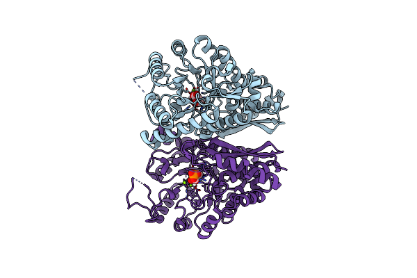
Aspergillus Fumigatus Enolase Bound To 2-Phosphoglycerate
Organism: Neosartorya fumigata (strain atcc mya-4609 / af293 / cbs 101355 / fgsc a1100)
Method: X-RAY DIFFRACTION Resolution:1.90 Å Release Date: 2022-03-16 Classification: LYASE Ligands: 2PG, MG |
Organism: Neosartorya fumigata (strain atcc mya-4609 / af293 / cbs 101355 / fgsc a1100)
Method: X-RAY DIFFRACTION
Release Date: 2022-03-16
Ligands: 2PG, MG
 |
Aspergillus Fumigatus Enolase Bound To Phosphoenolpyruvate And 2-Phosphoglycerate
Organism: Neosartorya fumigata (strain atcc mya-4609 / af293 / cbs 101355 / fgsc a1100)
Method: X-RAY DIFFRACTION Resolution:2.30 Å Release Date: 2022-03-16 Classification: LYASE Ligands: MG, 2PG, PEP |
Organism: Neosartorya fumigata (strain atcc mya-4609 / af293 / cbs 101355 / fgsc a1100)
Method: X-RAY DIFFRACTION
Release Date: 2022-03-16
Ligands: MG, 2PG, PEP
 |
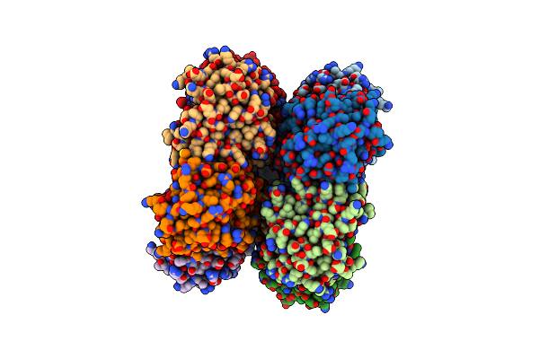
Class Ii Fructose-1,6-Bisphosphate Aldolase Of Escherichia Coli With D-Glucitol 1,6-Bisphosphate
Organism: Escherichia coli (strain k12)
Method: X-RAY DIFFRACTION Resolution:1.65 Å Release Date: 2018-04-11 Classification: LYASE |
Organism: Escherichia coli (strain k12)
Method: X-RAY DIFFRACTION
Release Date: 2018-04-11
 |
Structure Of An Archaeal Fructose-1,6-Bisphosphate Aldolase With The Catalytic Lys Covalently Bound To The Carbinolamine Intermediate Of The Substrate.
Organism: Thermoproteus tenax
Method: X-RAY DIFFRACTION Resolution:1.93 Å Release Date: 2011-04-27 Classification: LYASE Ligands: M2P |
Organism: Thermoproteus tenax
Method: X-RAY DIFFRACTION
Release Date: 2011-04-27
Ligands: M2P