

Search Count: 31
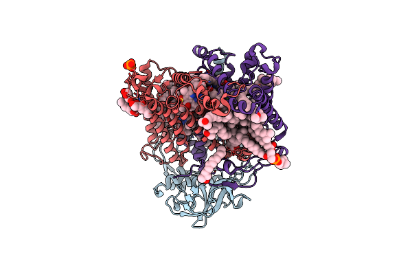 |
X-Ray High Resolution Structure Of The Photosynthetic Reaction Center From Rb. Sphaeroides At Ph 8 In The Neutral State
Organism: Rhodobacter sphaeroides
Method: X-RAY DIFFRACTION Resolution:1.87 Å Release Date: 2007-07-03 Classification: ELECTRON TRANSPORT Ligands: GOL, BCL, LDA, BPH, U10, PO4, HTO, FE, SPO, CDL, PC1, GGD |
Organism: Rhodobacter sphaeroides
Method: X-RAY DIFFRACTION
Release Date: 2007-07-03
Ligands: GOL, BCL, LDA, BPH, U10, PO4, HTO, FE, SPO, CDL, PC1, GGD
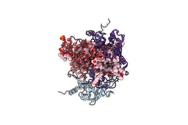 |
X-Ray High Resolution Structure Of The Photosynthetic Reaction Center From Rb. Sphaeroides At Ph 8 In The Charge-Separated State
Organism: Rhodobacter sphaeroides
Method: X-RAY DIFFRACTION Resolution:2.07 Å Release Date: 2007-07-03 Classification: ELECTRON TRANSPORT Ligands: GOL, BCL, LDA, BPH, U10, PO4, HTO, FE, SPO, CDL |
Organism: Rhodobacter sphaeroides
Method: X-RAY DIFFRACTION
Release Date: 2007-07-03
Ligands: GOL, BCL, LDA, BPH, U10, PO4, HTO, FE, SPO, CDL
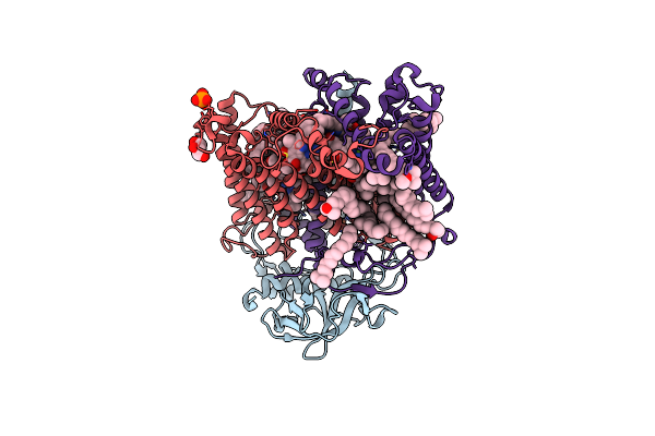 |
X-Ray High Resolution Structure Of The Photosynthetic Reaction Center From Rb. Sphaeroides At Ph 6.5 In The Charge-Separated State
Organism: Rhodobacter sphaeroides
Method: X-RAY DIFFRACTION Resolution:2.90 Å Release Date: 2007-07-03 Classification: PHOTOSYNTHESIS Ligands: GOL, LDA, BCL, BPH, UQ2, PO4, FE, U10, SPO |
Organism: Rhodobacter sphaeroides
Method: X-RAY DIFFRACTION
Release Date: 2007-07-03
Ligands: GOL, LDA, BCL, BPH, UQ2, PO4, FE, U10, SPO
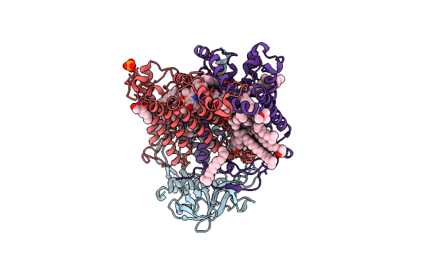 |
X-Ray High Resolution Structure Of The Photosynthetic Reaction Center From Rb. Sphaeroides At Ph 6.5 In The Charge-Separated State 2Nd Dataset
Organism: Rhodobacter sphaeroides
Method: X-RAY DIFFRACTION Resolution:2.50 Å Release Date: 2007-07-03 Classification: PHOTOSYNTHESIS Ligands: GOL, BCL, BPH, UQ2, PO4, HTO, LDA, FE, U10, SPO |
Organism: Rhodobacter sphaeroides
Method: X-RAY DIFFRACTION
Release Date: 2007-07-03
Ligands: GOL, BCL, BPH, UQ2, PO4, HTO, LDA, FE, U10, SPO
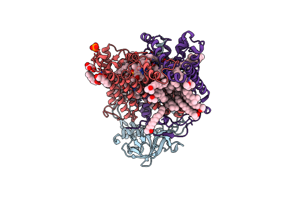 |
X-Ray High Resolution Structure Of The Photosynthetic Reaction Center From Rb. Sphaeroides At Ph 6.5 In The Neutral State, 2Nd Dataset
Organism: Rhodobacter sphaeroides
Method: X-RAY DIFFRACTION Resolution:2.04 Å Release Date: 2007-07-03 Classification: PHOTOSYNTHESIS Ligands: GOL, BCL, LDA, BPH, UQ2, PO4, HTO, FE, U10, SPO, CDL |
Organism: Rhodobacter sphaeroides
Method: X-RAY DIFFRACTION
Release Date: 2007-07-03
Ligands: GOL, BCL, LDA, BPH, UQ2, PO4, HTO, FE, U10, SPO, CDL
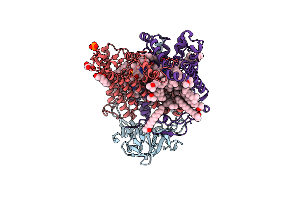 |
X-Ray High Resolution Structure Of The Photosynthetic Reaction Center From Rb. Sphaeroides At Ph 6.5 In The Charge-Separated State, 3Rd Dataset
Organism: Rhodobacter sphaeroides
Method: X-RAY DIFFRACTION Resolution:2.13 Å Release Date: 2007-07-03 Classification: PHOTOSYNTHESIS Ligands: GOL, BCL, LDA, BPH, UQ2, PO4, HTO, FE, U10, SPO |
Organism: Rhodobacter sphaeroides
Method: X-RAY DIFFRACTION
Release Date: 2007-07-03
Ligands: GOL, BCL, LDA, BPH, UQ2, PO4, HTO, FE, U10, SPO
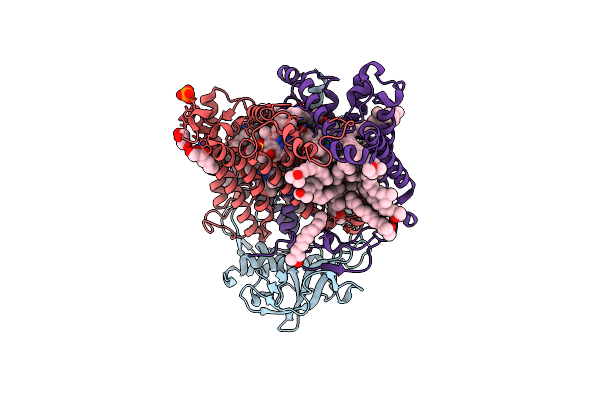 |
X-Ray High Resolution Structure Of The Photosynthetic Reaction Center From Rb. Sphaeroides At Ph 6.5 In The Neutral State
Organism: Rhodobacter sphaeroides
Method: X-RAY DIFFRACTION Resolution:2.05 Å Release Date: 2007-07-03 Classification: PHOTOSYNTHESIS Ligands: GOL, BCL, LDA, BPH, UQ2, PO4, HTO, FE, U10, SPO, CDL |
Organism: Rhodobacter sphaeroides
Method: X-RAY DIFFRACTION
Release Date: 2007-07-03
Ligands: GOL, BCL, LDA, BPH, UQ2, PO4, HTO, FE, U10, SPO, CDL
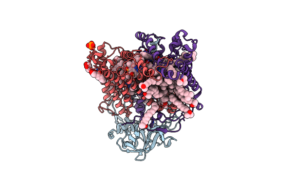 |
X-Ray High Resolution Structure Of The Photosynthetic Reaction Center From Rb. Sphaeroides At Ph 9 In The Neutral State
Organism: Rhodobacter sphaeroides
Method: X-RAY DIFFRACTION Resolution:2.50 Å Release Date: 2007-07-03 Classification: PHOTOSYNTHESIS Ligands: GOL, BCL, LDA, BPH, UQ2, PO4, HTO, FE, U10, SPO, CDL |
Organism: Rhodobacter sphaeroides
Method: X-RAY DIFFRACTION
Release Date: 2007-07-03
Ligands: GOL, BCL, LDA, BPH, UQ2, PO4, HTO, FE, U10, SPO, CDL
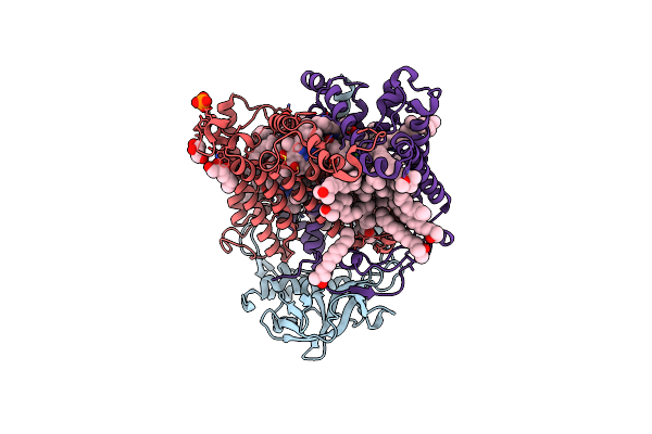 |
X-Ray High Resolution Structure Of The Photosynthetic Reaction Center From Rb. Sphaeroides At Ph 9 In The Charge-Separated State, 2Nd Dataset
Organism: Rhodobacter sphaeroides
Method: X-RAY DIFFRACTION Resolution:2.51 Å Release Date: 2007-07-03 Classification: PHOTOSYNTHESIS Ligands: GOL, BCL, LDA, BPH, UQ2, PO4, HTO, FE, U10, SPO, CDL |
Organism: Rhodobacter sphaeroides
Method: X-RAY DIFFRACTION
Release Date: 2007-07-03
Ligands: GOL, BCL, LDA, BPH, UQ2, PO4, HTO, FE, U10, SPO, CDL
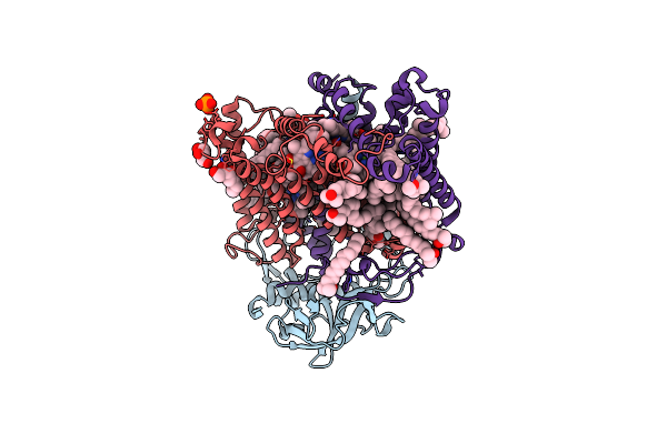 |
X-Ray High Resolution Structure Of The Photosynthetic Reaction Center From Rb. Sphaeroides At Ph 9 In The Charge-Separated State
Organism: Rhodobacter sphaeroides
Method: X-RAY DIFFRACTION Resolution:2.21 Å Release Date: 2007-07-03 Classification: PHOTOSYNTHESIS Ligands: GOL, BCL, LDA, BPH, UQ2, PO4, HTO, FE, U10, SPO, CDL |
Organism: Rhodobacter sphaeroides
Method: X-RAY DIFFRACTION
Release Date: 2007-07-03
Ligands: GOL, BCL, LDA, BPH, UQ2, PO4, HTO, FE, U10, SPO, CDL
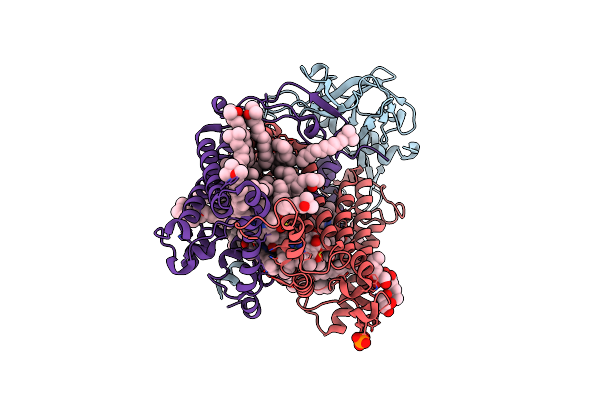 |
X-Ray High Resolution Structure Of The Photosynthetic Reaction Center From Rb. Sphaeroides At Ph 10 In The Neutral State
Organism: Rhodobacter sphaeroides
Method: X-RAY DIFFRACTION Resolution:2.25 Å Release Date: 2007-07-03 Classification: PHOTOSYNTHESIS Ligands: GOL, BCL, LDA, BPH, UQ2, PO4, HTO, FE, U10, SPO, CDL |
Organism: Rhodobacter sphaeroides
Method: X-RAY DIFFRACTION
Release Date: 2007-07-03
Ligands: GOL, BCL, LDA, BPH, UQ2, PO4, HTO, FE, U10, SPO, CDL
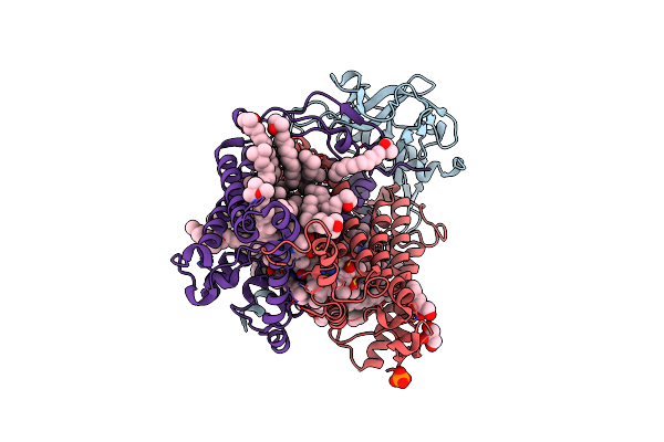 |
X-Ray High Resolution Structure Of The Photosynthetic Reaction Center From Rb. Sphaeroides At Ph 10 In The Charge-Separated State
Organism: Rhodobacter sphaeroides
Method: X-RAY DIFFRACTION Resolution:2.31 Å Release Date: 2007-07-03 Classification: PHOTOSYNTHESIS Ligands: GOL, BCL, LDA, BPH, UQ2, PO4, HTO, FE, U10, SPO, CDL |
Organism: Rhodobacter sphaeroides
Method: X-RAY DIFFRACTION
Release Date: 2007-07-03
Ligands: GOL, BCL, LDA, BPH, UQ2, PO4, HTO, FE, U10, SPO, CDL
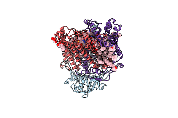 |
X-Ray High Resolution Structure Of The Photosynthetic Reaction Center From Rb. Sphaeroides At Ph 10 In The Neutral State, 2Nd Dataset
Organism: Rhodobacter sphaeroides
Method: X-RAY DIFFRACTION Resolution:2.88 Å Release Date: 2007-07-03 Classification: PHOTOSYNTHESIS Ligands: GOL, BCL, BPH, UQ2, HTO, LDA, FE, U10, SPO |
Organism: Rhodobacter sphaeroides
Method: X-RAY DIFFRACTION
Release Date: 2007-07-03
Ligands: GOL, BCL, BPH, UQ2, HTO, LDA, FE, U10, SPO
 |
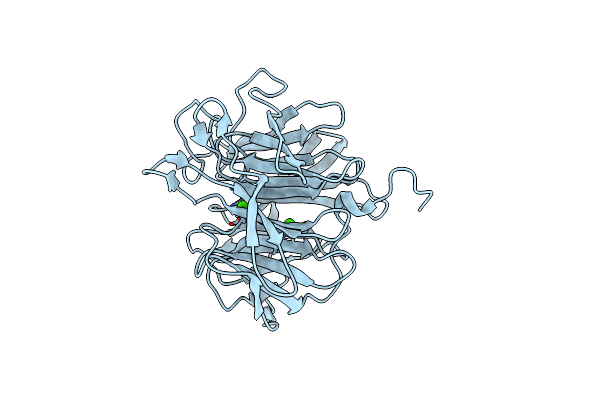
X-Ray High Resolution Structure Of The Photosynthetic Reaction Center From Rb. Sphaeroides At Ph 10 In The Charge-Separated State, 2Nd Dataset
Organism: Rhodobacter sphaeroides
Method: X-RAY DIFFRACTION Resolution:2.70 Å Release Date: 2007-07-03 Classification: PHOTOSYNTHESIS Ligands: GOL, BCL, LDA, BPH, UQ2, PO4, HTO, FE, U10, SPO |
Organism: Rhodobacter sphaeroides
Method: X-RAY DIFFRACTION
Release Date: 2007-07-03
Ligands: GOL, BCL, LDA, BPH, UQ2, PO4, HTO, FE, U10, SPO
 |
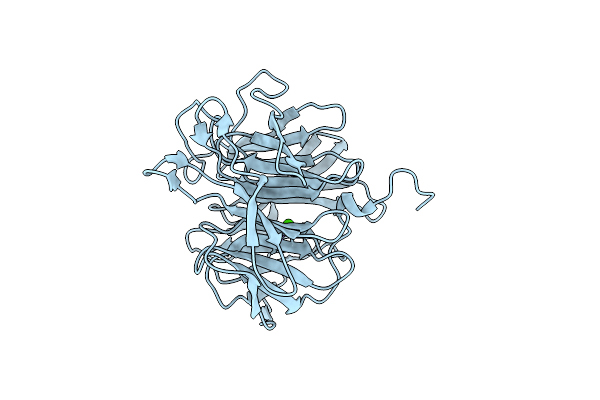
Crystal Structure Of Squid Ganglion Dfpase E37Q Mutant
Organism: Loligo vulgaris
Method: X-RAY DIFFRACTION Resolution:2.00 Å Release Date: 2006-09-26 Classification: HYDROLASE Ligands: CA |
Organism: Loligo vulgaris
Method: X-RAY DIFFRACTION
Release Date: 2006-09-26
Ligands: CA
 |
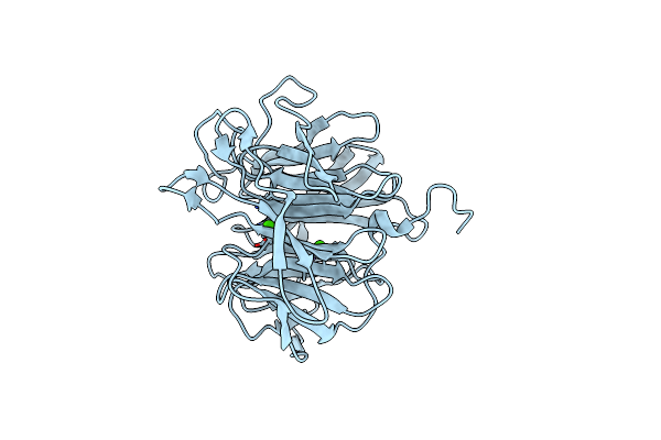
Crystal Structure Of Squid Ganglion Dfpase E21Q Mutant
Organism: Loligo vulgaris
Method: X-RAY DIFFRACTION Resolution:1.90 Å Release Date: 2006-09-26 Classification: HYDROLASE Ligands: CA |
Organism: Loligo vulgaris
Method: X-RAY DIFFRACTION
Release Date: 2006-09-26
Ligands: CA
 |
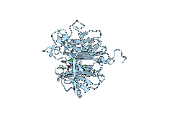
Crystal Structure Of Squid Ganglion Dfpase S271A Mutant
Organism: Loligo vulgaris
Method: X-RAY DIFFRACTION Resolution:2.10 Å Release Date: 2006-09-26 Classification: HYDROLASE Ligands: CA |
Organism: Loligo vulgaris
Method: X-RAY DIFFRACTION
Release Date: 2006-09-26
Ligands: CA
 |
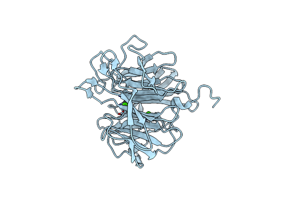
Crystal Structure Of Squid Ganglion Dfpase W244H Mutant
Organism: Loligo vulgaris
Method: X-RAY DIFFRACTION Resolution:1.90 Å Release Date: 2006-09-26 Classification: HYDROLASE Ligands: CA |
Organism: Loligo vulgaris
Method: X-RAY DIFFRACTION
Release Date: 2006-09-26
Ligands: CA
 |
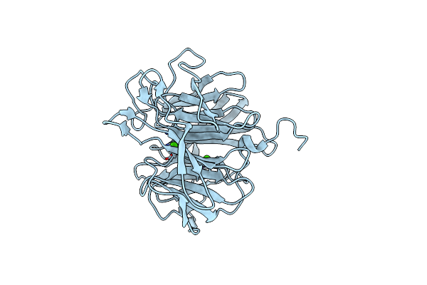
Crystal Structure Of Squid Ganglion Dfpase W244F Mutant
Organism: Loligo vulgaris
Method: X-RAY DIFFRACTION Resolution:2.00 Å Release Date: 2006-09-26 Classification: HYDROLASE Ligands: CA |
Organism: Loligo vulgaris
Method: X-RAY DIFFRACTION
Release Date: 2006-09-26
Ligands: CA
 |
Crystal Structure Of Squid Ganglion Dfpase W244L Mutant
Organism: Loligo vulgaris
Method: X-RAY DIFFRACTION Resolution:1.90 Å Release Date: 2006-09-26 Classification: HYDROLASE Ligands: CA |
Organism: Loligo vulgaris
Method: X-RAY DIFFRACTION
Release Date: 2006-09-26
Ligands: CA