

Search Count: 27
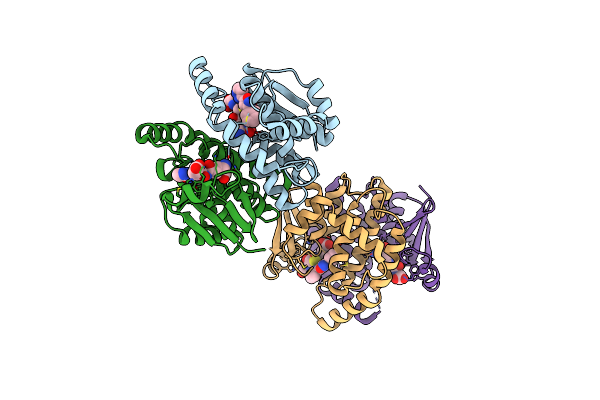 |
Complex Structure Of Prostaglandin D2 Synthase At 1.95A.
Organism: Homo sapiens
Method: X-RAY DIFFRACTION Resolution:1.95 Å Release Date: 2008-04-15 Classification: ISOMERASE Ligands: GSH, D25 |
Organism: Homo sapiens
Method: X-RAY DIFFRACTION
Release Date: 2008-04-15
Ligands: GSH, D25
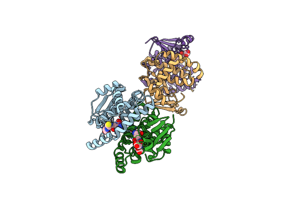 |
Complex Structure Of Prostaglandin D2 Synthase At 1.95A.
Organism: Homo sapiens
Method: X-RAY DIFFRACTION Resolution:1.95 Å Release Date: 2008-04-15 Classification: ISOMERASE Ligands: GSH, ZZA |
Organism: Homo sapiens
Method: X-RAY DIFFRACTION
Release Date: 2008-04-15
Ligands: GSH, ZZA
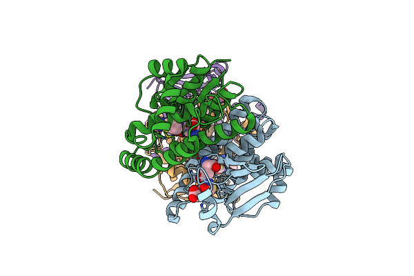 |
Complex Structure Of Prostaglandin D2 Synthase At 2.1A.
Organism: Homo sapiens
Method: X-RAY DIFFRACTION Resolution:2.10 Å Release Date: 2008-04-15 Classification: ISOMERASE Ligands: MG, GSH, D26 |
Organism: Homo sapiens
Method: X-RAY DIFFRACTION
Release Date: 2008-04-15
Ligands: MG, GSH, D26
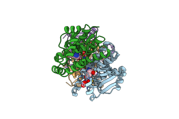 |
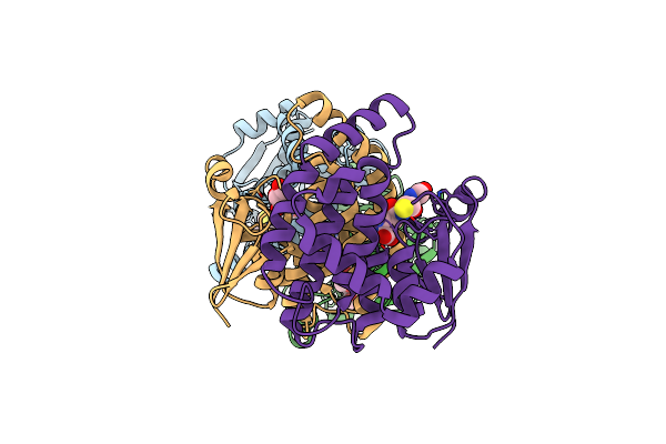
Complex Structure Of Prostaglandin D2 Synthase At 1.95A.
Organism: Homo sapiens
Method: X-RAY DIFFRACTION Resolution:1.95 Å Release Date: 2008-04-15 Classification: ISOMERASE Ligands: GSH, VC3 |
Organism: Homo sapiens
Method: X-RAY DIFFRACTION
Release Date: 2008-04-15
Ligands: GSH, VC3
 |
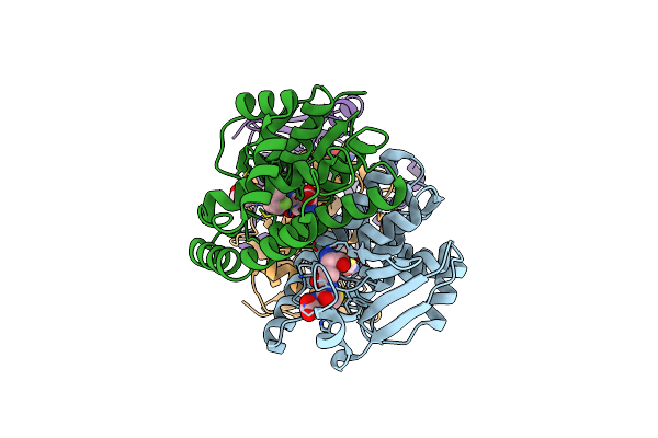
Complex Structure Of Prostaglandin D2 Synthase At 2.2A.
Organism: Homo sapiens
Method: X-RAY DIFFRACTION Resolution:2.20 Å Release Date: 2008-04-15 Classification: ISOMERASE Ligands: GSH, D27, GOL |
Organism: Homo sapiens
Method: X-RAY DIFFRACTION
Release Date: 2008-04-15
Ligands: GSH, D27, GOL
 |
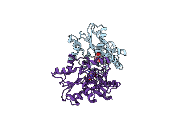
Complex Structure Of Prostaglandin D2 Synthase At 2.25A.
Organism: Homo sapiens
Method: X-RAY DIFFRACTION Resolution:2.25 Å Release Date: 2008-04-15 Classification: ISOMERASE Ligands: GSH, D28, MG |
Organism: Homo sapiens
Method: X-RAY DIFFRACTION
Release Date: 2008-04-15
Ligands: GSH, D28, MG
 |
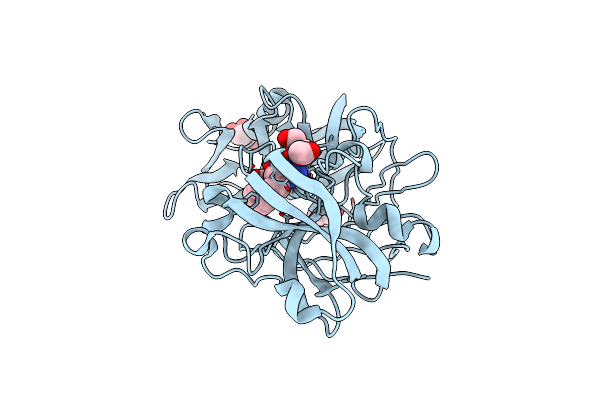
Crystal Structure Of Helicobacter Pylori Glutamate Racemase In Complex With D-Glutamate
Organism: Helicobacter pylori
Method: X-RAY DIFFRACTION Resolution:1.90 Å Release Date: 2008-02-19 Classification: ISOMERASE Ligands: DGL |
Organism: Helicobacter pylori
Method: X-RAY DIFFRACTION
Release Date: 2008-02-19
Ligands: DGL
 |
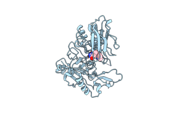
X-Ray Crystal Structure Of Endothiapepsin Complexed With Compound 1
Organism: Cryphonectria parasitica
Method: X-RAY DIFFRACTION Resolution:1.55 Å Release Date: 2007-12-04 Classification: HYDROLASE Ligands: GOL, ACT, V15 |
Organism: Cryphonectria parasitica
Method: X-RAY DIFFRACTION
Release Date: 2007-12-04
Ligands: GOL, ACT, V15
 |
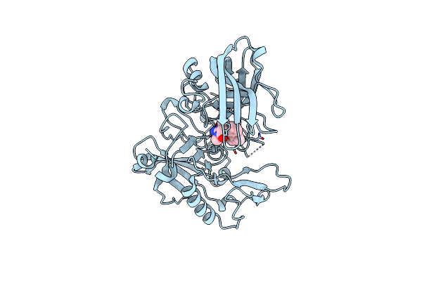
X-Ray Crystal Structure Of Beta Secretase Complexed With Compound 8C
Organism: Homo sapiens
Method: X-RAY DIFFRACTION Resolution:2.75 Å Release Date: 2007-11-13 Classification: HYDROLASE Ligands: IOD, C8C |
Organism: Homo sapiens
Method: X-RAY DIFFRACTION
Release Date: 2007-11-13
Ligands: IOD, C8C
 |
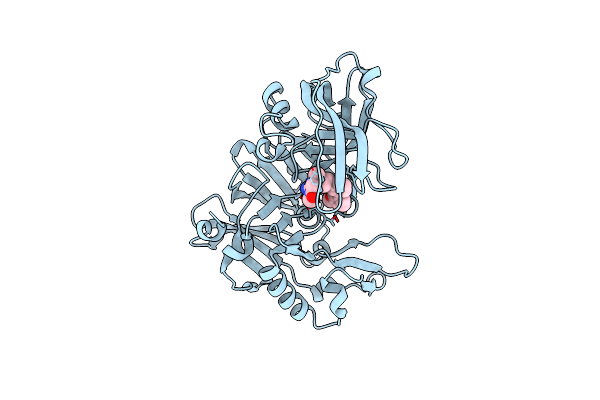
X-Ray Crystal Structure Of Beta Secretase Complexed With Compound 24
Organism: Homo sapiens
Method: X-RAY DIFFRACTION Resolution:2.50 Å Release Date: 2007-11-13 Classification: HYDROLASE Ligands: IOD, H24 |
Organism: Homo sapiens
Method: X-RAY DIFFRACTION
Release Date: 2007-11-13
Ligands: IOD, H24
 |
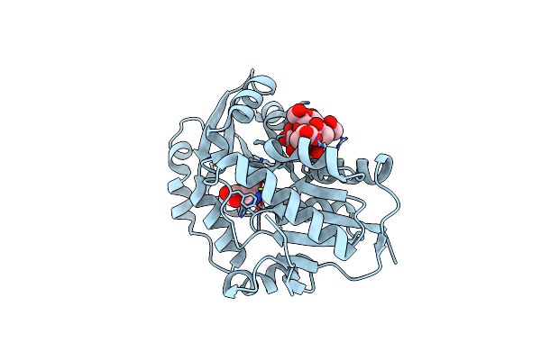
X-Ray Crystal Structure Of Beta Secretase Complexed With Compound 27
Organism: Homo sapiens
Method: X-RAY DIFFRACTION Resolution:2.20 Å Release Date: 2007-11-13 Classification: HYDROLASE Ligands: IOD, C27 |
Organism: Homo sapiens
Method: X-RAY DIFFRACTION
Release Date: 2007-11-13
Ligands: IOD, C27
 |
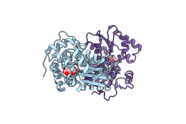
Crystal Structure Of Escherichia Coli Glutamate Racemase In Complex With L- Glutamate And Activator Udp-Murnac-Ala
Organism: Escherichia coli
Method: X-RAY DIFFRACTION Resolution:1.90 Å Release Date: 2007-07-03 Classification: ISOMERASE Ligands: GLU, UMA |
Organism: Escherichia coli
Method: X-RAY DIFFRACTION
Release Date: 2007-07-03
Ligands: GLU, UMA
 |
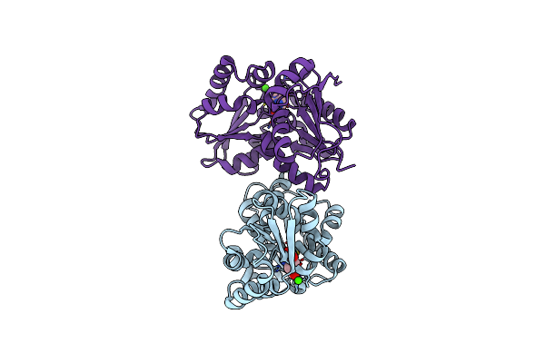
Crystal Structure Of Enterococcus Faecalis Glutamate Racemase In Complex With D- And L-Glutamate
Organism: Enterococcus faecalis
Method: X-RAY DIFFRACTION Resolution:2.50 Å Release Date: 2007-07-03 Classification: ISOMERASE Ligands: GLU, DGL |
Organism: Enterococcus faecalis
Method: X-RAY DIFFRACTION
Release Date: 2007-07-03
Ligands: GLU, DGL
 |
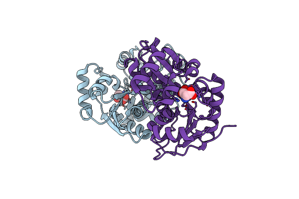
Crystal Structure Of Enterococcus Faecalis Glutamate Racemase In Complex With D- Glutamate
Organism: Enterococcus faecalis
Method: X-RAY DIFFRACTION Resolution:1.98 Å Release Date: 2007-07-03 Classification: ISOMERASE Ligands: DGL, CA |
Organism: Enterococcus faecalis
Method: X-RAY DIFFRACTION
Release Date: 2007-07-03
Ligands: DGL, CA
 |
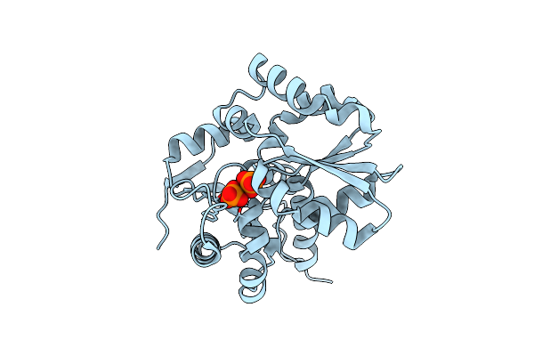
Crystal Structure Of Staphylococcus Aureus Glutamate Racemase In Complex With D- Glutamate
Organism: Staphylococcus aureus
Method: X-RAY DIFFRACTION Resolution:2.15 Å Release Date: 2007-07-03 Classification: ISOMERASE Ligands: DGL |
Organism: Staphylococcus aureus
Method: X-RAY DIFFRACTION
Release Date: 2007-07-03
Ligands: DGL
 |
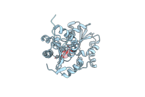
Crystal Structure Of Enterococcus Faecium Glutamate Racemase In Complex With Phosphate
Organism: Enterococcus faecium
Method: X-RAY DIFFRACTION Resolution:1.80 Å Release Date: 2007-07-03 Classification: ISOMERASE Ligands: PO4 |
Organism: Enterococcus faecium
Method: X-RAY DIFFRACTION
Release Date: 2007-07-03
Ligands: PO4
 |
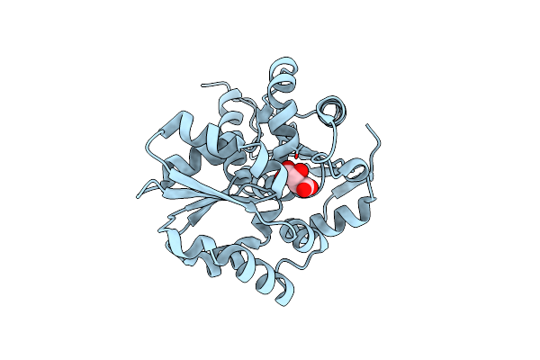
Crystal Structure Of Enterococcus Faecium Glutamate Racemase In Complex With Citrate
Organism: Enterococcus faecium
Method: X-RAY DIFFRACTION Resolution:1.80 Å Release Date: 2007-07-03 Classification: ISOMERASE Ligands: FLC |
Organism: Enterococcus faecium
Method: X-RAY DIFFRACTION
Release Date: 2007-07-03
Ligands: FLC
 |
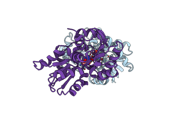
Crystal Structure Of Enterococcus Faecium Glutamate Racemase In Complex With Tartrate
Organism: Enterococcus faecium
Method: X-RAY DIFFRACTION Resolution:2.00 Å Release Date: 2007-07-03 Classification: ISOMERASE Ligands: TLA |
Organism: Enterococcus faecium
Method: X-RAY DIFFRACTION
Release Date: 2007-07-03
Ligands: TLA
 |
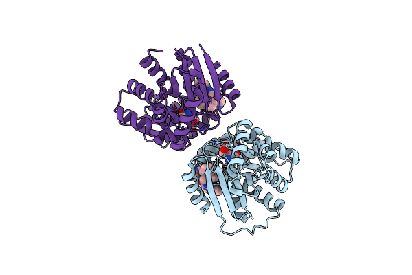
Crystal Structure Of Helicobacter Pylori Glutamate Racemase In Complex With D-Glutamate
Organism: Helicobacter pylori
Method: X-RAY DIFFRACTION Resolution:2.30 Å Release Date: 2007-07-03 Classification: ISOMERASE Ligands: DGL |
Organism: Helicobacter pylori
Method: X-RAY DIFFRACTION
Release Date: 2007-07-03
Ligands: DGL
 |
Crystal Structure Of Helicobacter Pylori Glutamate Racemase In Complex With D-Glutamate And An Inhibitor
Organism: Helicobacter pylori
Method: X-RAY DIFFRACTION Resolution:1.86 Å Release Date: 2007-07-03 Classification: ISOMERASE Ligands: 003, DGL |
Organism: Helicobacter pylori
Method: X-RAY DIFFRACTION
Release Date: 2007-07-03
Ligands: 003, DGL