

Search Count: 41
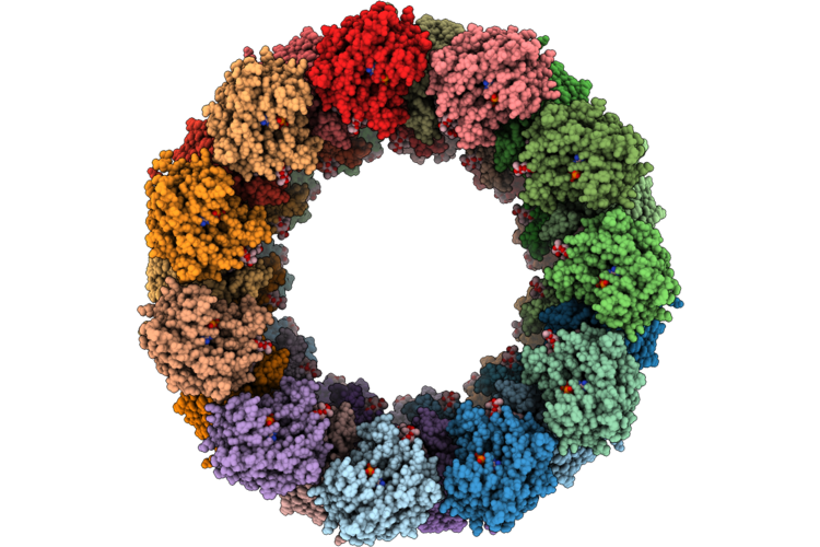 |
Cryo-Em Structure Of The Oligomeric Lpor:Chlide:Nadph Complexes Rf-23
Organism: Arabidopsis thaliana
Method: ELECTRON MICROSCOPY Release Date: 2026-08-26 Classification: PHOTOSYNTHESIS Ligands: NDP, LMG, A1JWG |
Organism: Arabidopsis thaliana
Method: ELECTRON MICROSCOPY
Release Date: 2026-08-26
Ligands: NDP, LMG, A1JWG
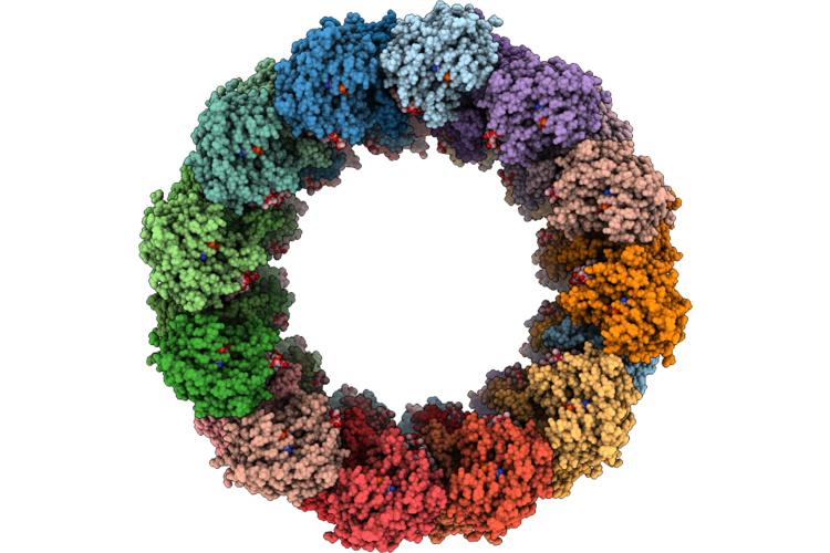 |
Cryo-Em Structure Of The Oligomeric Lpor:Chlide:Nadph Complexes Rf-25
Organism: Arabidopsis thaliana
Method: ELECTRON MICROSCOPY Release Date: 2026-08-19 Classification: PHOTOSYNTHESIS Ligands: NDP, LMG, A1JWG |
Organism: Arabidopsis thaliana
Method: ELECTRON MICROSCOPY
Release Date: 2026-08-19
Ligands: NDP, LMG, A1JWG
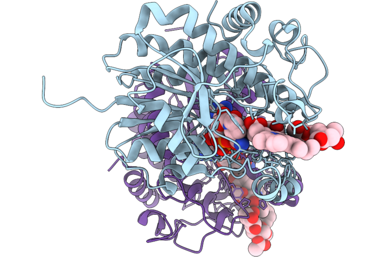 |
Cryo-Em Structure Of The Lpor:Chlide:Nadph Complexes (Rd-21) - Improved Resolution Of A Dimer Building Block Form Rf-21
Organism: Arabidopsis thaliana
Method: ELECTRON MICROSCOPY Resolution:2.64 Å Release Date: 2026-08-12 Classification: PHOTOSYNTHESIS Ligands: NDP, A1JWG, LMG |
Organism: Arabidopsis thaliana
Method: ELECTRON MICROSCOPY
Release Date: 2026-08-12
Ligands: NDP, A1JWG, LMG
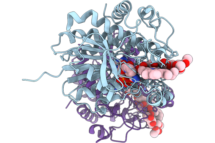 |
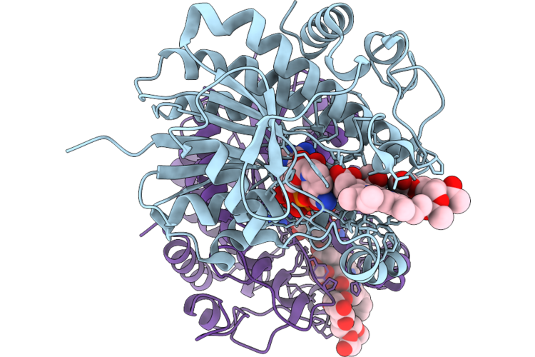
Cryo-Em Structure Of The Lpor:Chlide:Nadph Complexes (Rd-23) - Improved Resolution Of A Dimer Building Block Form Rf-23
Organism: Arabidopsis thaliana
Method: ELECTRON MICROSCOPY Release Date: 2026-08-12 Classification: PHOTOSYNTHESIS Ligands: NDP, LMG, A1JWG |
Organism: Arabidopsis thaliana
Method: ELECTRON MICROSCOPY
Release Date: 2026-08-12
Ligands: NDP, LMG, A1JWG
 |
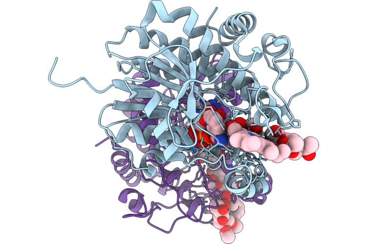
Cryo-Em Structure Of The Lpor:Chlide:Nadph Complexes (Rd-A) - Improved Resolution Of A Dimer Building Block Form Rf-21, Rf-23 And Rf-25
Organism: Arabidopsis thaliana
Method: ELECTRON MICROSCOPY Resolution:2.55 Å Release Date: 2026-08-12 Classification: PHOTOSYNTHESIS Ligands: NDP, A1JWG, LMG |
Organism: Arabidopsis thaliana
Method: ELECTRON MICROSCOPY
Release Date: 2026-08-12
Ligands: NDP, A1JWG, LMG
 |
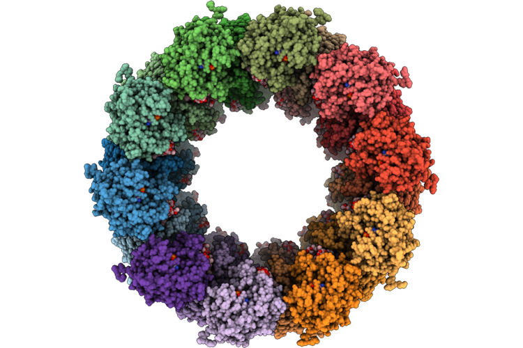
Cryo-Em Structure Of The Lpor:Chlide:Nadph Complexes (Rd-25) - Improved Resolution Of A Dimer Building Block Form Rf-25
Organism: Arabidopsis thaliana
Method: ELECTRON MICROSCOPY Resolution:2.58 Å Release Date: 2026-08-12 Classification: PHOTOSYNTHESIS Ligands: NDP, LMG, A1JWG |
Organism: Arabidopsis thaliana
Method: ELECTRON MICROSCOPY
Release Date: 2026-08-12
Ligands: NDP, LMG, A1JWG
 |
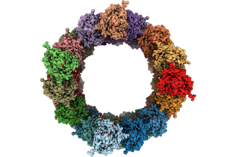
Cryo-Em Structure Of The Oligomeric Lpor:Chlide:Nadph Complexes Rf-21
Organism: Arabidopsis thaliana
Method: ELECTRON MICROSCOPY Release Date: 2026-08-12 Classification: PHOTOSYNTHESIS Ligands: NDP, A1JWG, LMG |
Organism: Arabidopsis thaliana
Method: ELECTRON MICROSCOPY
Release Date: 2026-08-12
Ligands: NDP, A1JWG, LMG
 |
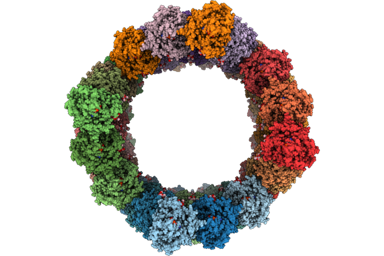
Cryo-Em Structure Of The Oligomeric Lpor:Chlide:Nadph Complexes Hf-23
Organism: Arabidopsis thaliana
Method: ELECTRON MICROSCOPY Release Date: 2026-08-12 Classification: PHOTOSYNTHESIS Ligands: NDP, LMG, A1JWG |
Organism: Arabidopsis thaliana
Method: ELECTRON MICROSCOPY
Release Date: 2026-08-12
Ligands: NDP, LMG, A1JWG
 |
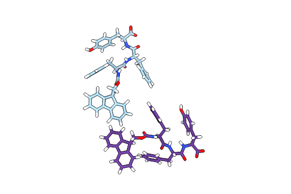
Cryo-Em Structure Of The Oligomeric Lpor:Chlide:Nadph Complexes Hf-25
Organism: Arabidopsis thaliana
Method: ELECTRON MICROSCOPY Release Date: 2026-08-12 Classification: PHOTOSYNTHESIS Ligands: NDP, LMG, A1JWG |
Organism: Arabidopsis thaliana
Method: ELECTRON MICROSCOPY
Release Date: 2026-08-12
Ligands: NDP, LMG, A1JWG
 |
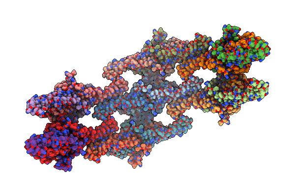
3D Cryo-Em Reveals The Structure Of A 3-Fmoc Zipper Motif Ensuring The Self-Assembly Of Tripeptide Nanofiber
Organism: Synthetic construct
Method: ELECTRON MICROSCOPY Release Date: 2024-11-06 Classification: BIOSYNTHETIC PROTEIN |
Organism: Synthetic construct
Method: ELECTRON MICROSCOPY
Release Date: 2024-11-06
 |
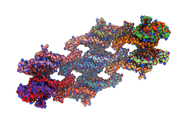
Pil1 In Native Eisosome Lattice Bound To Plasma Membrane Microdomain
Organism: Saccharomyces cerevisiae
Method: ELECTRON MICROSCOPY Release Date: 2024-07-24 Classification: LIPID BINDING PROTEIN |
Organism: Saccharomyces cerevisiae
Method: ELECTRON MICROSCOPY
Release Date: 2024-07-24
 |
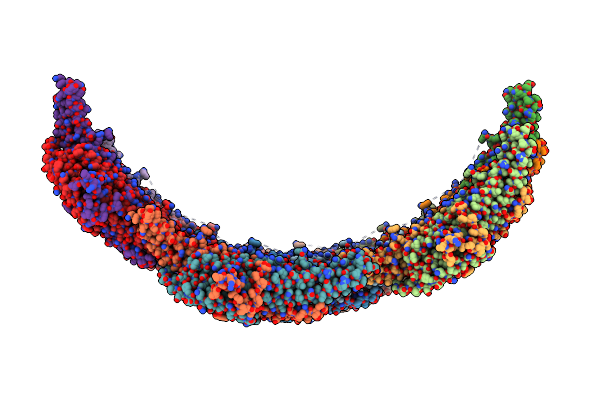
Lsp1 In Native Eisosome Lattice Bound To Plasma Membrane Microdomain
Organism: Saccharomyces cerevisiae
Method: ELECTRON MICROSCOPY Release Date: 2024-07-24 Classification: LIPID BINDING PROTEIN |
Organism: Saccharomyces cerevisiae
Method: ELECTRON MICROSCOPY
Release Date: 2024-07-24
 |
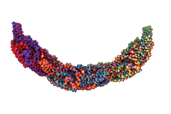
Helical Reconstruction Of Yeast Eisosome Protein Pil1 Bound To Membrane Composed Of Lipid Mixture -Pip2/+Sterol (Dopc, Dope, Dops, Cholesterol 30:20:20:30)
Organism: Saccharomyces cerevisiae
Method: ELECTRON MICROSCOPY Release Date: 2024-07-24 Classification: LIPID BINDING PROTEIN |
Organism: Saccharomyces cerevisiae
Method: ELECTRON MICROSCOPY
Release Date: 2024-07-24
 |
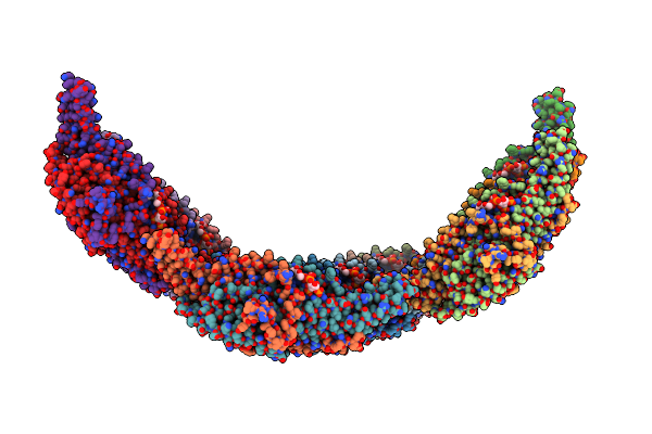
Helical Reconstruction Of Yeast Eisosome Protein Pil1 Bound To Membrane Composed Of Lipid Mixture +Pip2/-Sterol (Dopc, Dope, Dops, Pi(4,5)P2 50:20:20:10)
Organism: Saccharomyces cerevisiae
Method: ELECTRON MICROSCOPY Release Date: 2024-07-24 Classification: LIPID BINDING PROTEIN Ligands: I3P |
Organism: Saccharomyces cerevisiae
Method: ELECTRON MICROSCOPY
Release Date: 2024-07-24
Ligands: I3P
 |
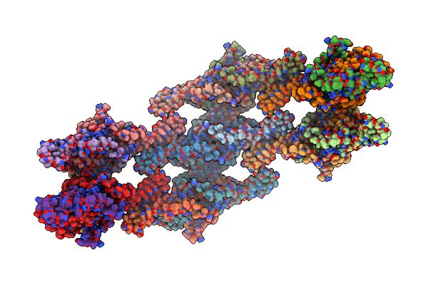
Helical Reconstruction Of Yeast Eisosome Protein Pil1 Bound To Membrane Composed Of Lipid Mixture +Pip2/+Sterol (Dopc, Dope, Dops, Cholesterol, Pi(4,5)P2 35:20:20:15:10)
Organism: Saccharomyces cerevisiae
Method: ELECTRON MICROSCOPY Release Date: 2024-07-24 Classification: LIPID BINDING PROTEIN Ligands: I3P, P5S |
Organism: Saccharomyces cerevisiae
Method: ELECTRON MICROSCOPY
Release Date: 2024-07-24
Ligands: I3P, P5S
 |
Compact State - Pil1 In Native Eisosome Lattice Bound To Plasma Membrane Microdomain
Organism: Saccharomyces cerevisiae
Method: ELECTRON MICROSCOPY Release Date: 2024-07-24 Classification: LIPID BINDING PROTEIN |
Organism: Saccharomyces cerevisiae
Method: ELECTRON MICROSCOPY
Release Date: 2024-07-24
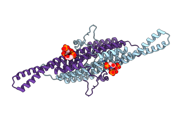 |
Compact State - Pil1 Dimer With Lipid Headgroups Fitted In Native Eisosome Lattice Bound To Plasma Membrane Microdomain
Organism: Saccharomyces cerevisiae
Method: ELECTRON MICROSCOPY Release Date: 2024-07-24 Classification: LIPID BINDING PROTEIN Ligands: I3P, SEP |
Organism: Saccharomyces cerevisiae
Method: ELECTRON MICROSCOPY
Release Date: 2024-07-24
Ligands: I3P, SEP
 |
Stretched State - Pil1 In Native Eisosome Lattice Bound To Plasma Membrane Microdomain
Organism: Saccharomyces cerevisiae
Method: ELECTRON MICROSCOPY Release Date: 2024-07-24 Classification: LIPID BINDING PROTEIN |
Organism: Saccharomyces cerevisiae
Method: ELECTRON MICROSCOPY
Release Date: 2024-07-24
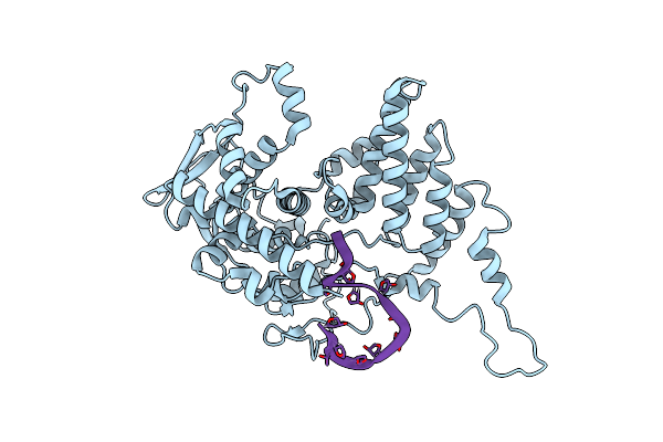 |
Model For Influenza A Virus Helical Ribonucleoprotein-Like Structure
Organism: Influenza a virus (a/wsn/1933(h1n1)), Synthetic construct
Method: ELECTRON MICROSCOPY Release Date: 2023-12-27 Classification: VIRAL PROTEIN |
Organism: Influenza a virus (a/wsn/1933(h1n1)), Synthetic construct
Method: ELECTRON MICROSCOPY
Release Date: 2023-12-27
 |
Model For Focused Reconstruction Of Influenza A Rnp-Like Particle
Organism: Influenza a virus, Synthetic construct
Method: ELECTRON MICROSCOPY Release Date: 2023-12-27 Classification: VIRAL PROTEIN |
Organism: Influenza a virus, Synthetic construct
Method: ELECTRON MICROSCOPY
Release Date: 2023-12-27