

Search Count: 3
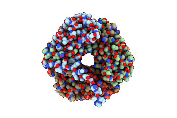 |
The Cryo-Electron Microscopy Structure Of The Type 1 Chaperone-Usher Pilus Rod
Organism: Escherichia coli j96
Method: ELECTRON MICROSCOPY Release Date: 2017-11-22 Classification: PROTEIN FIBRIL |
Organism: Escherichia coli j96
Method: ELECTRON MICROSCOPY
Release Date: 2017-11-22
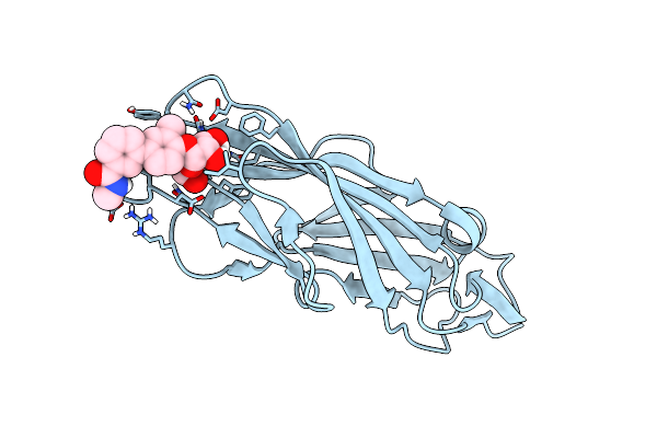 |
Crystal Structure Of Para-Biphenyl-2-Methyl-3'-Methyl Amide Mannoside Bound To Fimh Lectin Domain
Organism: Escherichia coli j96
Method: X-RAY DIFFRACTION Resolution:1.76 Å Release Date: 2016-12-07 Classification: SUGAR BINDING PROTEIN Ligands: 5US |
Organism: Escherichia coli j96
Method: X-RAY DIFFRACTION
Release Date: 2016-12-07
Ligands: 5US
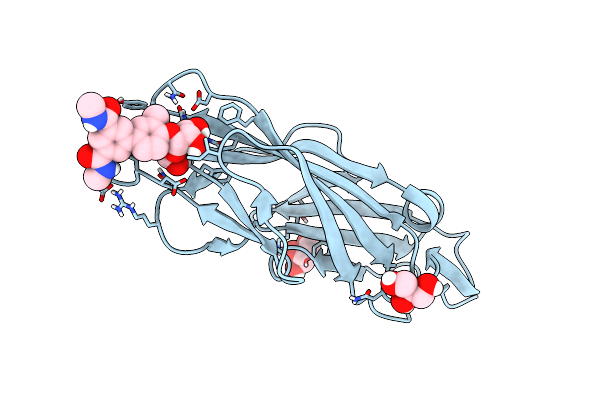 |
Crystal Structure Of Para-Biphenyl-2-Methyl-3', 5' Di-Methyl Amide Mannoside Bound To Fimh Lectin Domain
Organism: Escherichia coli j96
Method: X-RAY DIFFRACTION Resolution:1.67 Å Release Date: 2016-05-04 Classification: SUGAR BINDING PROTEIN Ligands: 5U7, GOL |
Organism: Escherichia coli j96
Method: X-RAY DIFFRACTION
Release Date: 2016-05-04
Ligands: 5U7, GOL