

Search Count: 18
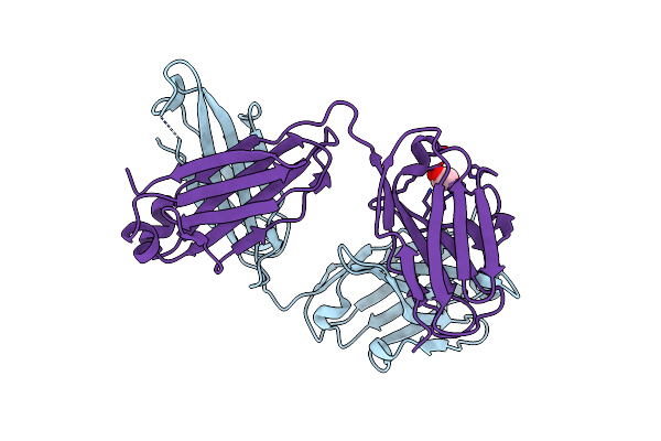 |
Monovalent Bispecific Igg Antibodies Through Novel Electrostatic Steering Mutations At The Ch1-Cl Interface
Organism: Homo sapiens
Method: X-RAY DIFFRACTION Resolution:2.10 Å Release Date: 2023-11-22 Classification: IMMUNE SYSTEM Ligands: GOL |
Organism: Homo sapiens
Method: X-RAY DIFFRACTION
Release Date: 2023-11-22
Ligands: GOL
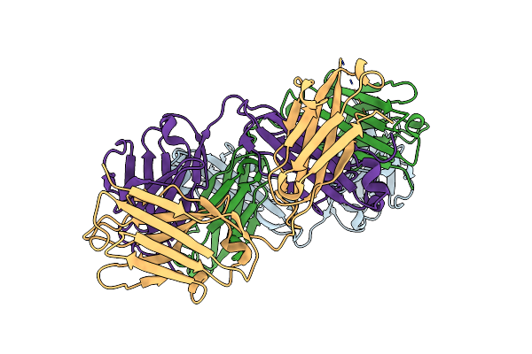 |
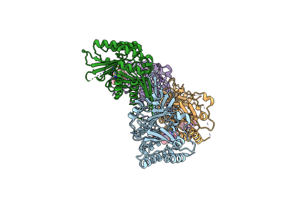
Monovalent Bispecific Igg Antibodies Through Novel Electrostatic Steering Mutations At The Ch1-Cl Interface
Organism: Homo sapiens
Method: X-RAY DIFFRACTION Resolution:2.30 Å Release Date: 2023-11-22 Classification: IMMUNE SYSTEM |
Organism: Homo sapiens
Method: X-RAY DIFFRACTION
Release Date: 2023-11-22
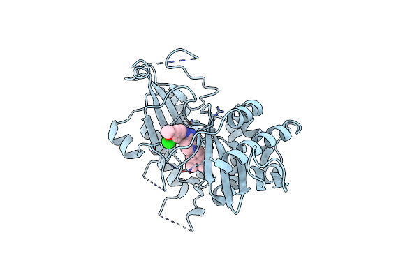 |
Crystal Structure Of Phosphatidylinositol 5-Phosphate 4-Kinase Type-2 Alpha (Pi5P4Ka) Bound To An Inhibitor
Organism: Homo sapiens
Method: X-RAY DIFFRACTION Resolution:2.10 Å Release Date: 2023-04-26 Classification: TRANSFERASE Ligands: U1U |
Organism: Homo sapiens
Method: X-RAY DIFFRACTION
Release Date: 2023-04-26
Ligands: U1U
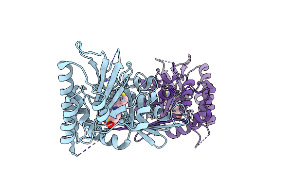 |
Crystal Structure Of Phosphatidylinositol 5-Phosphate 4-Kinase (Pi5P4K2C) Bound To An Inhibitor
Organism: Homo sapiens
Method: X-RAY DIFFRACTION Resolution:2.42 Å Release Date: 2022-12-28 Classification: TRANSFERASE Ligands: QZR |
Organism: Homo sapiens
Method: X-RAY DIFFRACTION
Release Date: 2022-12-28
Ligands: QZR
 |
Crystal Structure Of Phosphatidylinositol 5-Phosphate 4-Kinase (Pi5P4K2C) Bound To An Allosteric Inhibitor
Organism: Homo sapiens
Method: X-RAY DIFFRACTION Resolution:2.39 Å Release Date: 2022-05-18 Classification: TRANSFERASE Ligands: DVF |
Organism: Homo sapiens
Method: X-RAY DIFFRACTION
Release Date: 2022-05-18
Ligands: DVF
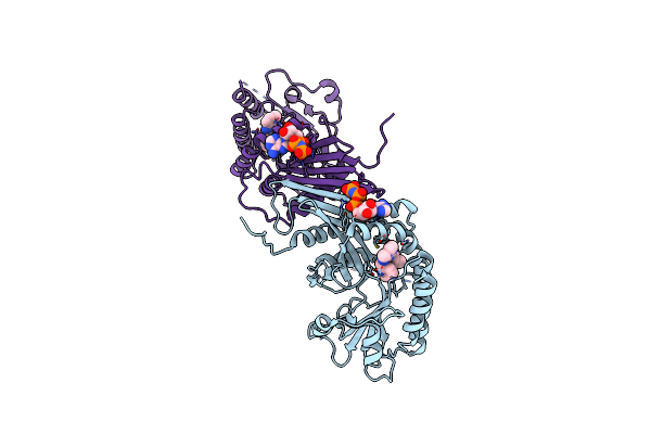 |
Crystal Structure Of Phosphatidylinositol 5-Phosphate 4-Kinase (Pi5P4K2C) Bound To An Allosteric Inhibitor And Amp-Pnp
Organism: Homo sapiens
Method: X-RAY DIFFRACTION Resolution:1.95 Å Release Date: 2022-05-18 Classification: TRANSFERASE Ligands: DVF, ANP |
Organism: Homo sapiens
Method: X-RAY DIFFRACTION
Release Date: 2022-05-18
Ligands: DVF, ANP
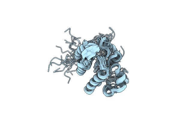 |
Trai (381-569)
|
Organism: Escherichia coli
Method: SOLUTION NMR
Release Date: 2012-01-11
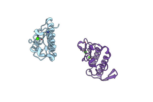 |
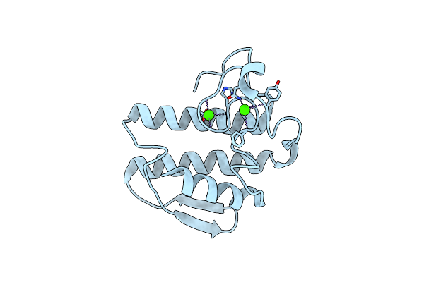
Crystal Structure Of The H48Q Mutant Of Human Group Iia Phospholipase A2
Organism: Homo sapiens
Method: X-RAY DIFFRACTION Resolution:1.50 Å Release Date: 2003-10-28 Classification: HYDROLASE Ligands: CA |
Organism: Homo sapiens
Method: X-RAY DIFFRACTION
Release Date: 2003-10-28
Ligands: CA
 |
Crystal Structure Of The N1A Mutant Of Human Group Iia Phospholipase A2
Organism: Homo sapiens
Method: X-RAY DIFFRACTION Resolution:2.60 Å Release Date: 2003-10-28 Classification: HYDROLASE Ligands: CA |
Organism: Homo sapiens
Method: X-RAY DIFFRACTION
Release Date: 2003-10-28
Ligands: CA
 |
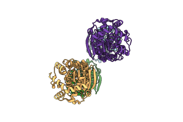
Crystal Structure Analysis Of The Anthranilate Phosphoribosyltransferase From Erwinia Carotovora (Current Name, Pectobacterium Carotovorum)
Organism: Pectobacterium carotovorum
Method: X-RAY DIFFRACTION Resolution:2.40 Å Release Date: 2002-10-23 Classification: TRANSFERASE Ligands: HG, MN, PRP |
Organism: Pectobacterium carotovorum
Method: X-RAY DIFFRACTION
Release Date: 2002-10-23
Ligands: HG, MN, PRP
 |
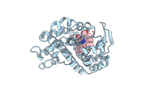
Crystal Structure Analysis Of The Anthranilate Phosphoribosyltransferase From Erwinia Carotovora At 1.9 Resolution (Current Name, Pectobacterium Carotovorum)
Organism: Pectobacterium carotovorum
Method: X-RAY DIFFRACTION Resolution:1.86 Å Release Date: 2002-10-23 Classification: TRANSFERASE |
Organism: Pectobacterium carotovorum
Method: X-RAY DIFFRACTION
Release Date: 2002-10-23
 |
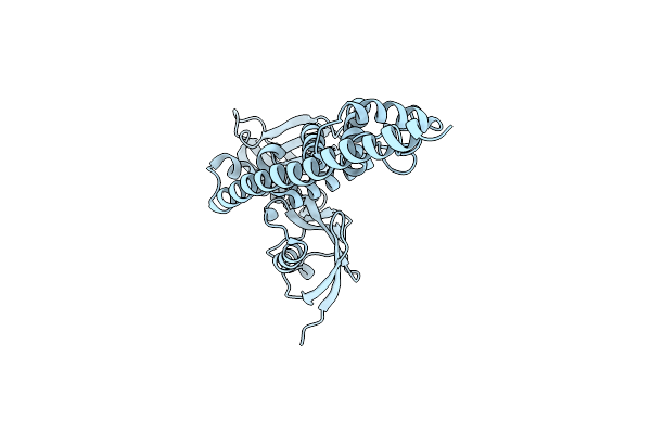
Laue Diffraction Study On The Structure Of Cytochrome C Peroxidase Compound I
Organism: Saccharomyces cerevisiae
Method: X-RAY DIFFRACTION Resolution:2.20 Å Release Date: 2001-07-26 Classification: OXIDOREDUCTASE Ligands: HEM, O |
Organism: Saccharomyces cerevisiae
Method: X-RAY DIFFRACTION
Release Date: 2001-07-26
Ligands: HEM, O
 |
Structure Of Isolated Ferm Domain And First Long Helix Of Moesin
Organism: Homo sapiens
Method: X-RAY DIFFRACTION Resolution:2.70 Å Release Date: 2001-06-27 Classification: MEMBRANE PROTEIN |
Organism: Homo sapiens
Method: X-RAY DIFFRACTION
Release Date: 2001-06-27
 |
Crystallographic Refinement Of Lignin Peroxidase At 2 Angstroms
Organism: Phanerochaete chrysosporium
Method: X-RAY DIFFRACTION Resolution:2.03 Å Release Date: 1993-10-31 Classification: OXIDOREDUCTASE Ligands: NAG, CA, HEM |
Organism: Phanerochaete chrysosporium
Method: X-RAY DIFFRACTION
Release Date: 1993-10-31
Ligands: NAG, CA, HEM
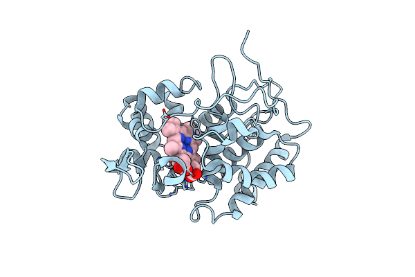 |
X-Ray Structures Of Recombinant Yeast Cytochrome C Peroxidase And Three Heme-Cleft Mutants Prepared By Site-Directed Mutagenesis
Organism: Saccharomyces cerevisiae
Method: X-RAY DIFFRACTION Resolution:2.20 Å Release Date: 1991-07-15 Classification: OXIDOREDUCTASE Ligands: HEM |
Organism: Saccharomyces cerevisiae
Method: X-RAY DIFFRACTION
Release Date: 1991-07-15
Ligands: HEM
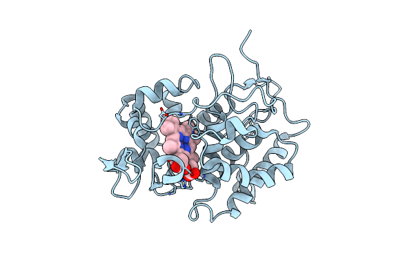 |
X-Ray Structures Of Recombinant Yeast Cytochrome C Peroxidase And Three Heme-Cleft Mutants Prepared By Site-Directed Mutagenesis
Organism: Saccharomyces cerevisiae
Method: X-RAY DIFFRACTION Resolution:2.20 Å Release Date: 1991-07-15 Classification: OXIDOREDUCTASE Ligands: HEM |
Organism: Saccharomyces cerevisiae
Method: X-RAY DIFFRACTION
Release Date: 1991-07-15
Ligands: HEM
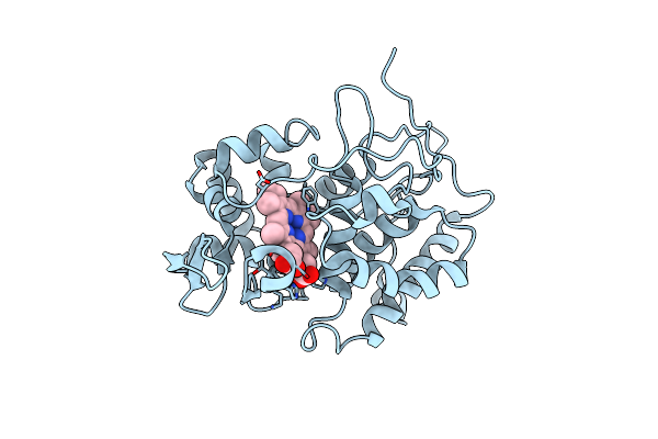 |
X-Ray Structures Of Recombinant Yeast Cytochrome C Peroxidase And Three Heme-Cleft Mutants Prepared By Site-Directed Mutagenesis
Organism: Saccharomyces cerevisiae
Method: X-RAY DIFFRACTION Resolution:2.20 Å Release Date: 1991-07-15 Classification: OXIDOREDUCTASE Ligands: HEM |
Organism: Saccharomyces cerevisiae
Method: X-RAY DIFFRACTION
Release Date: 1991-07-15
Ligands: HEM
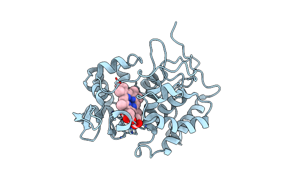 |
X-Ray Structures Of Recombinant Yeast Cytochrome C Peroxidase And Three Heme-Cleft Mutants Prepared By Site-Directed Mutagenesis
Organism: Saccharomyces cerevisiae
Method: X-RAY DIFFRACTION Resolution:2.20 Å Release Date: 1991-07-15 Classification: OXIDOREDUCTASE Ligands: HEM |
Organism: Saccharomyces cerevisiae
Method: X-RAY DIFFRACTION
Release Date: 1991-07-15
Ligands: HEM