

Search Count: 16
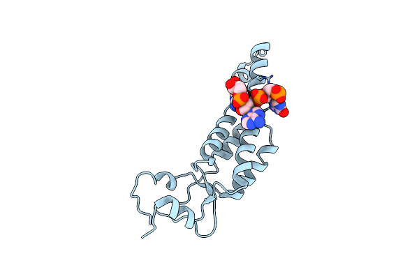 |
Cryo Idpc-Stem Structure Recorded With Csa 2.0
Organism: Tobacco mosaic virus (strain vulgare), Tobacco mosaic virus (vulgare)
Method: ELECTRON MICROSCOPY Release Date: 2022-09-21 Classification: VIRUS |
Organism: Tobacco mosaic virus (strain vulgare), Tobacco mosaic virus (vulgare)
Method: ELECTRON MICROSCOPY
Release Date: 2022-09-21
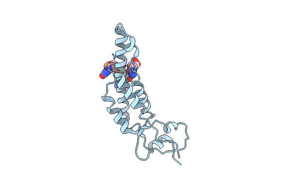 |
Cryo Idpc-Stem Structure Recorded With Csa 3.0
Organism: Tobacco mosaic virus (strain vulgare)
Method: ELECTRON MICROSCOPY Release Date: 2022-09-21 Classification: VIRUS |
Organism: Tobacco mosaic virus (strain vulgare)
Method: ELECTRON MICROSCOPY
Release Date: 2022-09-21
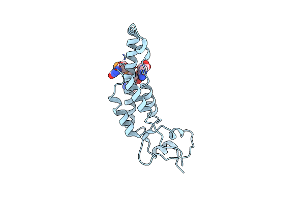 |
Cryo Idpc-Stem Structure Recorded With Csa 3.5
Organism: Tobacco mosaic virus (vulgare)
Method: ELECTRON MICROSCOPY Release Date: 2022-09-21 Classification: VIRUS |
Organism: Tobacco mosaic virus (vulgare)
Method: ELECTRON MICROSCOPY
Release Date: 2022-09-21
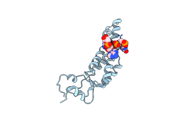 |
Cryo Idpc-Stem Structure Recorded With Csa 4.0
Organism: Tobacco mosaic virus (vulgare)
Method: ELECTRON MICROSCOPY Release Date: 2022-09-21 Classification: VIRUS |
Organism: Tobacco mosaic virus (vulgare)
Method: ELECTRON MICROSCOPY
Release Date: 2022-09-21
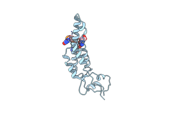 |
Cryo Idpc-Stem Structure Recorded With Csa 4.5
Organism: Tobacco mosaic virus (strain vulgare), Tobacco mosaic virus (vulgare)
Method: ELECTRON MICROSCOPY Release Date: 2022-09-21 Classification: VIRUS |
Organism: Tobacco mosaic virus (strain vulgare), Tobacco mosaic virus (vulgare)
Method: ELECTRON MICROSCOPY
Release Date: 2022-09-21
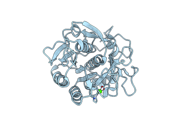 |
Crystal Structure Of Proteinase K Nanocrystals By Electron Diffraction With A 20 Micrometre C2 Condenser Aperture
Organism: Parengyodontium album
Method: ELECTRON CRYSTALLOGRAPHY Resolution:2.70 Å Release Date: 2020-10-14 Classification: HYDROLASE Ligands: CA |
Organism: Parengyodontium album
Method: ELECTRON CRYSTALLOGRAPHY
Release Date: 2020-10-14
Ligands: CA
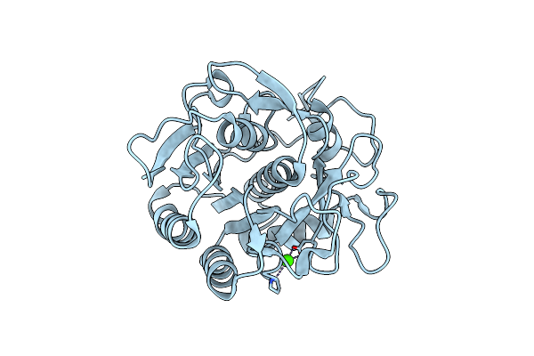 |
Crystal Structure Of Proteinase K Lamella By Electron Diffraction With A 50 Micrometre C2 Condenser Aperture
Organism: Parengyodontium album
Method: ELECTRON CRYSTALLOGRAPHY Resolution:2.00 Å Release Date: 2020-10-14 Classification: HYDROLASE Ligands: CA |
Organism: Parengyodontium album
Method: ELECTRON CRYSTALLOGRAPHY
Release Date: 2020-10-14
Ligands: CA
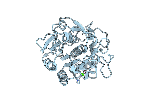 |
Crystal Structure Of Proteinase K Lamellae By Electron Diffraction With A 20 Micrometre C2 Condenser Aperture
Organism: Parengyodontium album
Method: ELECTRON CRYSTALLOGRAPHY Resolution:2.40 Å Release Date: 2020-10-14 Classification: HYDROLASE Ligands: CA |
Organism: Parengyodontium album
Method: ELECTRON CRYSTALLOGRAPHY
Release Date: 2020-10-14
Ligands: CA
 |
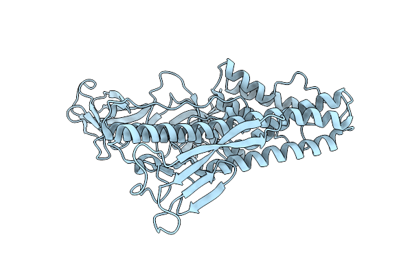
Atomic Models Of P1, P4 C-Terminal Fragment And P8 Fitted In The Bacteriophage Phi6 Nucleocapsid Reconstructed With Icosahedral Symmetry
Organism: Pseudomonas phage phi6
Method: ELECTRON MICROSCOPY Release Date: 2019-06-12 Classification: VIRUS |
Organism: Pseudomonas phage phi6
Method: ELECTRON MICROSCOPY
Release Date: 2019-06-12
 |
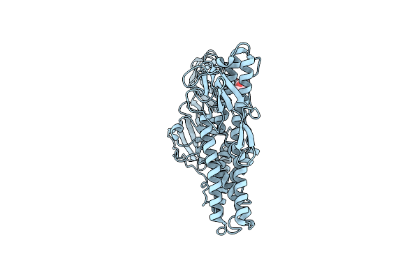
Structure Of Hrpv6 Vp5 Fitted In The Cryoem Density Of The Spike
Organism: Halorubrum pleomorphic virus 6
Method: ELECTRON MICROSCOPY Release Date: 2019-03-06 Classification: VIRAL PROTEIN |
Organism: Halorubrum pleomorphic virus 6
Method: ELECTRON MICROSCOPY
Release Date: 2019-03-06
 |
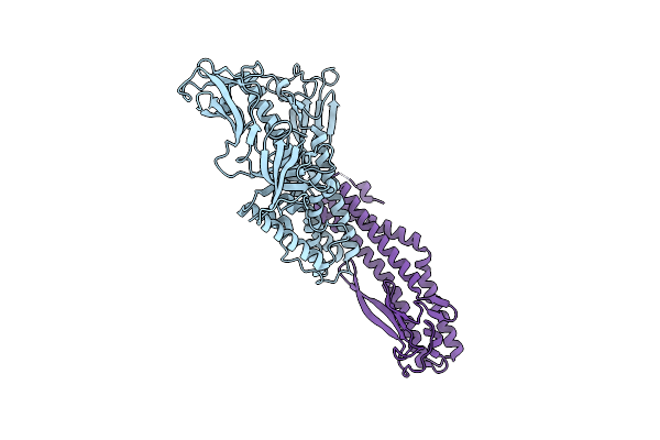
Crystal Structure Of Vp5 From Haloarchaeal Pleomorphic Virus 2
Organism: Halorubrum pleomorphic virus 2
Method: X-RAY DIFFRACTION Resolution:2.46 Å Release Date: 2019-02-27 Classification: VIRAL PROTEIN Ligands: NAG, CL |
Organism: Halorubrum pleomorphic virus 2
Method: X-RAY DIFFRACTION
Release Date: 2019-02-27
Ligands: NAG, CL
 |
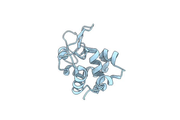
Crystal Structure Of Vp5 From Haloarchaeal Pleomorphic Virus 6
Organism: Halorubrum pleomorphic virus 6
Method: X-RAY DIFFRACTION Resolution:2.69 Å Release Date: 2019-02-27 Classification: VIRAL PROTEIN Ligands: BR |
Organism: Halorubrum pleomorphic virus 6
Method: X-RAY DIFFRACTION
Release Date: 2019-02-27
Ligands: BR
 |
Lysozyme: Machining Protein Microcrystals For Structure Determination By Electron Diffraction
Organism: Gallus gallus
Method: ELECTRON CRYSTALLOGRAPHY Resolution:1.90 Å Release Date: 2018-09-12 Classification: HYDROLASE Ligands: CL |
Organism: Gallus gallus
Method: ELECTRON CRYSTALLOGRAPHY
Release Date: 2018-09-12
Ligands: CL
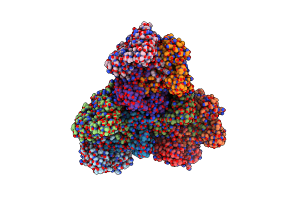 |
Quasi-Atomic Resolution Model Of Bacteriophage Prd1 P3-Shell, Obtained By Combined Cryo-Em And X-Ray Crystallography.
Organism: Bacteriophage prd1
Method: ELECTRON MICROSCOPY Resolution:12.00 Å Release Date: 2001-12-05 Classification: VIRUS |
Organism: Bacteriophage prd1
Method: ELECTRON MICROSCOPY
Release Date: 2001-12-05
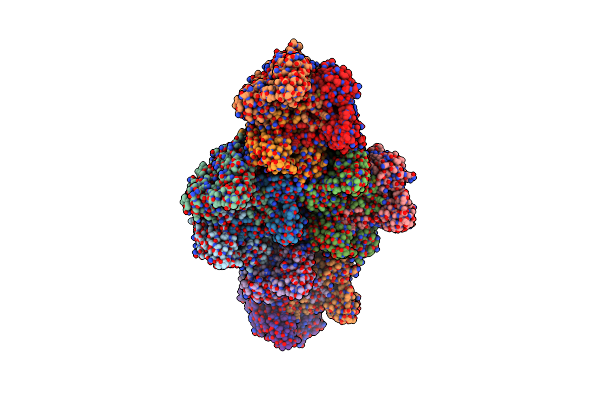 |
Quasi-Atomic Resolution Model Of Bacteriophage Prd1 Sus1 Mutant, Obtained By Combined Cryo-Em And X-Ray Crystallography.
Organism: Bacteriophage prd1
Method: ELECTRON MICROSCOPY Resolution:14.00 Å Release Date: 2001-12-05 Classification: VIRUS |
Organism: Bacteriophage prd1
Method: ELECTRON MICROSCOPY
Release Date: 2001-12-05
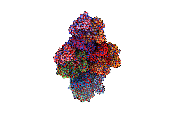 |
Quasi-Atomic Resolution Model Of Bacteriophage Prd1 Wild Type Virion, Obtained By Combined Cryo-Em And X-Ray Crystallography.
Organism: Bacteriophage prd1
Method: ELECTRON MICROSCOPY Resolution:25.00 Å Release Date: 2001-12-05 Classification: VIRUS |
Organism: Bacteriophage prd1
Method: ELECTRON MICROSCOPY
Release Date: 2001-12-05