

Search Count: 17
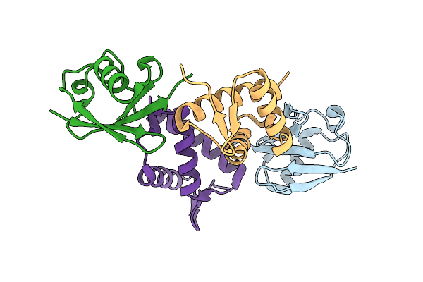 |
Ubiquitin Binding Variants
Organism: Homo sapiens
Method: X-RAY DIFFRACTION Resolution:2.20 Å Release Date: 2020-01-15 Classification: LIGASE |
Organism: Homo sapiens
Method: X-RAY DIFFRACTION
Release Date: 2020-01-15
 |
Ubiquitin Binding Variants
Organism: Homo sapiens
Method: X-RAY DIFFRACTION Resolution:2.80 Å Release Date: 2020-01-15 Classification: LIGASE |
Organism: Homo sapiens
Method: X-RAY DIFFRACTION
Release Date: 2020-01-15
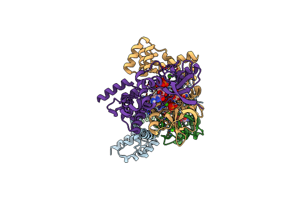 |
Crystal Structure Of Bacterial Parb Dimer Bound To Cdp
Organism: Bacillus subtilis (strain 168)
Method: X-RAY DIFFRACTION Resolution:1.81 Å Release Date: 2019-10-23 Classification: DNA BINDING PROTEIN Ligands: CDP, CA |
Organism: Bacillus subtilis (strain 168)
Method: X-RAY DIFFRACTION
Release Date: 2019-10-23
Ligands: CDP, CA
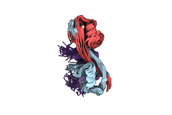 |
Structure Of Whb In Complex With Ubiquitin Variant
Organism: Homo sapiens
Method: SOLUTION NMR Release Date: 2019-08-14 Classification: PROTEIN BINDING |
Organism: Homo sapiens
Method: SOLUTION NMR
Release Date: 2019-08-14
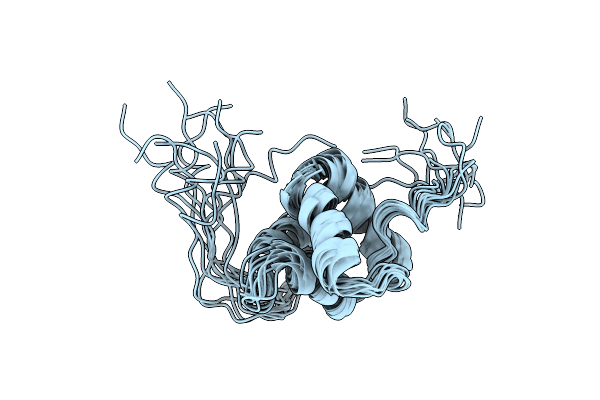 |
Solution Structure Of Avr3A_60-147 From Phytophthora Infestans
Organism: Phytophthora infestans
Method: SOLUTION NMR Release Date: 2017-01-11 Classification: PROTEIN BINDING |
Organism: Phytophthora infestans
Method: SOLUTION NMR
Release Date: 2017-01-11
 |
Model Of Human Anaphase-Promoting Complex/Cyclosome (Apc/C-Cdh1) With E2 Ube2S Poised For Polyubiquitination Where Ube2S, Apc2, And Apc11 Are Modeled Into Low Resolution Density
Organism: Homo sapiens, Saccharomyces cerevisiae (strain atcc 204508 / s288c)
Method: ELECTRON MICROSCOPY Release Date: 2016-10-26 Classification: CELL CYCLE |
Organism: Homo sapiens, Saccharomyces cerevisiae (strain atcc 204508 / s288c)
Method: ELECTRON MICROSCOPY
Release Date: 2016-10-26
 |
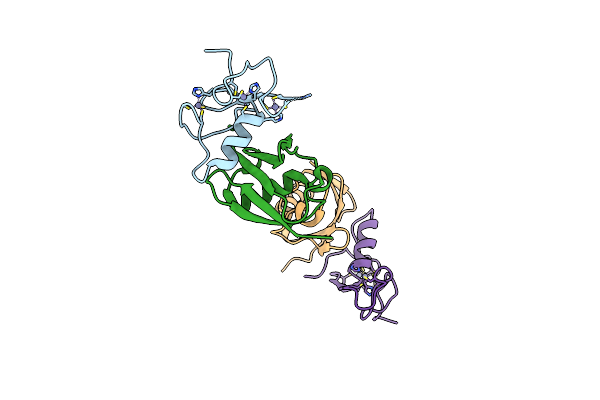
Model Of Human Anaphase-Promoting Complex/Cyclosome (Apc/C-Cdh1) With A Cross Linked Ubiquitin Variant-Substrate-Ube2C (Ubch10) Complex Representing Key Features Of Multiubiquitination
Organism: Homo sapiens, Saccharomyces cerevisiae, Rattus norvegicus
Method: ELECTRON MICROSCOPY Release Date: 2016-09-14 Classification: SIGNALING PROTEIN |
Organism: Homo sapiens, Saccharomyces cerevisiae, Rattus norvegicus
Method: ELECTRON MICROSCOPY
Release Date: 2016-09-14
 |
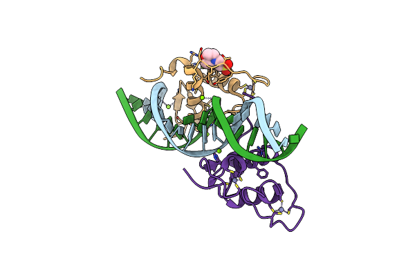
Apc11-Ubv Shows Role Of Noncovalent Ring-Ubiquitin Interactions In Processive Multiubiquitination And Ubiquitin Chain Elongation By Apc/C
Organism: Homo sapiens
Method: X-RAY DIFFRACTION Resolution:2.00 Å Release Date: 2016-06-15 Classification: CELL CYCLE Ligands: ZN |
Organism: Homo sapiens
Method: X-RAY DIFFRACTION
Release Date: 2016-06-15
Ligands: ZN
 |
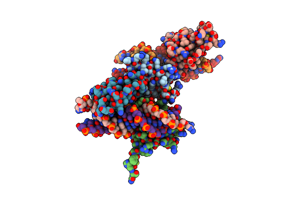
Crystal Structure Of The Human Retinoid X Receptor Dna-Binding Domain Bound To The Human Ramp2 Response Element
Organism: Homo sapiens
Method: X-RAY DIFFRACTION Resolution:2.07 Å Release Date: 2015-02-18 Classification: TRANSCRIPTION/DNA Ligands: MG, ZN, MES, CL |
Organism: Homo sapiens
Method: X-RAY DIFFRACTION
Release Date: 2015-02-18
Ligands: MG, ZN, MES, CL
 |
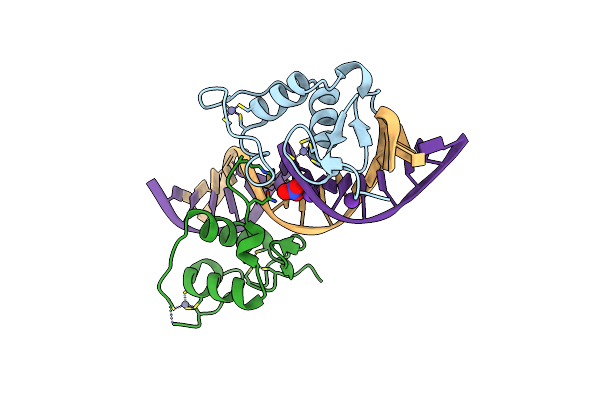
Crystal Structure Of The Human Retinoid X Receptor Dna-Binding Domain Bound To The Human Gde1Spa Response Element
Organism: Homo sapiens
Method: X-RAY DIFFRACTION Resolution:2.35 Å Release Date: 2015-02-18 Classification: TRANSCRIPTION/DNA Ligands: ZN |
Organism: Homo sapiens
Method: X-RAY DIFFRACTION
Release Date: 2015-02-18
Ligands: ZN
 |
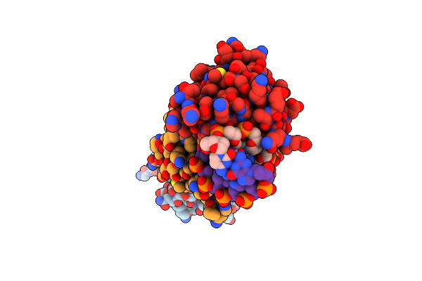
Crystal Structure Of The Human Retinoid X Receptor Dna-Binding Domain Bound To The Human Nr1D1 Response Element
Organism: Homo sapiens
Method: X-RAY DIFFRACTION Resolution:2.00 Å Release Date: 2015-02-18 Classification: TRANSCRIPTION/DNA Ligands: ZN, CL, K, NO3 |
Organism: Homo sapiens
Method: X-RAY DIFFRACTION
Release Date: 2015-02-18
Ligands: ZN, CL, K, NO3
 |
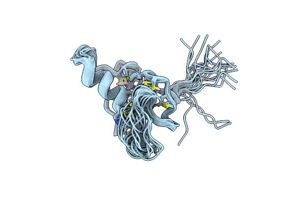
Crystal Structure Of The Human Retinoid X Receptor Dna-Binding Domain Bound To An Idealized Dr1 Response Element
Organism: Homo sapiens
Method: X-RAY DIFFRACTION Resolution:2.34 Å Release Date: 2015-02-18 Classification: TRANSCRIPTION/DNA Ligands: ZN, CL |
Organism: Homo sapiens
Method: X-RAY DIFFRACTION
Release Date: 2015-02-18
Ligands: ZN, CL
 |
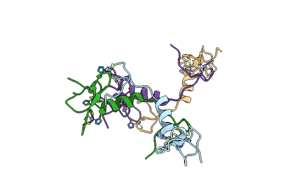
Isolated Ring Domain
Organism: Homo sapiens
Method: SOLUTION NMR Release Date: 2014-10-29 Classification: METAL BINDING PROTEIN Ligands: ZN |
Organism: Homo sapiens
Method: SOLUTION NMR
Release Date: 2014-10-29
Ligands: ZN
 |
Crystal Structure Of Apc11 Ring Domain
Organism: Homo sapiens
Method: X-RAY DIFFRACTION Resolution:1.76 Å Release Date: 2014-10-29 Classification: LIGASE Ligands: ZN |
Organism: Homo sapiens
Method: X-RAY DIFFRACTION
Release Date: 2014-10-29
Ligands: ZN
 |
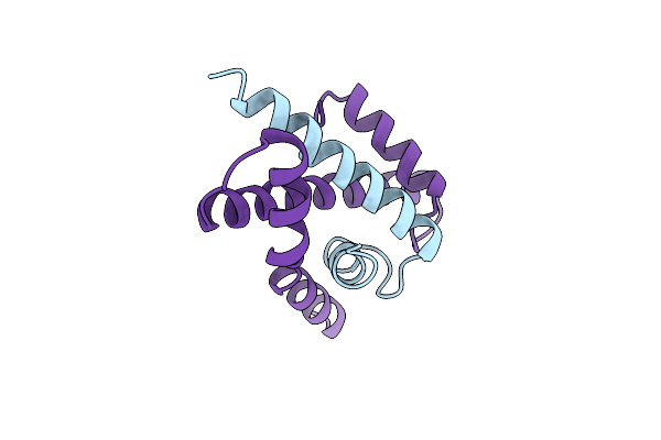
Crystal Structure Of The Human Taf4-Taf12 (Tafii135-Tafii20) Complex
Organism: Homo sapiens
Method: X-RAY DIFFRACTION Resolution:2.30 Å Release Date: 2002-09-26 Classification: TRANSCRIPTION/TBP-ASSOCIATED FACTORS |
Organism: Homo sapiens
Method: X-RAY DIFFRACTION
Release Date: 2002-09-26
 |
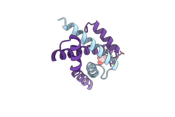
Htafii18/Htafii28 Heterodimer Crystal Structure
Organism: Homo sapiens
Method: X-RAY DIFFRACTION Resolution:3.00 Å Release Date: 1999-06-22 Classification: TRANSCRIPTION REGULATION COMPLEX |
Organism: Homo sapiens
Method: X-RAY DIFFRACTION
Release Date: 1999-06-22
 |
Htafii18/Htafii28 Heterodimer Crystal Structure With Bound Pcmbs
Organism: Homo sapiens
Method: X-RAY DIFFRACTION Resolution:2.60 Å Release Date: 1999-06-22 Classification: TRANSCRIPTION REGULATION COMPLEX Ligands: PMB |
Organism: Homo sapiens
Method: X-RAY DIFFRACTION
Release Date: 1999-06-22
Ligands: PMB