
Search Count: 4
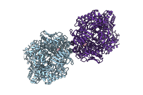 |
Organism: Glutamicibacter protophormiae
Method: ELECTRON MICROSCOPY Release Date: 2025-10-15 Classification: HYDROLASE Ligands: CA, DUC |
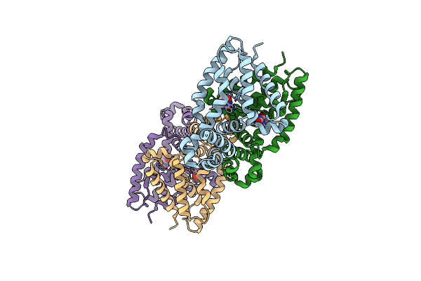 |
Organism: Escherichia coli (strain k12)
Method: X-RAY DIFFRACTION Resolution:2.27 Å Release Date: 2015-05-13 Classification: Transcription Regulator Ligands: DUC |
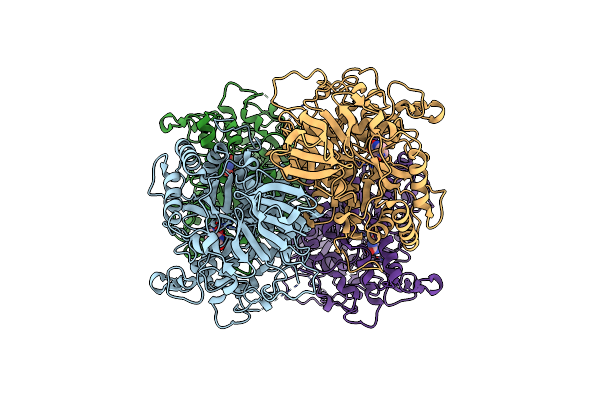 |
Crystal Structure Of Dihydropyrimidinase From Saccharomyces Kluyveri In Complex With The Substrate Dihydrouracil
Organism: Lachancea kluyveri
Method: X-RAY DIFFRACTION Resolution:2.40 Å Release Date: 2006-03-14 Classification: HYDROLASE Ligands: ZN, DUC |
 |
Organism: Saccharomyces cerevisiae
Method: X-RAY DIFFRACTION Resolution:1.60 Å Release Date: 2003-04-29 Classification: HYDROLASE Ligands: ZN, DUC |