
Search Count: 42
All
Selected
 |
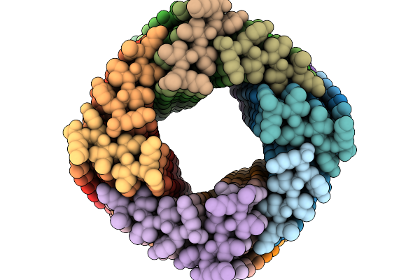
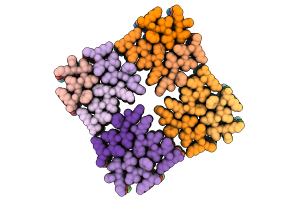
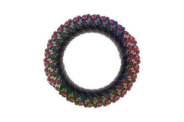
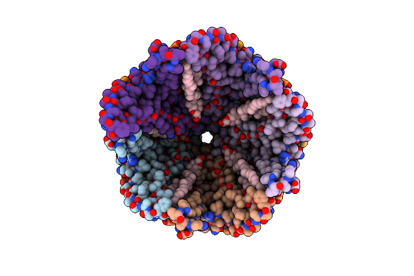
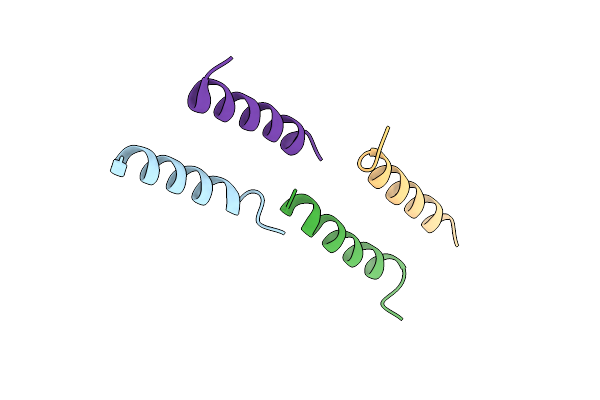
Cryoem Structure Of Computationally Designed Bundlemer Peptide Nanotube (14 Protofilament)
Organism: Synthetic construct
Method: ELECTRON MICROSCOPY Release Date: 2026-03-25 Classification: PROTEIN FIBRIL |
 |
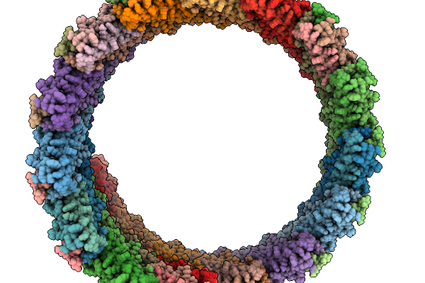
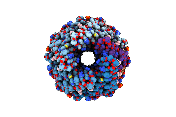
Cryoem Structure Of Computationally Designed Bundlemer Peptide Nanotube (15 Protofilament)
Organism: Synthetic construct
Method: ELECTRON MICROSCOPY Release Date: 2026-03-25 Classification: PROTEIN FIBRIL |
 |
Organism: Synthetic construct
Method: ELECTRON MICROSCOPY Release Date: 2026-02-25 Classification: PROTEIN FIBRIL |
 |
Organism: Synthetic construct
Method: ELECTRON MICROSCOPY Release Date: 2026-02-25 Classification: PROTEIN FIBRIL |
 |
Organism: Synthetic construct
Method: ELECTRON MICROSCOPY Resolution:3.93 Å Release Date: 2026-02-18 Classification: PROTEIN FIBRIL |
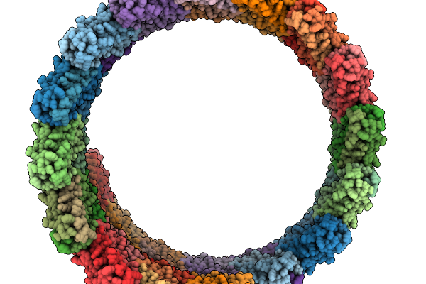 |
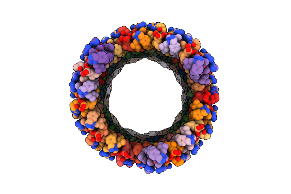
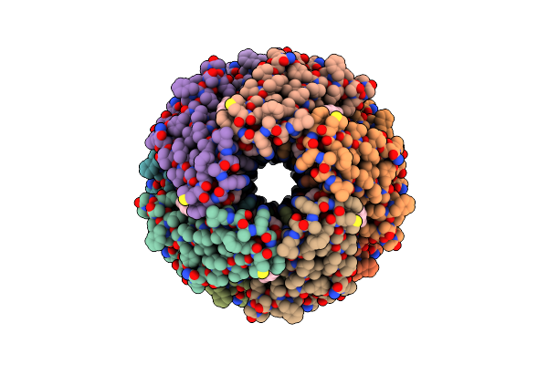
Nanotube Of Computationally Designed Peptide Assembly R3K (16 Protofilament)
Organism: Synthetic construct
Method: ELECTRON MICROSCOPY Resolution:3.58 Å Release Date: 2026-02-18 Classification: PROTEIN FIBRIL |
 |
Organism: Pyrodictium abyssi
Method: X-RAY DIFFRACTION Resolution:1.95 Å Release Date: 2025-08-27 Classification: CELL ADHESION |
 |
Organism: Synthetic construct
Method: ELECTRON MICROSCOPY Release Date: 2025-08-13 Classification: PROTEIN FIBRIL |
 |
Organism: Hyperthermus sp.
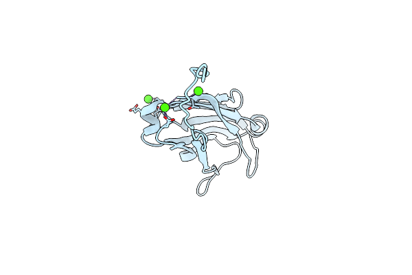
Method: ELECTRON MICROSCOPY Release Date: 2025-04-09 Classification: PROTEIN FIBRIL Ligands: CA |
 |
Organism: Hyperthermus sp.
Method: ELECTRON MICROSCOPY Release Date: 2025-04-09 Classification: PROTEIN FIBRIL Ligands: CA |
 |
Organism: Synthetic construct
Method: ELECTRON MICROSCOPY Resolution:3.80 Å Release Date: 2025-03-26 Classification: PROTEIN FIBRIL |
 |
Organism: Synthetic construct
Method: ELECTRON MICROSCOPY Release Date: 2025-02-26 Classification: PROTEIN FIBRIL |
 |
Organism: Pyrodictium abyssi dsm 6158
Method: ELECTRON MICROSCOPY Release Date: 2024-12-18 Classification: PROTEIN FIBRIL Ligands: CA, NAG |
 |
Organism: Pyrodictium abyssi
Method: ELECTRON MICROSCOPY Release Date: 2023-04-12 Classification: PROTEIN FIBRIL Ligands: CA |
 |
Organism: Pyrobaculum calidifontis
Method: ELECTRON MICROSCOPY Release Date: 2023-03-22 Classification: PROTEIN FIBRIL Ligands: TT0 |
 |
Organism: Aeropyrum pernix
Method: ELECTRON MICROSCOPY Release Date: 2023-03-22 Classification: PROTEIN FIBRIL Ligands: RHR |
 |
Organism: Agrobacterium fabrum (strain c58 / atcc 33970)
Method: ELECTRON MICROSCOPY Release Date: 2023-03-22 Classification: PROTEIN FIBRIL Ligands: X3D |
 |
Organism: Staphylococcus aureus
Method: ELECTRON MICROSCOPY Release Date: 2022-05-18 Classification: PROTEIN FIBRIL |
 |
Organism: Staphylococcus aureus
Method: ELECTRON MICROSCOPY Release Date: 2022-05-18 Classification: PROTEIN FIBRIL |
 |
Organism: Staphylococcus aureus
Method: ELECTRON MICROSCOPY Release Date: 2022-05-18 Classification: PROTEIN FIBRIL |