

Search Count: 30
 |
Crystal Structure Of Gallus Gallus C-Src Kinase Domain With Point Mutation Y416D And Deletion Of Residues N414, T417, And R419 Bound To Amp-Pnp
Organism: Gallus gallus
Method: X-RAY DIFFRACTION Resolution:1.78 Å Release Date: 2026-06-10 Classification: TRANSFERASE Ligands: ANP, MG |
Organism: Gallus gallus
Method: X-RAY DIFFRACTION
Release Date: 2026-06-10
Ligands: ANP, MG
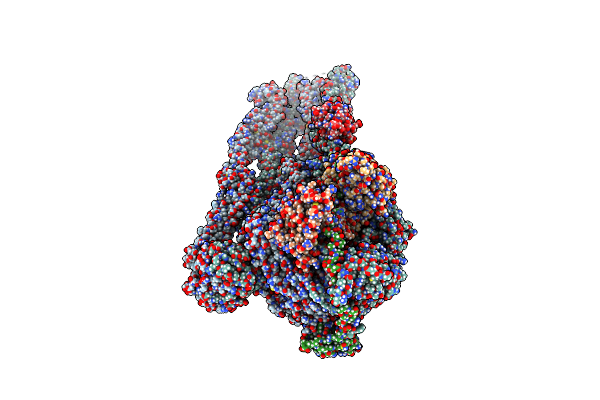 |
Mukbef In A Nucleotide-Bound State With Open Neck Gate (Heads Core)
Organism: Photorhabdus thracensis, Escherichia coli
Method: ELECTRON MICROSCOPY Release Date: 2025-03-26 Classification: DNA BINDING PROTEIN Ligands: MG, ATP |
Organism: Photorhabdus thracensis, Escherichia coli
Method: ELECTRON MICROSCOPY
Release Date: 2025-03-26
Ligands: MG, ATP
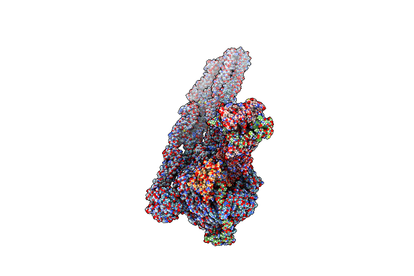 |
Mukbef In A Nucleotide-Bound State With Open Neck Gate (Monomer)
Organism: Photorhabdus thracensis, Escherichia coli
Method: ELECTRON MICROSCOPY Release Date: 2025-03-26 Classification: DNA BINDING PROTEIN Ligands: MG, ATP |
Organism: Photorhabdus thracensis, Escherichia coli
Method: ELECTRON MICROSCOPY
Release Date: 2025-03-26
Ligands: MG, ATP
 |
Mukbef In A Nucleotide-Bound State With Open Neck Gate
Organism: Photorhabdus thracensis, Escherichia coli
Method: ELECTRON MICROSCOPY Release Date: 2025-03-26 Classification: DNA BINDING PROTEIN Ligands: MG, ATP |
Organism: Photorhabdus thracensis, Escherichia coli
Method: ELECTRON MICROSCOPY
Release Date: 2025-03-26
Ligands: MG, ATP
 |
Mukbef In A Dna Capture State
Organism: Photorhabdus thracensis, Escherichia coli
Method: ELECTRON MICROSCOPY Release Date: 2025-03-26 Classification: DNA BINDING PROTEIN Ligands: MG, ATP |
Organism: Photorhabdus thracensis, Escherichia coli
Method: ELECTRON MICROSCOPY
Release Date: 2025-03-26
Ligands: MG, ATP
 |
Mukbef In A Dna Capture State (Dimer)
Organism: Photorhabdus thracensis, Escherichia coli
Method: ELECTRON MICROSCOPY Release Date: 2025-03-26 Classification: DNA BINDING PROTEIN Ligands: MG, ATP |
Organism: Photorhabdus thracensis, Escherichia coli
Method: ELECTRON MICROSCOPY
Release Date: 2025-03-26
Ligands: MG, ATP
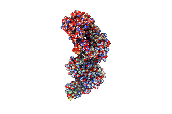 |
Mukef In Complex With The Phage Protein Gp5.9
Organism: Escherichia coli, Escherichia phage t7
Method: ELECTRON MICROSCOPY Release Date: 2025-03-26 Classification: DNA BINDING PROTEIN |
Organism: Escherichia coli, Escherichia phage t7
Method: ELECTRON MICROSCOPY
Release Date: 2025-03-26
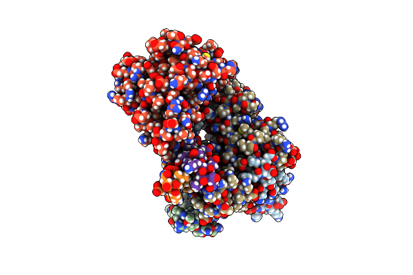 |
Mukef In Complex With The Phage Protein Gp5.9 (Focus)
Organism: Escherichia coli, Escherichia phage t7
Method: ELECTRON MICROSCOPY Release Date: 2025-03-26 Classification: DNA BINDING PROTEIN |
Organism: Escherichia coli, Escherichia phage t7
Method: ELECTRON MICROSCOPY
Release Date: 2025-03-26
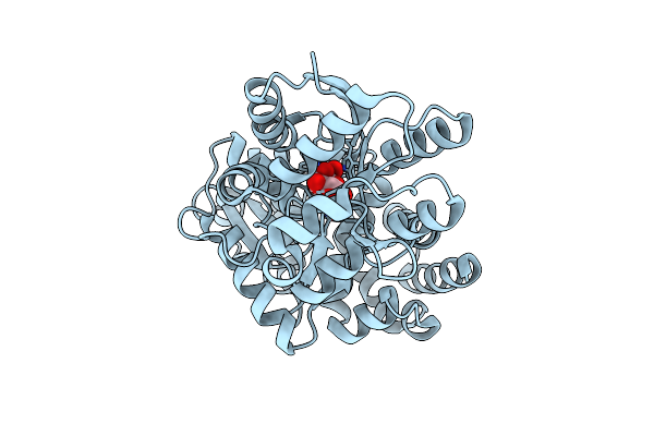 |
Crystal Structure Of Sar11_0769 From 'Candidatus Pelagibacter Ubique' Htcc1062 Bound To A Co-Purified Ligand, Beta-Galactopyranose
Organism: Candidatus pelagibacter ubique htcc1062
Method: X-RAY DIFFRACTION Resolution:1.68 Å Release Date: 2024-07-17 Classification: TRANSPORT PROTEIN Ligands: GAL |
Organism: Candidatus pelagibacter ubique htcc1062
Method: X-RAY DIFFRACTION
Release Date: 2024-07-17
Ligands: GAL
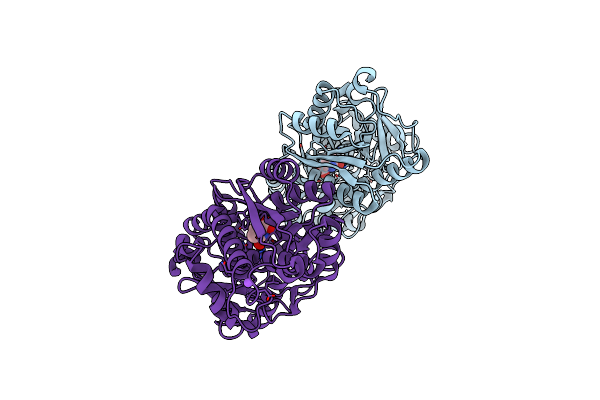 |
Crystal Structure Of Sar11_0655 Bound To A Co-Purified Ligand, L-Pyroglutamate
Organism: Candidatus pelagibacter ubique htcc1062
Method: X-RAY DIFFRACTION Resolution:1.52 Å Release Date: 2024-07-17 Classification: TRANSPORT PROTEIN Ligands: PCA, NA |
Organism: Candidatus pelagibacter ubique htcc1062
Method: X-RAY DIFFRACTION
Release Date: 2024-07-17
Ligands: PCA, NA
 |
Crystal Structure Of The Glucose-Binding Protein Sar11_0769 From "Candidatus Pelagibacter Ubique" Htcc1062 Bound To Glucose
Organism: Candidatus pelagibacter ubique htcc1062
Method: X-RAY DIFFRACTION Resolution:1.86 Å Release Date: 2023-12-27 Classification: TRANSPORT PROTEIN Ligands: BGC |
Organism: Candidatus pelagibacter ubique htcc1062
Method: X-RAY DIFFRACTION
Release Date: 2023-12-27
Ligands: BGC
 |
Crystal Structure Of The Arginine-/Lysine-Binding Protein Sar11_1210 From 'Candidatus Pelagibacter Ubique' Htcc1062 Bound To Arginine
Organism: Candidatus pelagibacter ubique htcc1062
Method: X-RAY DIFFRACTION Resolution:1.32 Å Release Date: 2023-12-27 Classification: TRANSPORT PROTEIN Ligands: ARG |
Organism: Candidatus pelagibacter ubique htcc1062
Method: X-RAY DIFFRACTION
Release Date: 2023-12-27
Ligands: ARG
 |
X-Ray Crystal Structure Of Plycb Mutant Y28H
Organism: Streptococcus virus c1
Method: X-RAY DIFFRACTION Resolution:1.79 Å Release Date: 2021-04-07 Classification: ANTIMICROBIAL PROTEIN |
Organism: Streptococcus virus c1
Method: X-RAY DIFFRACTION
Release Date: 2021-04-07
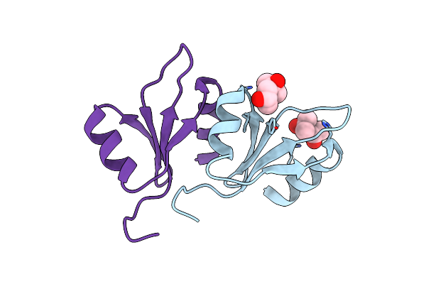 |
X-Ray Crystal Structure Of Plycb Mutant K59H
Organism: Streptococcus virus c1
Method: X-RAY DIFFRACTION Resolution:1.80 Å Release Date: 2021-04-07 Classification: ANTIMICROBIAL PROTEIN Ligands: MPD |
Organism: Streptococcus virus c1
Method: X-RAY DIFFRACTION
Release Date: 2021-04-07
Ligands: MPD
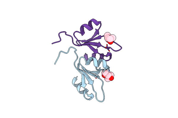 |
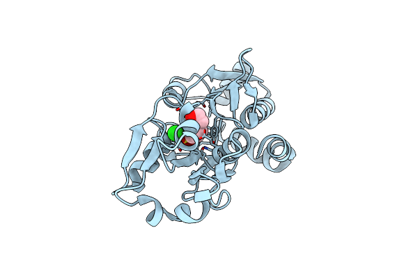
X-Ray Crystal Structure Of Plycb Mutant R66K
Organism: Streptococcus virus c1
Method: X-RAY DIFFRACTION Resolution:1.70 Å Release Date: 2021-04-07 Classification: ANTIMICROBIAL PROTEIN Ligands: MPD |
Organism: Streptococcus virus c1
Method: X-RAY DIFFRACTION
Release Date: 2021-04-07
Ligands: MPD
 |
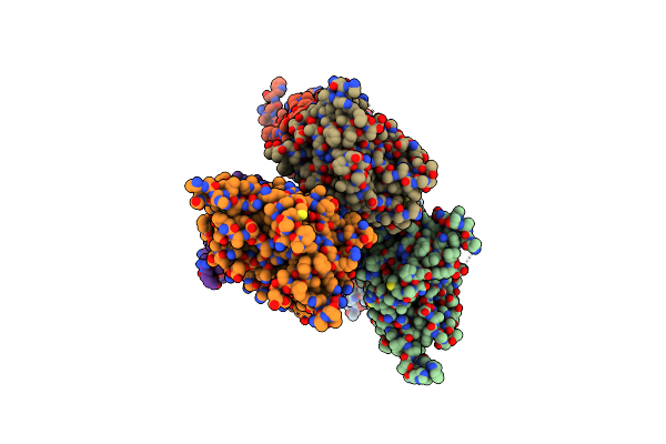
Crystal Structure Of An Ancestral Cyclohexadienyl Dehydratase, Anccdt-5
Organism: Synthetic construct
Method: X-RAY DIFFRACTION Resolution:1.49 Å Release Date: 2020-05-13 Classification: LYASE Ligands: CL, EPE |
Organism: Synthetic construct
Method: X-RAY DIFFRACTION
Release Date: 2020-05-13
Ligands: CL, EPE
 |
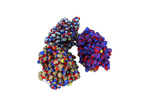
Human A20 Otu Domain (Wt) With Acetamidylated C103
Organism: Homo sapiens
Method: X-RAY DIFFRACTION Resolution:3.00 Å Release Date: 2018-03-21 Classification: HYDROLASE,LIGASE |
Organism: Homo sapiens
Method: X-RAY DIFFRACTION
Release Date: 2018-03-21
 |
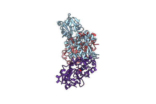
Human A20 Otu Domain (I325N) With Acetamidylated C103
Organism: Homo sapiens
Method: X-RAY DIFFRACTION Resolution:2.50 Å Release Date: 2018-03-21 Classification: HYDROLASE,LIGASE |
Organism: Homo sapiens
Method: X-RAY DIFFRACTION
Release Date: 2018-03-21
 |
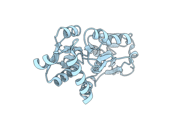
Low-Resolution Structure Of Cyclohexadienyl Dehydratase From Pseudomonas Aeruginosa In Space Group P4322.
Organism: Pseudomonas aeruginosa
Method: X-RAY DIFFRACTION Resolution:3.20 Å Release Date: 2017-12-13 Classification: LYASE Ligands: ACT |
Organism: Pseudomonas aeruginosa
Method: X-RAY DIFFRACTION
Release Date: 2017-12-13
Ligands: ACT
 |
Ancestral Cationic Amino Acid Solute Binding Protein (Anccdt-1)
Organism: Unidentified
Method: X-RAY DIFFRACTION Resolution:3.35 Å Release Date: 2017-12-06 Classification: Solute Binding Protein |
Organism: Unidentified
Method: X-RAY DIFFRACTION
Release Date: 2017-12-06