

Search Count: 66
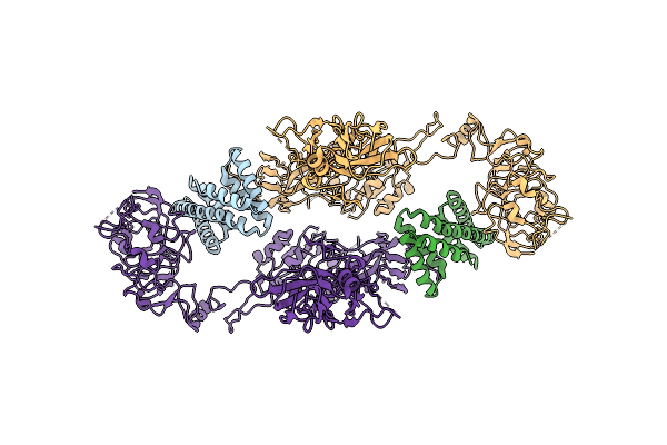 |
Insulin Receptor Bound With De Novo Designed Agonist Called "Rf-405".
Organism: Synthetic construct, Homo sapiens
Method: ELECTRON MICROSCOPY Release Date: 2025-10-01 Classification: SIGNALING PROTEIN |
Organism: Synthetic construct, Homo sapiens
Method: ELECTRON MICROSCOPY
Release Date: 2025-10-01
 |
Insulin Receptor Bound With De Novo Designed Agonist Called "S2-F1-S1"
Organism: Synthetic construct, Homo sapiens
Method: ELECTRON MICROSCOPY Release Date: 2025-10-01 Classification: SIGNALING PROTEIN |
Organism: Synthetic construct, Homo sapiens
Method: ELECTRON MICROSCOPY
Release Date: 2025-10-01
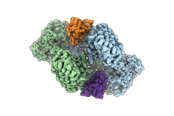 |
Insulin Receptor In Complex With Both Insulin And De Novo Designed Site-2 Binder "S2B".
Organism: Mus musculus, Homo sapiens, Synthetic construct
Method: ELECTRON MICROSCOPY Release Date: 2025-10-01 Classification: SIGNALING PROTEIN |
Organism: Mus musculus, Homo sapiens, Synthetic construct
Method: ELECTRON MICROSCOPY
Release Date: 2025-10-01
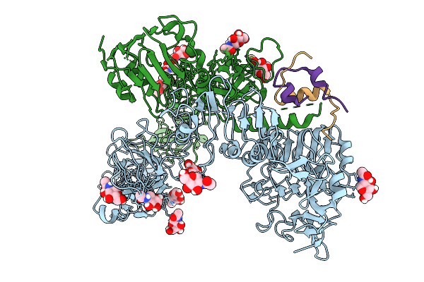 |
Cryo-Em Structure Of Drosophila Melanogaster Insulin Receptor (Dmir) Bound With One Dilp1, Asymmetric Conformation
Organism: Drosophila melanogaster
Method: ELECTRON MICROSCOPY Resolution:4.40 Å Release Date: 2025-10-01 Classification: STRUCTURAL PROTEIN Ligands: NAG |
Organism: Drosophila melanogaster
Method: ELECTRON MICROSCOPY
Release Date: 2025-10-01
Ligands: NAG
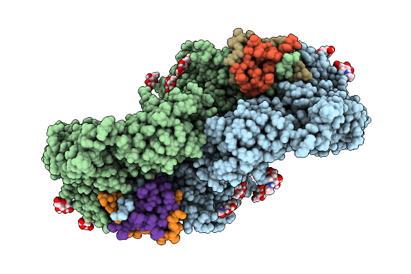 |
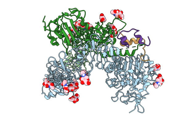
Cryo-Em Structure Of Drosophila Melanogaster Insulin Receptor (Dmir) Bound With Two Dilp1, Symmetric Conformation
Organism: Drosophila melanogaster
Method: ELECTRON MICROSCOPY Resolution:3.90 Å Release Date: 2025-10-01 Classification: STRUCTURAL PROTEIN Ligands: NAG |
Organism: Drosophila melanogaster
Method: ELECTRON MICROSCOPY
Release Date: 2025-10-01
Ligands: NAG
 |
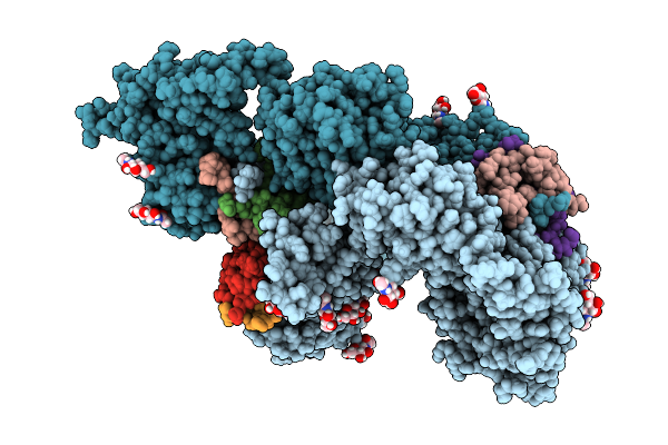
Cryo-Em Structure Of Drosophila Melanogaster Insulin Receptor (Dmir) Bound With One Dilp2, Asymmetric Conformation
Organism: Drosophila melanogaster
Method: ELECTRON MICROSCOPY Resolution:3.70 Å Release Date: 2025-10-01 Classification: STRUCTURAL PROTEIN Ligands: NAG |
Organism: Drosophila melanogaster
Method: ELECTRON MICROSCOPY
Release Date: 2025-10-01
Ligands: NAG
 |
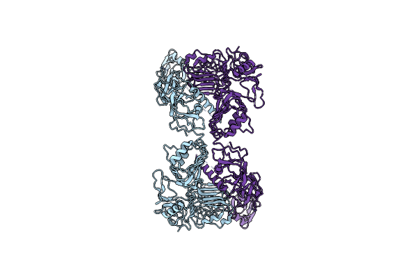
Cryo-Em Structure Of Drosophila Melanogaster Insulin Receptor (Dmir) Bound With Three Dilp5, Asymmetric Conformation
Organism: Drosophila melanogaster
Method: ELECTRON MICROSCOPY Resolution:3.60 Å Release Date: 2025-10-01 Classification: STRUCTURAL PROTEIN Ligands: NAG |
Organism: Drosophila melanogaster
Method: ELECTRON MICROSCOPY
Release Date: 2025-10-01
Ligands: NAG
 |
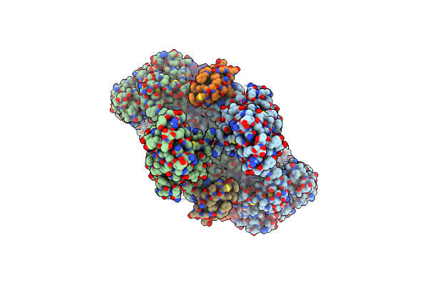
Cryo-Em Structure Of Long Form Insulin Receptor (Ir-B) In The Apo State
Organism: Homo sapiens
Method: ELECTRON MICROSCOPY Release Date: 2024-03-27 Classification: SIGNALING PROTEIN |
Organism: Homo sapiens
Method: ELECTRON MICROSCOPY
Release Date: 2024-03-27
 |
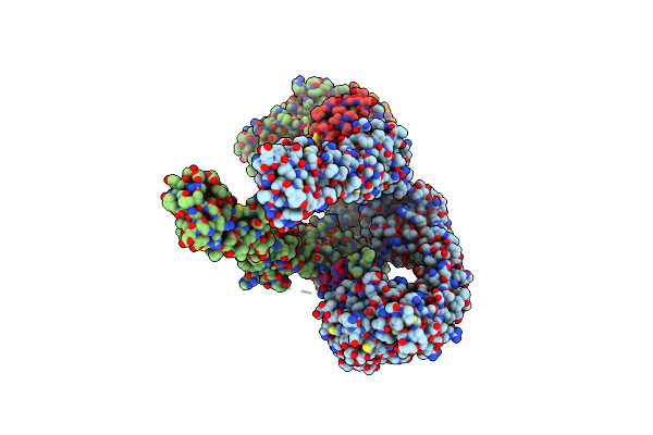
Cryo-Em Structure Of Long Form Insulin Receptor (Ir-B) With Four Igf2 Bound, Symmetric Conformation.
Organism: Homo sapiens
Method: ELECTRON MICROSCOPY Release Date: 2024-03-27 Classification: SIGNALING PROTEIN |
Organism: Homo sapiens
Method: ELECTRON MICROSCOPY
Release Date: 2024-03-27
 |
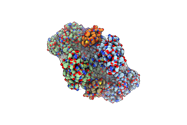
Cryo-Em Structure Of Long Form Insulin Receptor (Ir-B) With Three Igf2 Bound, Asymmetric Conformation.
Organism: Homo sapiens
Method: ELECTRON MICROSCOPY Release Date: 2024-03-27 Classification: SIGNALING PROTEIN |
Organism: Homo sapiens
Method: ELECTRON MICROSCOPY
Release Date: 2024-03-27
 |
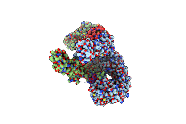
Cryo-Em Structure Of Short Form Insulin Receptor (Ir-A) With Four Igf2 Bound, Symmetric Conformation.
Organism: Homo sapiens
Method: ELECTRON MICROSCOPY Release Date: 2024-03-27 Classification: SIGNALING PROTEIN |
Organism: Homo sapiens
Method: ELECTRON MICROSCOPY
Release Date: 2024-03-27
 |
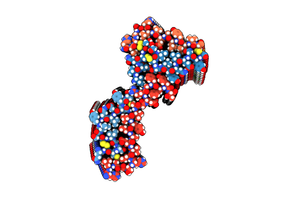
Cryo-Em Structure Of Short Form Insulin Receptor (Ir-A) With Three Igf2 Bound, Asymmetric Conformation.
Organism: Homo sapiens
Method: ELECTRON MICROSCOPY Release Date: 2024-03-27 Classification: SIGNALING PROTEIN |
Organism: Homo sapiens
Method: ELECTRON MICROSCOPY
Release Date: 2024-03-27
 |
Cryo-Em Structure Of Insulin Amyloid-Like Fibril That Is Composed Of Two Antiparallel Protofilaments
Organism: Homo sapiens
Method: ELECTRON MICROSCOPY Release Date: 2023-08-30 Classification: SIGNALING PROTEIN |
Organism: Homo sapiens
Method: ELECTRON MICROSCOPY
Release Date: 2023-08-30
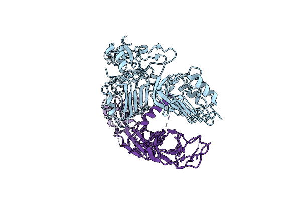 |
Cryo-Em Structure Of Insulin Receptor-Related Receptor (Irr) In Apo-State Captured At Ph 7. The 3D Refinement Was Focused On One Of Two Halves With C1 Symmetry Applied
Organism: Homo sapiens
Method: ELECTRON MICROSCOPY Release Date: 2023-02-15 Classification: SIGNALING PROTEIN |
Organism: Homo sapiens
Method: ELECTRON MICROSCOPY
Release Date: 2023-02-15
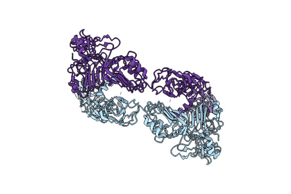 |
Cryo-Em Structure Of Insulin Receptor-Related Receptor (Irr) In Apo-State Captured At Ph 7. The 3D Refinement Was Applied With C2 Symmetry
Organism: Homo sapiens
Method: ELECTRON MICROSCOPY Release Date: 2023-02-15 Classification: SIGNALING PROTEIN |
Organism: Homo sapiens
Method: ELECTRON MICROSCOPY
Release Date: 2023-02-15
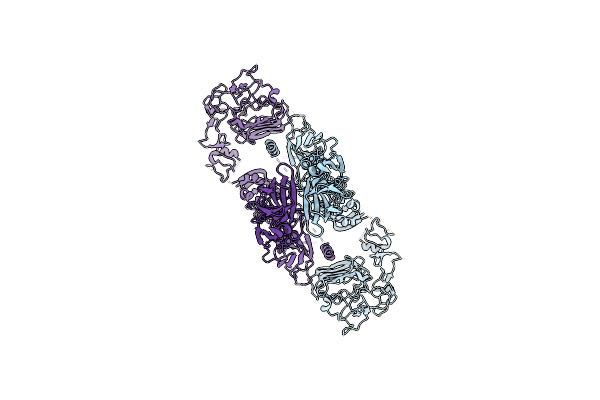 |
Cryo-Em Structure Of Insulin Receptor-Related Receptor (Irr) In Active-State Captured At Ph 9. The 3D Refinement Was Applied With C2 Symmetry
Organism: Homo sapiens
Method: ELECTRON MICROSCOPY Release Date: 2023-02-15 Classification: SIGNALING PROTEIN |
Organism: Homo sapiens
Method: ELECTRON MICROSCOPY
Release Date: 2023-02-15
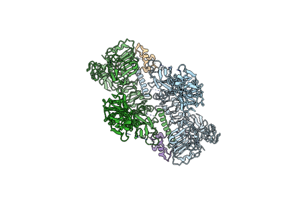 |
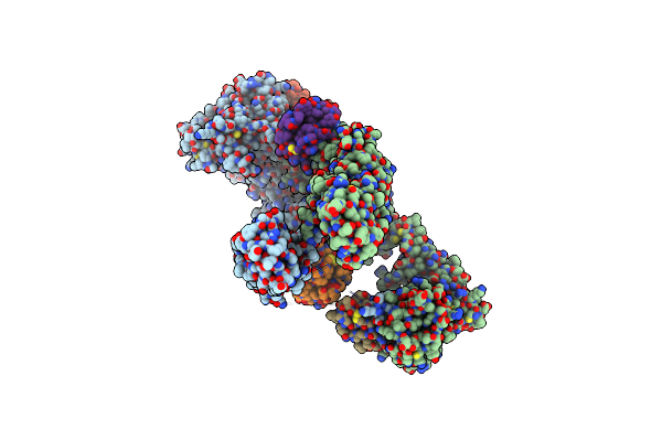
Cryo-Em Structure Of Two Igf1 Bound Full-Length Mouse Igf1R Mutant (Four Glycine Residues Inserted In The Alpha-Ct; Igf1R-P674G4): Symmetric Conformation
Organism: Mus musculus, Homo sapiens
Method: ELECTRON MICROSCOPY Release Date: 2022-11-09 Classification: SIGNALING PROTEIN |
Organism: Mus musculus, Homo sapiens
Method: ELECTRON MICROSCOPY
Release Date: 2022-11-09
 |
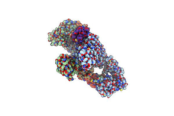
Cryo-Em Structure Of 4 Insulins Bound Full-Length Mouse Ir Mutant With Physically Decoupled Alpha Cts (C684S/C685S/C687S; Denoted As Ir-3Cs) Asymmetric Conformation 1
Organism: Mus musculus, Homo sapiens
Method: ELECTRON MICROSCOPY Release Date: 2022-11-09 Classification: SIGNALING PROTEIN |
Organism: Mus musculus, Homo sapiens
Method: ELECTRON MICROSCOPY
Release Date: 2022-11-09
 |
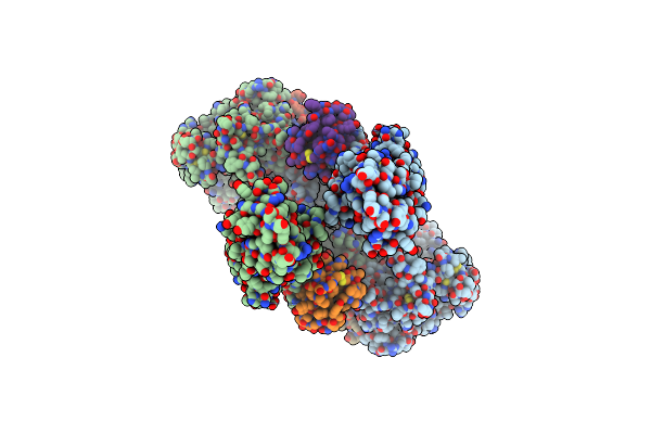
Cryo-Em Structure Of 4 Insulins Bound Full-Length Mouse Ir Mutant With Physically Decoupled Alpha Cts (C684S/C685S/C687S, Denoted As Ir-3Cs) Asymmetric Conformation 2
Organism: Mus musculus, Homo sapiens
Method: ELECTRON MICROSCOPY Release Date: 2022-11-09 Classification: SIGNALING PROTEIN |
Organism: Mus musculus, Homo sapiens
Method: ELECTRON MICROSCOPY
Release Date: 2022-11-09
 |
Cryo-Em Structure Of 4 Insulins Bound Full-Length Mouse Ir Mutant With Physically Decoupled Alpha Cts (C684S/C685S/C687S; Denoted As Ir-3Cs) Symmetric Conformation
Organism: Mus musculus, Homo sapiens
Method: ELECTRON MICROSCOPY Release Date: 2022-11-09 Classification: SIGNALING PROTEIN |
Organism: Mus musculus, Homo sapiens
Method: ELECTRON MICROSCOPY
Release Date: 2022-11-09