

Search Count: 52
 |
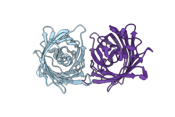
Structure Of Gfp Variant
Organism: Aequorea victoria
Method: X-RAY DIFFRACTION Resolution:1.50 Å Release Date: 2024-12-04 Classification: BIOSYNTHETIC PROTEIN |
Organism: Aequorea victoria
Method: X-RAY DIFFRACTION
Release Date: 2024-12-04
 |
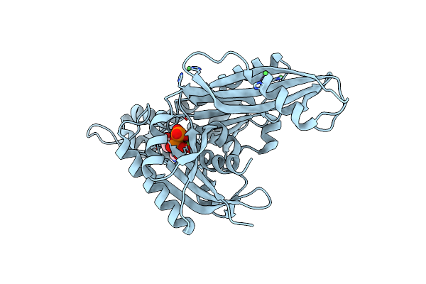
Structure Of Analogue Of Superfolded Gfp
Organism: Aequorea victoria
Method: X-RAY DIFFRACTION Resolution:1.54 Å Release Date: 2024-01-10 Classification: TRANSLATION |
Organism: Aequorea victoria
Method: X-RAY DIFFRACTION
Release Date: 2024-01-10
 |
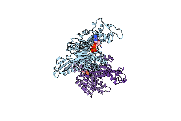
E. Coli Ftsa 1-405 Atp 3 Ni
Organism: Escherichia coli (strain k12)
Method: X-RAY DIFFRACTION Resolution:2.80 Å Release Date: 2022-09-21 Classification: CELL CYCLE Ligands: NI, MG, ATP |
Organism: Escherichia coli (strain k12)
Method: X-RAY DIFFRACTION
Release Date: 2022-09-21
Ligands: NI, MG, ATP
 |
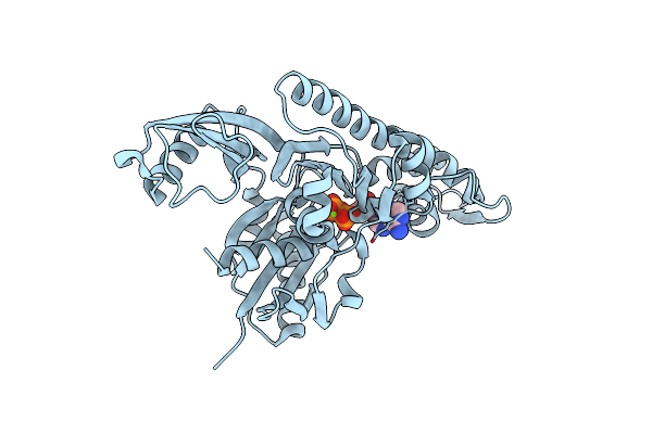
Vibrio Maritimus Ftsa 1-396 Atp, Double Filament
Organism: Vibrio maritimus
Method: X-RAY DIFFRACTION Resolution:3.31 Å Release Date: 2022-09-21 Classification: CELL CYCLE Ligands: ATP, MG |
Organism: Vibrio maritimus
Method: X-RAY DIFFRACTION
Release Date: 2022-09-21
Ligands: ATP, MG
 |
Xenorhabdus Poinarii Ftsa 1-396 Adp
Organism: Xenorhabdus poinarii g6
Method: X-RAY DIFFRACTION Resolution:2.80 Å Release Date: 2022-09-21 Classification: CELL CYCLE Ligands: ADP, MG |
Organism: Xenorhabdus poinarii g6
Method: X-RAY DIFFRACTION
Release Date: 2022-09-21
Ligands: ADP, MG
 |
Vibrio Maritimus Ftsa 1-396 Atp And Ftsn 1-29, Bent Tetramers In Double Filament Arrangement
Organism: Vibrio maritimus
Method: X-RAY DIFFRACTION Resolution:3.60 Å Release Date: 2022-09-21 Classification: CELL CYCLE Ligands: ATP, MG |
Organism: Vibrio maritimus
Method: X-RAY DIFFRACTION
Release Date: 2022-09-21
Ligands: ATP, MG
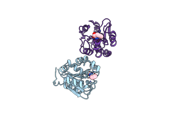 |
Crystal Structure Of Rbbp9 In Complex With Phenylalanine
Organism: Homo sapiens
Method: X-RAY DIFFRACTION Resolution:1.51 Å Release Date: 2021-12-22 Classification: HYDROLASE Ligands: PHE |
Organism: Homo sapiens
Method: X-RAY DIFFRACTION
Release Date: 2021-12-22
Ligands: PHE
 |
Cryo-Em Structure Of The Mukbef-Matp-Dna Head Module
Organism: Photorhabdus thracensis, Escherichia coli bl21(de3)
Method: ELECTRON MICROSCOPY Release Date: 2021-07-07 Classification: DNA BINDING PROTEIN Ligands: MG, ATP, PNS |
Organism: Photorhabdus thracensis, Escherichia coli bl21(de3)
Method: ELECTRON MICROSCOPY
Release Date: 2021-07-07
Ligands: MG, ATP, PNS
 |
Cryo-Em Structure Of The Mukbef-Matp-Dna Monomer (Closed Conformation)
Organism: Photorhabdus thracensis, Synthetic construct, Escherichia coli bl21(de3)
Method: ELECTRON MICROSCOPY Release Date: 2021-07-07 Classification: DNA BINDING PROTEIN Ligands: MG, ATP, PNS |
Organism: Photorhabdus thracensis, Synthetic construct, Escherichia coli bl21(de3)
Method: ELECTRON MICROSCOPY
Release Date: 2021-07-07
Ligands: MG, ATP, PNS
 |
Cryo-Em Structure Of The Mukbef Monomer
Organism: Photorhabdus thracensis, Escherichia coli bl21(de3)
Method: ELECTRON MICROSCOPY Release Date: 2021-07-07 Classification: DNA BINDING PROTEIN Ligands: PNS |
Organism: Photorhabdus thracensis, Escherichia coli bl21(de3)
Method: ELECTRON MICROSCOPY
Release Date: 2021-07-07
Ligands: PNS
 |
Cryo-Em Structure Of The Mukbef-Matp-Dna Monomer (Partially Open Conformation)
Organism: Photorhabdus thracensis, Escherichia coli bl21(de3)
Method: ELECTRON MICROSCOPY Release Date: 2021-07-07 Classification: DNA BINDING PROTEIN Ligands: MG, ATP, PNS |
Organism: Photorhabdus thracensis, Escherichia coli bl21(de3)
Method: ELECTRON MICROSCOPY
Release Date: 2021-07-07
Ligands: MG, ATP, PNS
 |
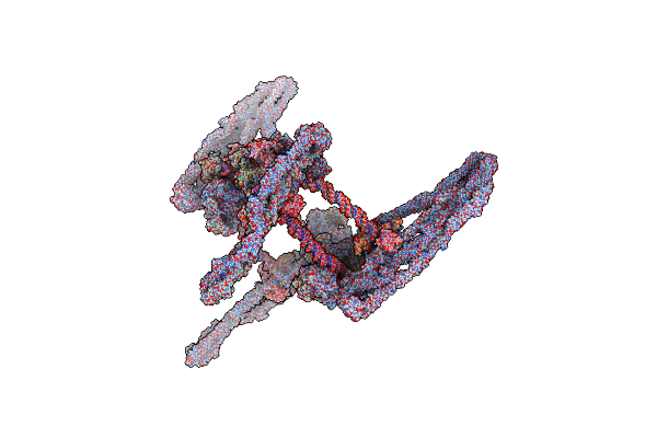
Cryo-Em Structure Of The Mukbef-Matp-Dna Monomer (Open Conformation)
Organism: Photorhabdus thracensis, Escherichia coli bl21(de3)
Method: ELECTRON MICROSCOPY Release Date: 2021-07-07 Classification: DNA BINDING PROTEIN Ligands: MG, ATP, PNS |
Organism: Photorhabdus thracensis, Escherichia coli bl21(de3)
Method: ELECTRON MICROSCOPY
Release Date: 2021-07-07
Ligands: MG, ATP, PNS
 |
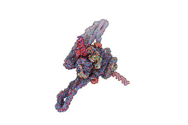
Cryo-Em Structure Of The Mukbef-Matp-Dna Tetrad
Organism: Photorhabdus thracensis, Escherichia coli bl21(de3)
Method: ELECTRON MICROSCOPY Release Date: 2021-07-07 Classification: DNA BINDING PROTEIN Ligands: MG, ATP, PNS |
Organism: Photorhabdus thracensis, Escherichia coli bl21(de3)
Method: ELECTRON MICROSCOPY
Release Date: 2021-07-07
Ligands: MG, ATP, PNS
 |
Cryo-Em Structure Of Apposed Mukbef-Matp Monomers On Dna
Organism: Photorhabdus thracensis, Escherichia coli bl21(de3)
Method: ELECTRON MICROSCOPY Release Date: 2021-07-07 Classification: DNA BINDING PROTEIN Ligands: MG, ATP, PNS |
Organism: Photorhabdus thracensis, Escherichia coli bl21(de3)
Method: ELECTRON MICROSCOPY
Release Date: 2021-07-07
Ligands: MG, ATP, PNS
 |
Cryo-Em Structure Of The Mukbef Dimer
Organism: Photorhabdus thracensis, Escherichia coli bl21(de3)
Method: ELECTRON MICROSCOPY Release Date: 2021-07-07 Classification: DNA BINDING PROTEIN Ligands: PNS |
Organism: Photorhabdus thracensis, Escherichia coli bl21(de3)
Method: ELECTRON MICROSCOPY
Release Date: 2021-07-07
Ligands: PNS
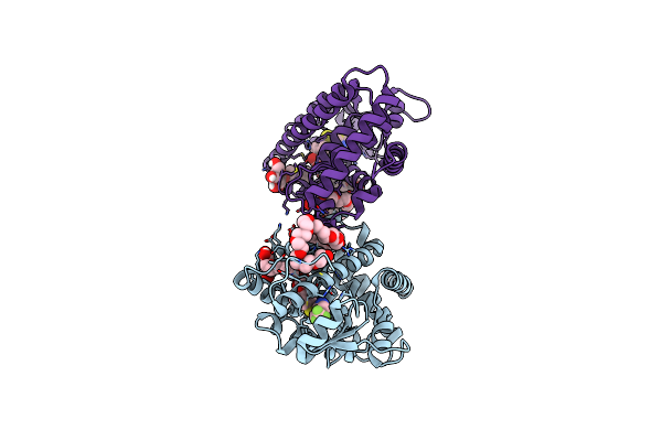 |
Crystal Structure Of Peroxisome Proliferator-Activated Receptor Delta (Ppard)Lbd In Complex With Dn003316
Organism: Homo sapiens
Method: X-RAY DIFFRACTION Resolution:2.10 Å Release Date: 2019-07-10 Classification: STRUCTURAL PROTEIN Ligands: 9RF, B7G, PE3 |
Organism: Homo sapiens
Method: X-RAY DIFFRACTION
Release Date: 2019-07-10
Ligands: 9RF, B7G, PE3
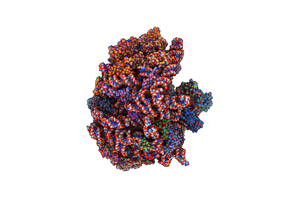 |
E. Coli 70S D2D8 Stapled Ribosome
Organism: Escherichia coli
Method: ELECTRON MICROSCOPY Release Date: 2018-12-19 Classification: RIBOSOME Ligands: MG, ZN |
Organism: Escherichia coli
Method: ELECTRON MICROSCOPY
Release Date: 2018-12-19
Ligands: MG, ZN
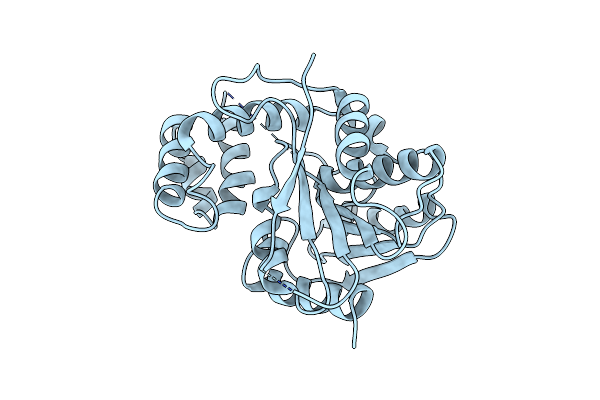 |
Vlm2 Thioesterase Domain Wild Type Structure 1
Organism: Streptomyces tsusimaensis
Method: X-RAY DIFFRACTION Resolution:1.70 Å Release Date: 2018-12-12 Classification: HYDROLASE |
Organism: Streptomyces tsusimaensis
Method: X-RAY DIFFRACTION
Release Date: 2018-12-12
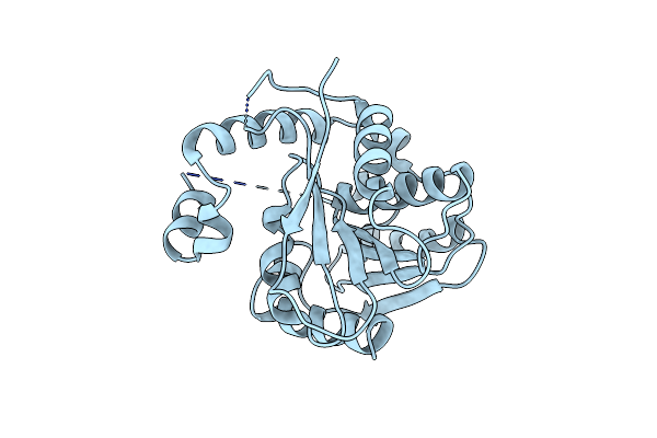 |
Vlm2 Thioesterase Domain Wild Type Structure 2
Organism: Streptomyces tsusimaensis
Method: X-RAY DIFFRACTION Resolution:1.80 Å Release Date: 2018-12-12 Classification: HYDROLASE |
Organism: Streptomyces tsusimaensis
Method: X-RAY DIFFRACTION
Release Date: 2018-12-12
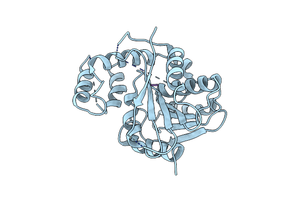 |
Vlm2 Thioesterase Domain With Genetically Encoded 2,3-Diaminopropionic Acid Bound With A Tetradepsipeptide
Organism: Streptomyces tsusimaensis, Synthetic construct
Method: X-RAY DIFFRACTION Resolution:1.90 Å Release Date: 2018-12-12 Classification: HYDROLASE |
Organism: Streptomyces tsusimaensis, Synthetic construct
Method: X-RAY DIFFRACTION
Release Date: 2018-12-12