

Search Count: 11
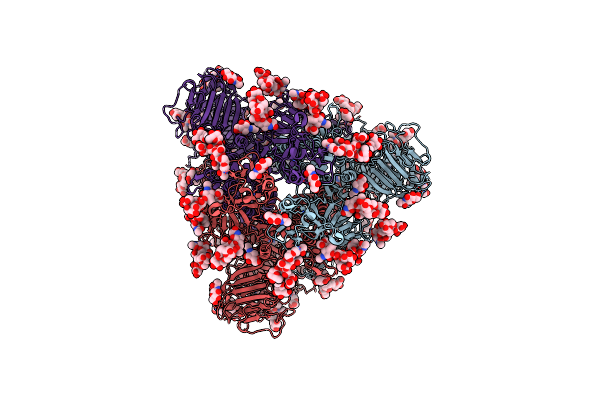 |
Cryo-Em Map Of Pedv (Pintung 52) S Protein With All Three Protomers In The D0-Down Conformation Determined In Situ On Intact Viral Particles.
Organism: Porcine epidemic diarrhea virus
Method: ELECTRON MICROSCOPY Release Date: 2022-08-03 Classification: VIRAL PROTEIN Ligands: NAG |
Organism: Porcine epidemic diarrhea virus
Method: ELECTRON MICROSCOPY
Release Date: 2022-08-03
Ligands: NAG
 |
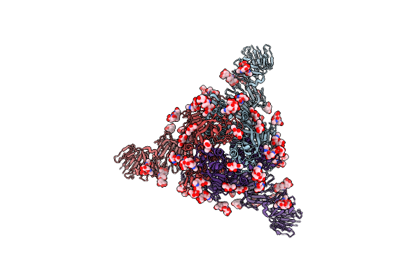
Cryo-Em Map Of Pedv S Protein With One Protomer In The D0-Up Conformation While The Other Two In The D0-Down Conformation
Organism: Porcine epidemic diarrhea virus
Method: ELECTRON MICROSCOPY Release Date: 2022-08-03 Classification: VIRAL PROTEIN Ligands: NAG |
Organism: Porcine epidemic diarrhea virus
Method: ELECTRON MICROSCOPY
Release Date: 2022-08-03
Ligands: NAG
 |
Cryo-Em Map Of Ipec-J2 Cell-Derived Pedv Pt52 S Protein With Three D0-Up
Organism: Porcine epidemic diarrhea virus
Method: ELECTRON MICROSCOPY Release Date: 2022-08-03 Classification: VIRAL PROTEIN Ligands: NAG |
Organism: Porcine epidemic diarrhea virus
Method: ELECTRON MICROSCOPY
Release Date: 2022-08-03
Ligands: NAG
 |
Cryo-Em Map Of Ipec-J2 Cell-Derived Pedv Pt52 S Protein One D0-Down And Two D0-Up
Organism: Porcine epidemic diarrhea virus
Method: ELECTRON MICROSCOPY Release Date: 2022-08-03 Classification: VIRAL PROTEIN Ligands: NAG |
Organism: Porcine epidemic diarrhea virus
Method: ELECTRON MICROSCOPY
Release Date: 2022-08-03
Ligands: NAG
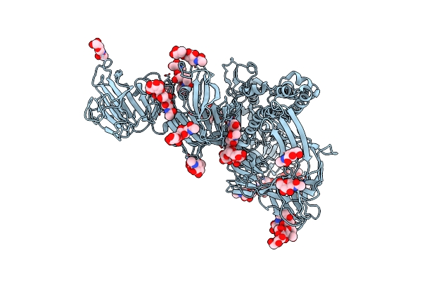 |
Symmetry-Expanded And Locally Refined Protomer Structure Of Ipec-J2 Cell-Derived Pedv Pt52 S With A Ctd-Close Conformation
Organism: Porcine epidemic diarrhea virus
Method: ELECTRON MICROSCOPY Release Date: 2022-08-03 Classification: VIRAL PROTEIN Ligands: NAG |
Organism: Porcine epidemic diarrhea virus
Method: ELECTRON MICROSCOPY
Release Date: 2022-08-03
Ligands: NAG
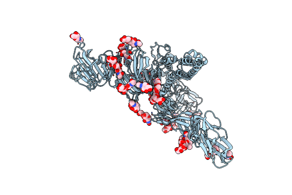 |
Symmetry-Expanded And Locally Refined Protomer Structure Of Ipec-J2 Cell-Derived Pedv Pt52 S With A Ctd-Open Conformation
Organism: Porcine epidemic diarrhea virus
Method: ELECTRON MICROSCOPY Resolution:3.30 Å Release Date: 2022-08-03 Classification: VIRAL PROTEIN Ligands: NAG |
Organism: Porcine epidemic diarrhea virus
Method: ELECTRON MICROSCOPY
Release Date: 2022-08-03
Ligands: NAG
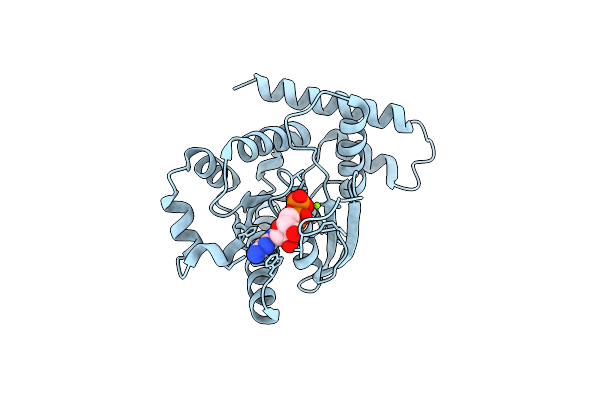 |
Crystal Structure Of Gdp-Bound Human Gimap5, Amino Acid Residues 1-276
Organism: Homo sapiens
Method: X-RAY DIFFRACTION Resolution:2.80 Å Release Date: 2021-12-01 Classification: HYDROLASE Ligands: MG, GDP |
Organism: Homo sapiens
Method: X-RAY DIFFRACTION
Release Date: 2021-12-01
Ligands: MG, GDP
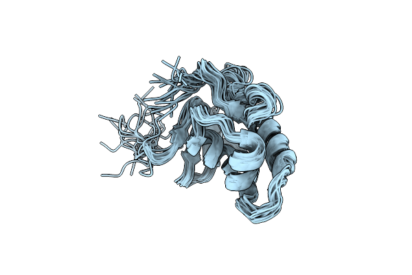 |
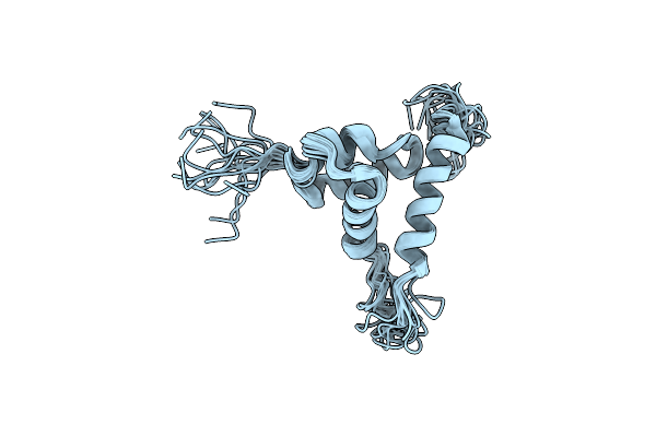
Three-Dimensional Structure Of The C-Terminal Dna-Binding Domain Of Rsta Protein From Klebsiella Pneumoniae
Organism: Klebsiella pneumoniae
Method: SOLUTION NMR Release Date: 2014-07-16 Classification: SIGNALING PROTEIN |
Organism: Klebsiella pneumoniae
Method: SOLUTION NMR
Release Date: 2014-07-16
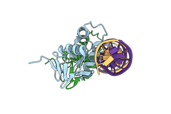 |
Crystal Structure Of Klebsiella Pneumoniae Rsta Dna-Binding Domain In Complex With Rsta Box
Organism: Klebsiella pneumoniae
Method: X-RAY DIFFRACTION Resolution:2.70 Å Release Date: 2014-07-16 Classification: TRANSCRIPTION REGULATOR/DNA |
Organism: Klebsiella pneumoniae
Method: X-RAY DIFFRACTION
Release Date: 2014-07-16
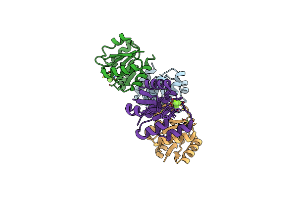 |
Crystal Structure Of Klebsiella Pneumoniae Rsta Bef3-Activated N-Terminal Receiver Domain
Organism: Klebsiella pneumoniae
Method: X-RAY DIFFRACTION Resolution:3.18 Å Release Date: 2014-07-16 Classification: TRANSCRIPTION REGULATOR Ligands: MG, BEF |
Organism: Klebsiella pneumoniae
Method: X-RAY DIFFRACTION
Release Date: 2014-07-16
Ligands: MG, BEF
 |
Solution Structure Of The Arabidopsis Thaliana Telomeric Repeat-Binding Protein Dna Binding Domain
Organism: Arabidopsis thaliana
Method: SOLUTION NMR Release Date: 2006-07-04 Classification: DNA BINDING PROTEIN |
Organism: Arabidopsis thaliana
Method: SOLUTION NMR
Release Date: 2006-07-04