

Search Count: 13
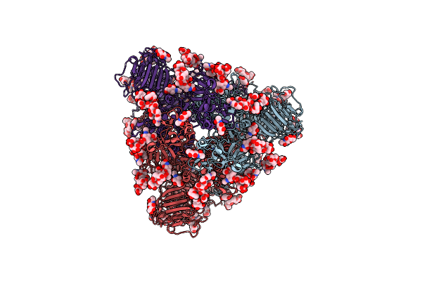 |
Cryo-Em Map Of Pedv (Pintung 52) S Protein With All Three Protomers In The D0-Down Conformation Determined In Situ On Intact Viral Particles.
Organism: Porcine epidemic diarrhea virus
Method: ELECTRON MICROSCOPY Release Date: 2022-08-03 Classification: VIRAL PROTEIN Ligands: NAG |
Organism: Porcine epidemic diarrhea virus
Method: ELECTRON MICROSCOPY
Release Date: 2022-08-03
Ligands: NAG
 |
Cryo-Em Map Of Pedv S Protein With One Protomer In The D0-Up Conformation While The Other Two In The D0-Down Conformation
Organism: Porcine epidemic diarrhea virus
Method: ELECTRON MICROSCOPY Release Date: 2022-08-03 Classification: VIRAL PROTEIN Ligands: NAG |
Organism: Porcine epidemic diarrhea virus
Method: ELECTRON MICROSCOPY
Release Date: 2022-08-03
Ligands: NAG
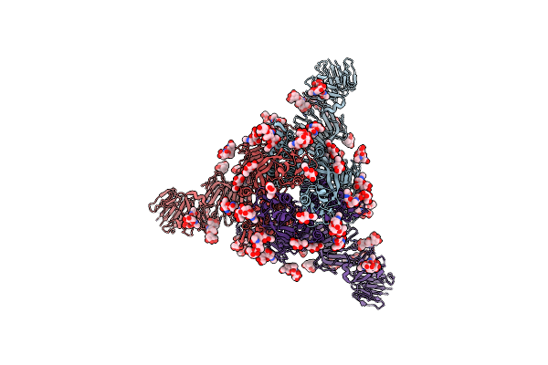 |
Cryo-Em Map Of Ipec-J2 Cell-Derived Pedv Pt52 S Protein With Three D0-Up
Organism: Porcine epidemic diarrhea virus
Method: ELECTRON MICROSCOPY Release Date: 2022-08-03 Classification: VIRAL PROTEIN Ligands: NAG |
Organism: Porcine epidemic diarrhea virus
Method: ELECTRON MICROSCOPY
Release Date: 2022-08-03
Ligands: NAG
 |
Cryo-Em Map Of Ipec-J2 Cell-Derived Pedv Pt52 S Protein One D0-Down And Two D0-Up
Organism: Porcine epidemic diarrhea virus
Method: ELECTRON MICROSCOPY Release Date: 2022-08-03 Classification: VIRAL PROTEIN Ligands: NAG |
Organism: Porcine epidemic diarrhea virus
Method: ELECTRON MICROSCOPY
Release Date: 2022-08-03
Ligands: NAG
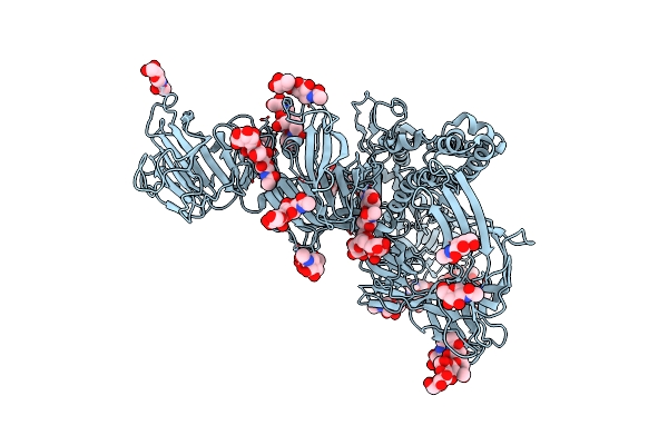 |
Symmetry-Expanded And Locally Refined Protomer Structure Of Ipec-J2 Cell-Derived Pedv Pt52 S With A Ctd-Close Conformation
Organism: Porcine epidemic diarrhea virus
Method: ELECTRON MICROSCOPY Release Date: 2022-08-03 Classification: VIRAL PROTEIN Ligands: NAG |
Organism: Porcine epidemic diarrhea virus
Method: ELECTRON MICROSCOPY
Release Date: 2022-08-03
Ligands: NAG
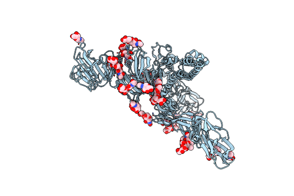 |
Symmetry-Expanded And Locally Refined Protomer Structure Of Ipec-J2 Cell-Derived Pedv Pt52 S With A Ctd-Open Conformation
Organism: Porcine epidemic diarrhea virus
Method: ELECTRON MICROSCOPY Resolution:3.30 Å Release Date: 2022-08-03 Classification: VIRAL PROTEIN Ligands: NAG |
Organism: Porcine epidemic diarrhea virus
Method: ELECTRON MICROSCOPY
Release Date: 2022-08-03
Ligands: NAG
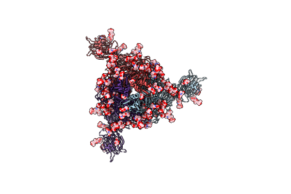 |
Cryo-Em Structure Of Spike Protein Of Feline Infectious Peritonitis Virus Strain Uu4
Organism: Feline infectious peritonitis virus
Method: ELECTRON MICROSCOPY Release Date: 2020-01-15 Classification: VIRAL PROTEIN Ligands: NAG, MAN |
Organism: Feline infectious peritonitis virus
Method: ELECTRON MICROSCOPY
Release Date: 2020-01-15
Ligands: NAG, MAN
 |
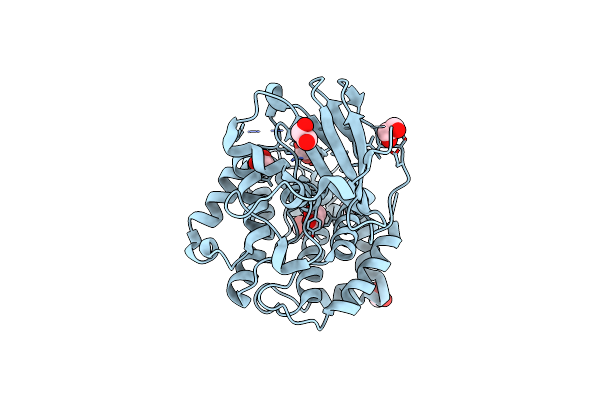
Crystal Structure Of Bacillus Subtilis Rsbs
Organism: Bacillus subtilis
Method: X-RAY DIFFRACTION Resolution:3.10 Å Release Date: 2019-09-11 Classification: PROTEIN BINDING |
Organism: Bacillus subtilis
Method: X-RAY DIFFRACTION
Release Date: 2019-09-11
 |
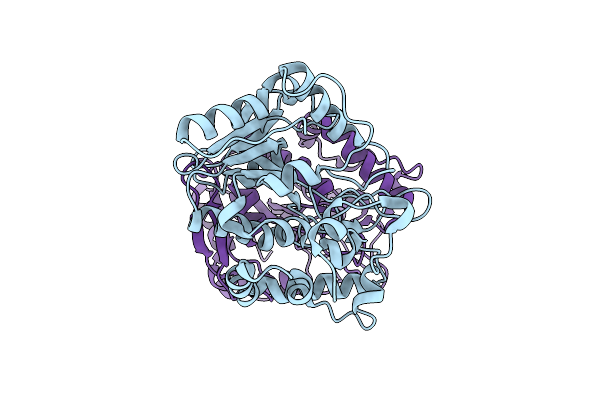
The Crystal Structure Of Vaccinia Virus A26 (Residues 1-397)
Organism: Vaccinia virus (strain western reserve)
Method: X-RAY DIFFRACTION Resolution:1.18 Å Release Date: 2019-06-12 Classification: VIRAL PROTEIN Ligands: EDO |
Organism: Vaccinia virus (strain western reserve)
Method: X-RAY DIFFRACTION
Release Date: 2019-06-12
Ligands: EDO
 |
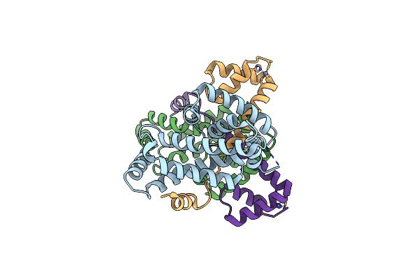
Crystal Structure Of Mimivirus Uracil-Dna Glycosylase
Organism: Acanthamoeba polyphaga mimivirus
Method: X-RAY DIFFRACTION Resolution:2.30 Å Release Date: 2017-08-09 Classification: HYDROLASE |
Organism: Acanthamoeba polyphaga mimivirus
Method: X-RAY DIFFRACTION
Release Date: 2017-08-09
 |
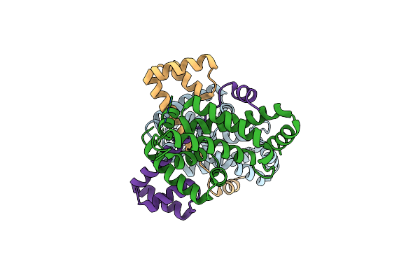
Crystal Structure Of Sigw In Complex With Its Anti-Sigma Rsiw, A Zinc Binding Form
Organism: Bacillus subtilis subsp. subtilis str. 168
Method: X-RAY DIFFRACTION Resolution:2.80 Å Release Date: 2017-03-29 Classification: METAL BINDING PROTEIN Ligands: ZN |
Organism: Bacillus subtilis subsp. subtilis str. 168
Method: X-RAY DIFFRACTION
Release Date: 2017-03-29
Ligands: ZN
 |
Crystal Structure Of Sigw In Complex With Its Anti-Sigma Rsiw, An Oxdized Form
Organism: Bacillus subtilis subsp. subtilis str. 168
Method: X-RAY DIFFRACTION Resolution:2.60 Å Release Date: 2017-03-29 Classification: METAL BINDING PROTEIN |
Organism: Bacillus subtilis subsp. subtilis str. 168
Method: X-RAY DIFFRACTION
Release Date: 2017-03-29
 |
Ggvlvn Segment From Human Prostatic Acid Phosphatase Residues 260-265, Involved In Semen-Derived Enhancer Of Viral Infection
Organism: Homo sapiens
Method: X-RAY DIFFRACTION Resolution:1.50 Å Release Date: 2011-06-29 Classification: PROTEIN FIBRIL Ligands: ZN, ACY |
Organism: Homo sapiens
Method: X-RAY DIFFRACTION
Release Date: 2011-06-29
Ligands: ZN, ACY