

Search Count: 66
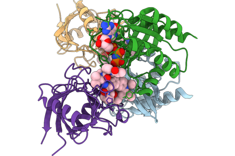 |
Tri-Complex Of Compound 1, Kras G13C, And Cypa
Organism: Homo sapiens
Method: X-RAY DIFFRACTION Resolution:1.47 Å Release Date: 2026-07-01 Classification: SIGNALING PROTEIN Ligands: GNP, MG, A1AOL |
Organism: Homo sapiens
Method: X-RAY DIFFRACTION
Release Date: 2026-07-01
Ligands: GNP, MG, A1AOL
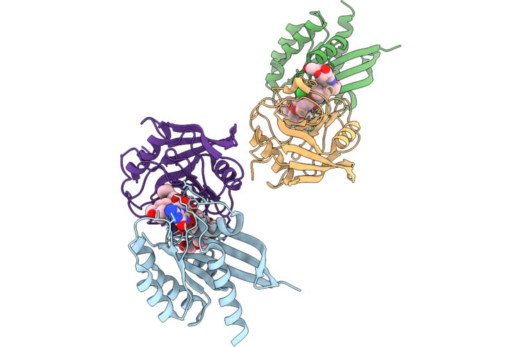 |
Tri-Complex Of Compound 2, Kras G13C, And Cypa
Organism: Homo sapiens
Method: X-RAY DIFFRACTION Resolution:1.74 Å Release Date: 2026-07-01 Classification: SIGNALING PROTEIN Ligands: GNP, MG, A1DKX, CL, PEG |
Organism: Homo sapiens
Method: X-RAY DIFFRACTION
Release Date: 2026-07-01
Ligands: GNP, MG, A1DKX, CL, PEG
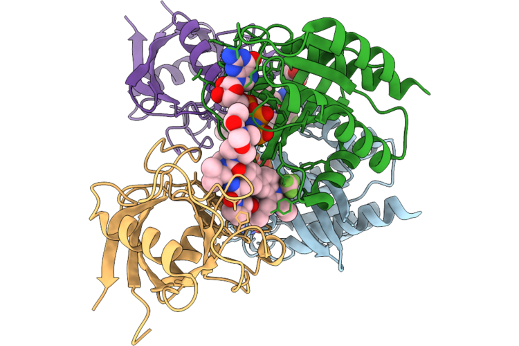 |
Tri-Complex Of Rmc-8839, Kras G13C, And Cypa
Organism: Homo sapiens
Method: X-RAY DIFFRACTION Resolution:1.21 Å Release Date: 2026-07-01 Classification: SIGNALING PROTEIN Ligands: GNP, MG, CL, A1DKY, PEG |
Organism: Homo sapiens
Method: X-RAY DIFFRACTION
Release Date: 2026-07-01
Ligands: GNP, MG, CL, A1DKY, PEG
 |
Crystal Structure Of Kras Wt Complexed With Gmppnp
Organism: Homo sapiens
Method: X-RAY DIFFRACTION Resolution:2.05 Å Release Date: 2026-07-01 Classification: SIGNALING PROTEIN Ligands: GNP, MG, EDO |
Organism: Homo sapiens
Method: X-RAY DIFFRACTION
Release Date: 2026-07-01
Ligands: GNP, MG, EDO
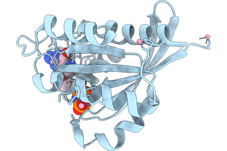 |
Crystal Structure Of Kras G13C Complexed With Gmppnp
Organism: Homo sapiens
Method: X-RAY DIFFRACTION Resolution:1.25 Å Release Date: 2026-07-01 Classification: SIGNALING PROTEIN Ligands: GNP, MG, CO |
Organism: Homo sapiens
Method: X-RAY DIFFRACTION
Release Date: 2026-07-01
Ligands: GNP, MG, CO
 |
Crystal Structure Of The Aaa+ Domain Of Vibrio Cholerae Flra
Organism: Vibrio cholerae o395
Method: X-RAY DIFFRACTION Resolution:3.20 Å Release Date: 2026-06-03 Classification: TRANSCRIPTION Ligands: EDO |
Organism: Vibrio cholerae o395
Method: X-RAY DIFFRACTION
Release Date: 2026-06-03
Ligands: EDO
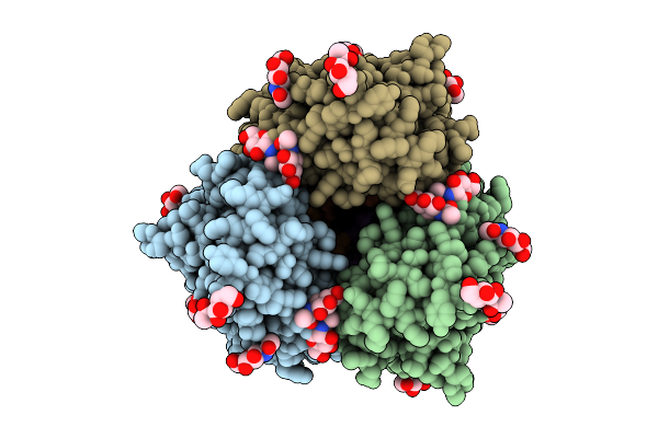 |
Map Of Endoh-Treated Hemagglutinin A/Sing/Infimh/16
Organism: Influenza a virus, Aequorea victoria
Method: ELECTRON MICROSCOPY Release Date: 2025-08-13 Classification: VIRAL PROTEIN Ligands: NAG |
Organism: Influenza a virus, Aequorea victoria
Method: ELECTRON MICROSCOPY
Release Date: 2025-08-13
Ligands: NAG
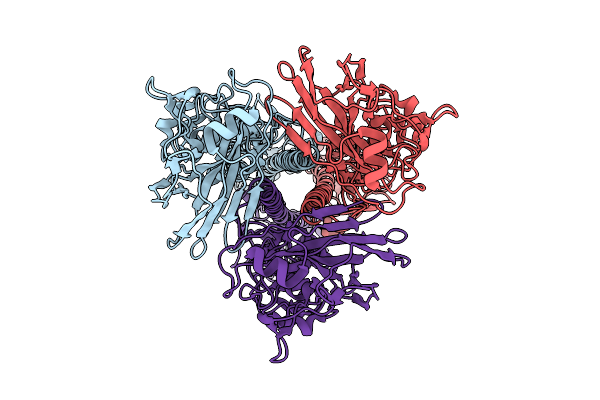 |
Map Of Influenza Hemagglutinin A/Sing/Infimh/16 Expressed In Gnti- Cells
Organism: Influenza a virus
Method: ELECTRON MICROSCOPY Release Date: 2025-08-13 Classification: VIRAL PROTEIN |
Organism: Influenza a virus
Method: ELECTRON MICROSCOPY
Release Date: 2025-08-13
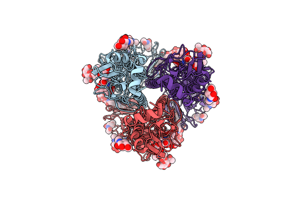 |
Map Of Hemagglutinin A/Sing/Infimh/16 Expressed In 293F Cells
Organism: Influenza a virus
Method: ELECTRON MICROSCOPY Release Date: 2025-08-13 Classification: VIRAL PROTEIN Ligands: NAG |
Organism: Influenza a virus
Method: ELECTRON MICROSCOPY
Release Date: 2025-08-13
Ligands: NAG
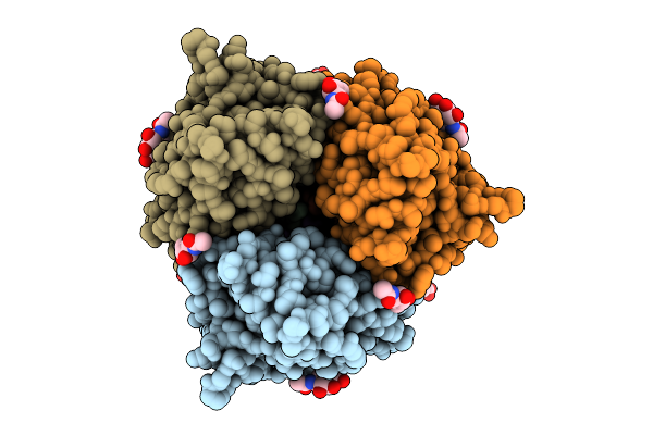 |
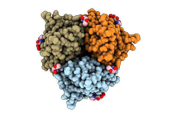
Hemagglutinin A/Hong Kong/1/68 Produced In Gnti- Cells
Organism: Influenza a virus (strain a/hong kong/1/1968 h3n2), Aequorea victoria
Method: ELECTRON MICROSCOPY Release Date: 2025-08-06 Classification: VIRAL PROTEIN Ligands: NAG |
Organism: Influenza a virus (strain a/hong kong/1/1968 h3n2), Aequorea victoria
Method: ELECTRON MICROSCOPY
Release Date: 2025-08-06
Ligands: NAG
 |
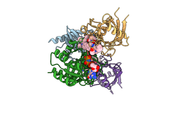
Endo H-Treated Hemagglutinin A/Hong Kong/1/68
Organism: Influenza a virus (strain a/hong kong/1/1968 h3n2), Aequorea victoria
Method: ELECTRON MICROSCOPY Release Date: 2025-08-06 Classification: VIRAL PROTEIN Ligands: NAG |
Organism: Influenza a virus (strain a/hong kong/1/1968 h3n2), Aequorea victoria
Method: ELECTRON MICROSCOPY
Release Date: 2025-08-06
Ligands: NAG
 |
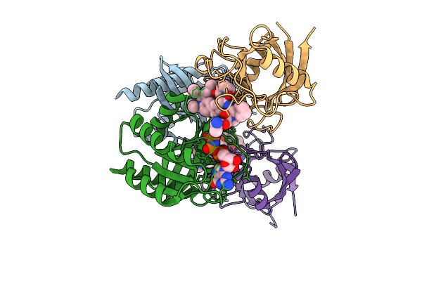
Tricomplex Of Compound 1, Kras G12D, And Cypa
Organism: Homo sapiens
Method: X-RAY DIFFRACTION Resolution:1.42 Å Release Date: 2025-07-23 Classification: HYDROLASE Ligands: GNP, MG, CL, A1AZV |
Organism: Homo sapiens
Method: X-RAY DIFFRACTION
Release Date: 2025-07-23
Ligands: GNP, MG, CL, A1AZV
 |
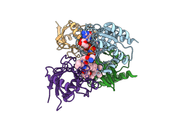
Tricomplex Of Compound 2, Kras G12D, And Cypa
Organism: Homo sapiens
Method: X-RAY DIFFRACTION Resolution:1.28 Å Release Date: 2025-07-23 Classification: HYDROLASE/HYDROLASE INHIBITOR Ligands: GNP, MG, A1AZX, CL |
Organism: Homo sapiens
Method: X-RAY DIFFRACTION
Release Date: 2025-07-23
Ligands: GNP, MG, A1AZX, CL
 |
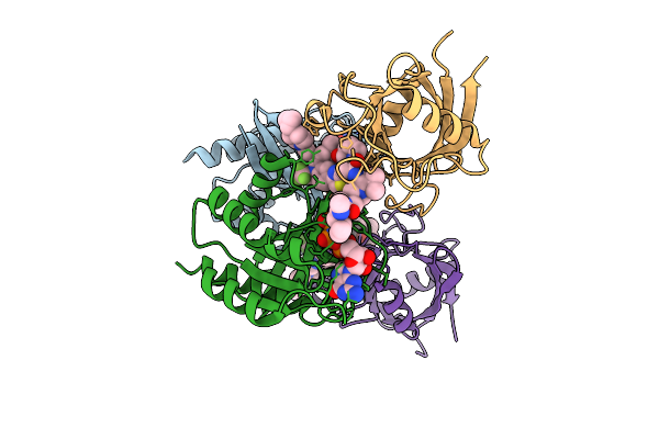
Tricomplex Of Compound 3, Kras G12D, And Cypa
Organism: Homo sapiens
Method: X-RAY DIFFRACTION Resolution:1.35 Å Release Date: 2025-07-23 Classification: HYDROLASE/HYDROLASE INHIBITOR Ligands: GNP, MG, CL, A1AZY |
Organism: Homo sapiens
Method: X-RAY DIFFRACTION
Release Date: 2025-07-23
Ligands: GNP, MG, CL, A1AZY
 |
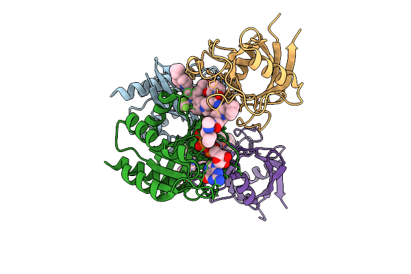
Tricomplex Of Rmc-9945, Kras G12D, And Cypa
Organism: Homo sapiens
Method: X-RAY DIFFRACTION Resolution:1.29 Å Release Date: 2025-07-23 Classification: HYDROLASE/HYDROLASE INHIBITOR Ligands: GNP, MG, A1AZW |
Organism: Homo sapiens
Method: X-RAY DIFFRACTION
Release Date: 2025-07-23
Ligands: GNP, MG, A1AZW
 |
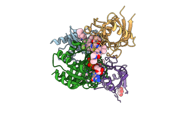
Tri-Complex Of Zoldonrasib (Rmc-9805), Kras G12D, And Cypa
Organism: Homo sapiens
Method: X-RAY DIFFRACTION Resolution:1.29 Å Release Date: 2025-07-23 Classification: HYDROLASE/HYDROLASE INHIBITOR Ligands: GNP, MG, CL, A1AZZ |
Organism: Homo sapiens
Method: X-RAY DIFFRACTION
Release Date: 2025-07-23
Ligands: GNP, MG, CL, A1AZZ
 |
Tricomplex Of Rmc-9945, Kras G12N, And Cypa
Organism: Homo sapiens
Method: X-RAY DIFFRACTION Resolution:1.08 Å Release Date: 2025-07-23 Classification: SIGNALING PROTEIN Ligands: GNP, MG, CL, A1BEA, GOL |
Organism: Homo sapiens
Method: X-RAY DIFFRACTION
Release Date: 2025-07-23
Ligands: GNP, MG, CL, A1BEA, GOL
 |
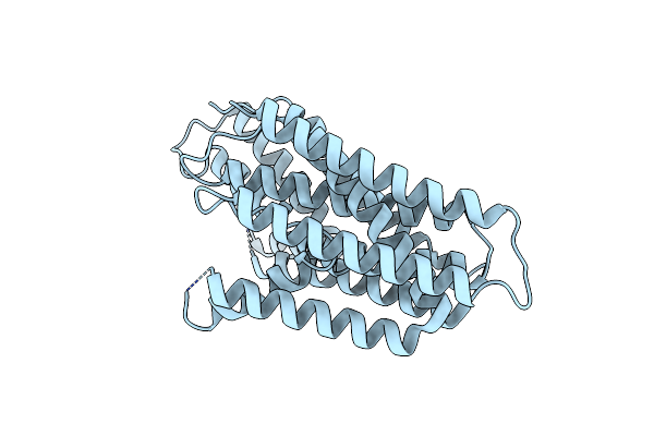
Myxococcus Xanthus Romx
Organism: Myxococcus xanthus dk 1622
Method: X-RAY DIFFRACTION Resolution:1.89 Å Release Date: 2025-06-04 Classification: SIGNALING PROTEIN Ligands: EDO |
Organism: Myxococcus xanthus dk 1622
Method: X-RAY DIFFRACTION
Release Date: 2025-06-04
Ligands: EDO
 |
Structure Of Human Xk-Related Protein 4
Organism: Homo sapiens
Method: ELECTRON MICROSCOPY Resolution:3.50 Å Release Date: 2025-05-14 Classification: MEMBRANE PROTEIN |
Organism: Homo sapiens
Method: ELECTRON MICROSCOPY
Release Date: 2025-05-14
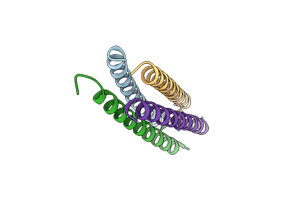 |
X-Ray Structure Of A De Novo Designed Self Assembled Peptide Tetramer Featuring A Cu(His)4(H2O) Coordination Motif
Organism: Synthetic construct
Method: X-RAY DIFFRACTION Resolution:1.36 Å Release Date: 2025-04-16 Classification: DE NOVO PROTEIN Ligands: CU |
Organism: Synthetic construct
Method: X-RAY DIFFRACTION
Release Date: 2025-04-16
Ligands: CU