

Search Count: 11
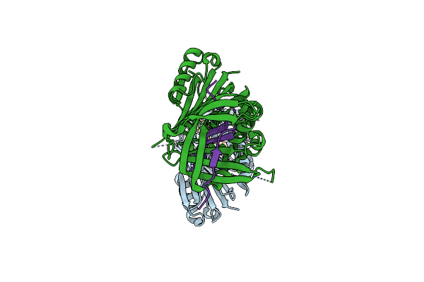 |
Cleaved Human Neuroserpin
Organism: Homo sapiens
Method: X-RAY DIFFRACTION Resolution:1.80 Å Release Date: 2009-05-26 Classification: HYDROLASE INHIBITOR |
Organism: Homo sapiens
Method: X-RAY DIFFRACTION
Release Date: 2009-05-26
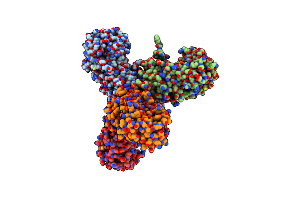 |
Structure Of Native Human Neuroserpin
Organism: Homo sapiens
Method: X-RAY DIFFRACTION Resolution:3.15 Å Release Date: 2009-05-26 Classification: HYDROLASE INHIBITOR |
Organism: Homo sapiens
Method: X-RAY DIFFRACTION
Release Date: 2009-05-26
 |
Crystal Structure Of The Potent Anticoagulant Thrombin Mutant W215A/E217A In Free Form
Organism: Homo sapiens
Method: X-RAY DIFFRACTION Resolution:2.80 Å Release Date: 2004-08-03 Classification: BLOOD CLOTTING |
Organism: Homo sapiens
Method: X-RAY DIFFRACTION
Release Date: 2004-08-03
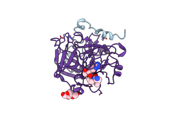 |
Crystal Structure Of The Anticoagulant Thrombin Mutant W215A/E217A Bound To Ppack
Organism: Homo sapiens
Method: X-RAY DIFFRACTION Resolution:2.40 Å Release Date: 2004-08-03 Classification: HYDROLASE/HYDROLASE INHIBITOR Ligands: ZN, 0G6, NAG |
Organism: Homo sapiens
Method: X-RAY DIFFRACTION
Release Date: 2004-08-03
Ligands: ZN, 0G6, NAG
 |
Fast Form Of Thrombin Mutant R(77A)A Bound To Ppack
Organism: Homo sapiens
Method: X-RAY DIFFRACTION Resolution:1.91 Å Release Date: 2004-06-08 Classification: HYDROLASE/hydrolase inhibitor Ligands: 0G6, NAG, NA |
Organism: Homo sapiens
Method: X-RAY DIFFRACTION
Release Date: 2004-06-08
Ligands: 0G6, NAG, NA
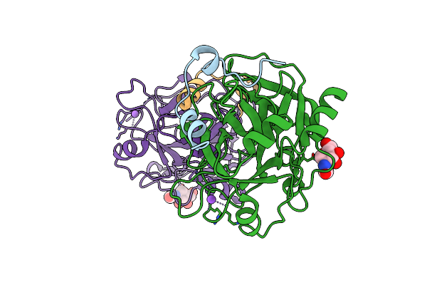 |
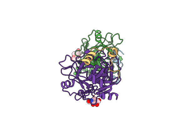
Crystal Structure Of The Procoagulant Fast Form Of Thrombin
Organism: Homo sapiens
Method: X-RAY DIFFRACTION Resolution:2.30 Å Release Date: 2004-06-08 Classification: HYDROLASE Ligands: NAG, NA |
Organism: Homo sapiens
Method: X-RAY DIFFRACTION
Release Date: 2004-06-08
Ligands: NAG, NA
 |
Crystal Structure Of The Anticoagulant Slow Form Of Thrombin
Organism: Homo sapiens
Method: X-RAY DIFFRACTION Resolution:2.30 Å Release Date: 2004-06-08 Classification: HYDROLASE Ligands: NAG |
Organism: Homo sapiens
Method: X-RAY DIFFRACTION
Release Date: 2004-06-08
Ligands: NAG
 |
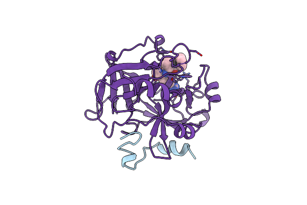
Slow Form Of Thrombin Bound With Ppack
Organism: Homo sapiens
Method: X-RAY DIFFRACTION Resolution:1.55 Å Release Date: 2004-06-08 Classification: HYDROLASE/hydrolase inhibitor Ligands: 0G6, NAG |
Organism: Homo sapiens
Method: X-RAY DIFFRACTION
Release Date: 2004-06-08
Ligands: 0G6, NAG
 |
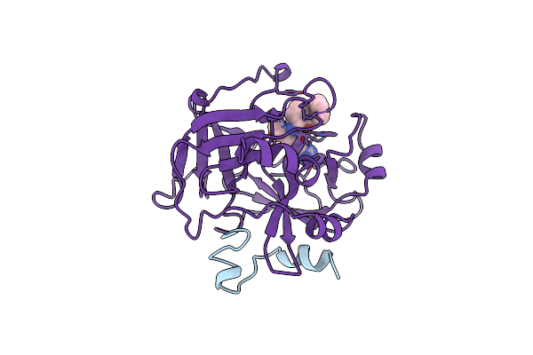
Structure Of Human Alpha-Thrombin Y225F Mutant Bound To D-Phe-Pro-Arg-Chloromethylketone
Organism: Homo sapiens
Method: X-RAY DIFFRACTION Resolution:2.10 Å Release Date: 1999-03-07 Classification: HYDROLASE/HYDROLASE INHIBITOR Ligands: 0G6, NA |
Organism: Homo sapiens
Method: X-RAY DIFFRACTION
Release Date: 1999-03-07
Ligands: 0G6, NA
 |
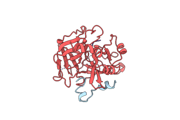
Structure Of Human Alpha-Thrombin Y225P Mutant Bound To D-Phe-Pro-Arg-Chloromethylketone
Organism: Homo sapiens
Method: X-RAY DIFFRACTION Resolution:2.10 Å Release Date: 1999-03-03 Classification: HYDROLASE/HYDROLASE INHIBITOR Ligands: 0G6 |
Organism: Homo sapiens
Method: X-RAY DIFFRACTION
Release Date: 1999-03-03
Ligands: 0G6
 |
Structure Of Human Alpha-Thrombin Y225I Mutant Bound To D-Phe-Pro-Arg-Chloromethylketone
Organism: Homo sapiens
Method: X-RAY DIFFRACTION Resolution:2.10 Å Release Date: 1999-03-02 Classification: HYDROLASE/HYDROLASE INHIBITOR |
Organism: Homo sapiens
Method: X-RAY DIFFRACTION
Release Date: 1999-03-02