

Search Count: 1,171
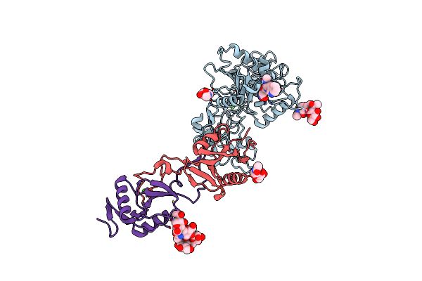 |
Crystal Structure Of Russell'S Viper Venom Metalloproteinase
Organism: Daboia russellii siamensis
Method: X-RAY DIFFRACTION Resolution:2.91 Å Release Date: 2007-12-11 Classification: HYDROLASE, BLOOD CLOTTING, TOXIN Ligands: NAG, ZN, CA, GM6 |
Organism: Daboia russellii siamensis
Method: X-RAY DIFFRACTION
Release Date: 2007-12-11
Ligands: NAG, ZN, CA, GM6
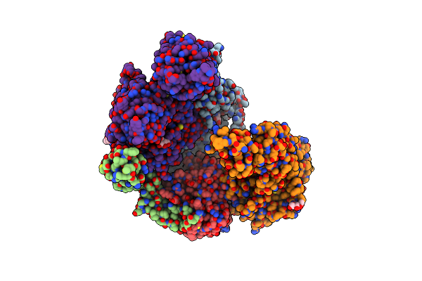 |
Preliminary Map Of The Prothrombin-Prothrombinase Complex On Nano Discs
Organism: Homo sapiens
Method: ELECTRON MICROSCOPY Release Date: 2024-08-07 Classification: BLOOD CLOTTING Ligands: NAG |
Organism: Homo sapiens
Method: ELECTRON MICROSCOPY
Release Date: 2024-08-07
Ligands: NAG
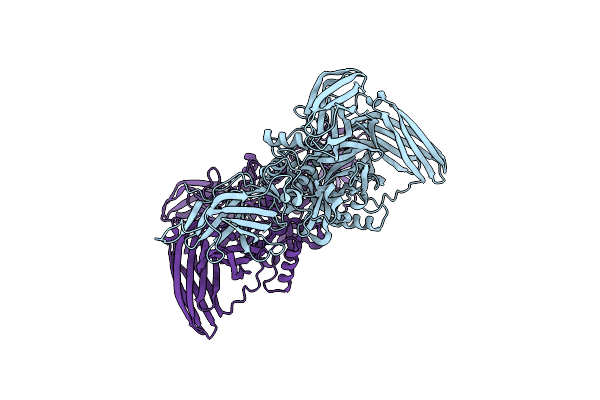 |
Recombinant Human Cellular Coagulation Factor Xiii
Organism: Homo sapiens
Method: X-RAY DIFFRACTION Resolution:2.10 Å Release Date: 1998-08-12 Classification: COAGULATION FACTOR |
Organism: Homo sapiens
Method: X-RAY DIFFRACTION
Release Date: 1998-08-12
 |
Cryo-Em Complex Of Meizothrombindesf1, Factor Xa, And Factor Va
Organism: Homo sapiens
Method: ELECTRON MICROSCOPY Release Date: 2026-05-20 Classification: BLOOD CLOTTING |
Organism: Homo sapiens
Method: ELECTRON MICROSCOPY
Release Date: 2026-05-20
 |
Human Coagulation Factor Ixa In Complex With P-Amino Benzamidine
Organism: Homo sapiens
Method: X-RAY DIFFRACTION Resolution:2.80 Å Release Date: 1999-09-01 Classification: COAGULATION FACTOR Ligands: CA, PBZ, TBU |
Organism: Homo sapiens
Method: X-RAY DIFFRACTION
Release Date: 1999-09-01
Ligands: CA, PBZ, TBU
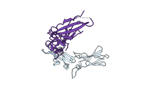 |
Extracellular Domain Of Human Tissue Factor
Organism: Homo sapiens
Method: X-RAY DIFFRACTION Resolution:2.40 Å Release Date: 1998-02-25 Classification: COAGULATION FACTOR |
Organism: Homo sapiens
Method: X-RAY DIFFRACTION
Release Date: 1998-02-25
 |
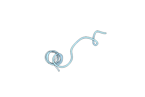
Structural Basis Of Thrombin Mediated Factor V Activation: Essential Role Of The Hirudin-Like Sequence Glu666-Glu672 For Processing At The Heavy Chain-B Domain Junction
Organism: Homo sapiens
Method: X-RAY DIFFRACTION Resolution:1.70 Å Release Date: 2011-06-15 Classification: HYDROLASE/HYDROLASE INHIBITOR |
Organism: Homo sapiens
Method: X-RAY DIFFRACTION
Release Date: 2011-06-15
 |
Coagulation Factor Viii, Nmr, 1 Structure
Organism: Homo sapiens
Method: SOLUTION NMR Release Date: 1997-01-11 Classification: COAGULATION FACTOR |
Organism: Homo sapiens
Method: SOLUTION NMR
Release Date: 1997-01-11
 |
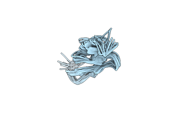
Three-Dimensional Structure Of A Transglutaminase: Human Blood Coagulation Factor Xiii
Organism: Homo sapiens
Method: X-RAY DIFFRACTION Resolution:2.65 Å Release Date: 1995-07-31 Classification: BLOOD COAGULATION |
Organism: Homo sapiens
Method: X-RAY DIFFRACTION
Release Date: 1995-07-31
 |
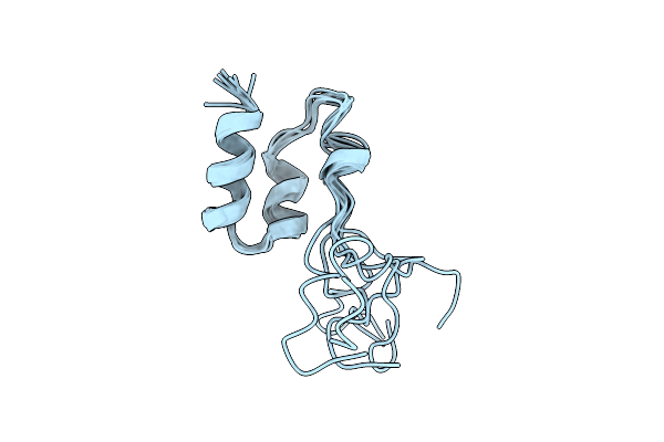
How An Epidermal Growth Factor (Egf)-Like Domain Binds Calcium-High Resolution Nmr Structure Of The Calcium Form Of The Nh2-Terminal Egf-Like Domain In Coagulation Factor X
Organism: Bos taurus
Method: SOLUTION NMR Release Date: 1994-05-31 Classification: COAGULATION FACTOR |
Organism: Bos taurus
Method: SOLUTION NMR
Release Date: 1994-05-31
 |
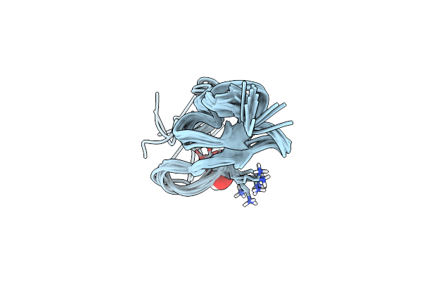
Coagulation Factor, Mg(Ii), Nmr, 7 Structures (Backbone Atoms Only)
Organism: Homo sapiens
Method: SOLUTION NMR Release Date: 1996-11-08 Classification: COAGULATION FACTOR |
Organism: Homo sapiens
Method: SOLUTION NMR
Release Date: 1996-11-08
 |
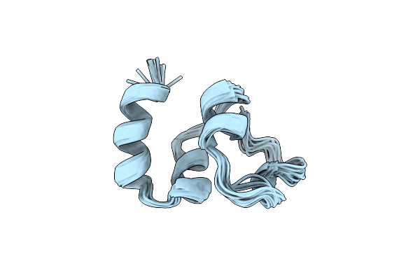
Three-Dimensional Structure Of The Apo Form Of The N-Terminal Egf-Like Module Of Blood Coagulation Factor X As Determined By Nmr Spectroscopy And Simulated Folding
Organism: Bos taurus
Method: SOLUTION NMR Release Date: 1994-01-31 Classification: COAGULATION FACTOR Ligands: OH |
Organism: Bos taurus
Method: SOLUTION NMR
Release Date: 1994-01-31
Ligands: OH
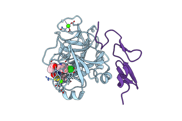 |
Factor Xa In Complex With A Cis-1,2-Diaminocyclohexane Derivative
Organism: Homo sapiens
Method: X-RAY DIFFRACTION Resolution:1.80 Å Release Date: 2010-08-04 Classification: HYDROLASE Ligands: CA, D14 |
Organism: Homo sapiens
Method: X-RAY DIFFRACTION
Release Date: 2010-08-04
Ligands: CA, D14
 |
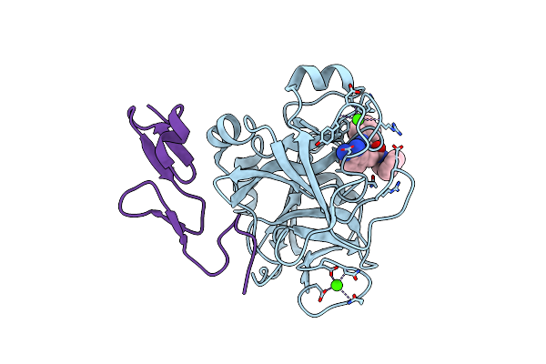
Nmr Structure Of Calcium Ion-Bound Gamma-Carboxy-Glutamic Acid-Rich Domain Of Factor Ix
Organism: Homo sapiens
Method: SOLUTION NMR Release Date: 1996-06-20 Classification: COAGULATION FACTOR |
Organism: Homo sapiens
Method: SOLUTION NMR
Release Date: 1996-06-20
 |
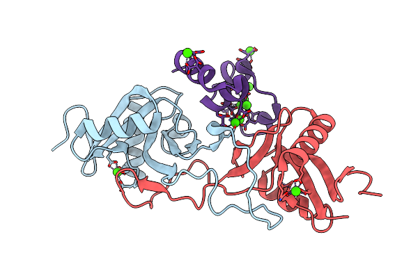
Factor Xa In Complex With (R)-2-(1-Adamantylcarbamoylamino)-3-(3-Carbamidoyl-Phenyl)-N-Phenethyl-Propionic Acid Amide
Organism: Homo sapiens
Method: X-RAY DIFFRACTION Resolution:2.22 Å Release Date: 2010-04-07 Classification: HYDROLASE Ligands: CA, RUP |
Organism: Homo sapiens
Method: X-RAY DIFFRACTION
Release Date: 2010-04-07
Ligands: CA, RUP
 |
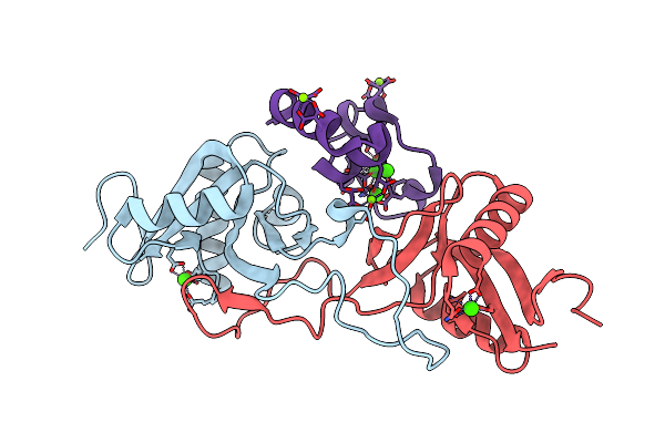
Crystal Structure Of Ca(Ii)-Bound Gla Domain Of Factor Ix Complexed With Binding Protein
Organism: Trimeresurus flavoviridis, Bos taurus
Method: X-RAY DIFFRACTION Resolution:1.80 Å Release Date: 2003-07-08 Classification: Protein binding/Blood clotting Ligands: CA |
Organism: Trimeresurus flavoviridis, Bos taurus
Method: X-RAY DIFFRACTION
Release Date: 2003-07-08
Ligands: CA
 |
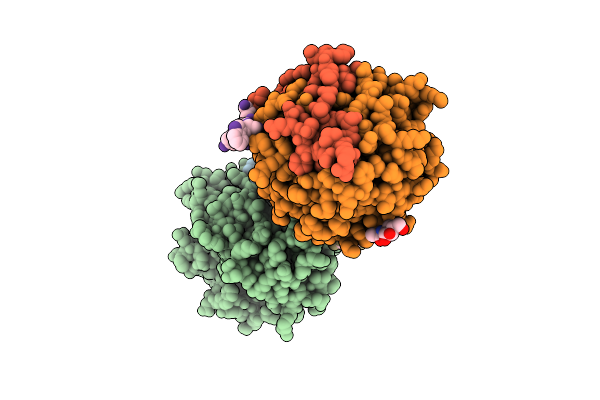
Crystal Structure Of Mg(Ii)-And Ca(Ii)-Bound Gla Domain Of Factor Ix Complexed With Binding Protein
Organism: Trimeresurus flavoviridis, Bos taurus
Method: X-RAY DIFFRACTION Resolution:1.55 Å Release Date: 2003-07-08 Classification: Protein binding/Blood clotting Ligands: CA, MG |
Organism: Trimeresurus flavoviridis, Bos taurus
Method: X-RAY DIFFRACTION
Release Date: 2003-07-08
Ligands: CA, MG
 |
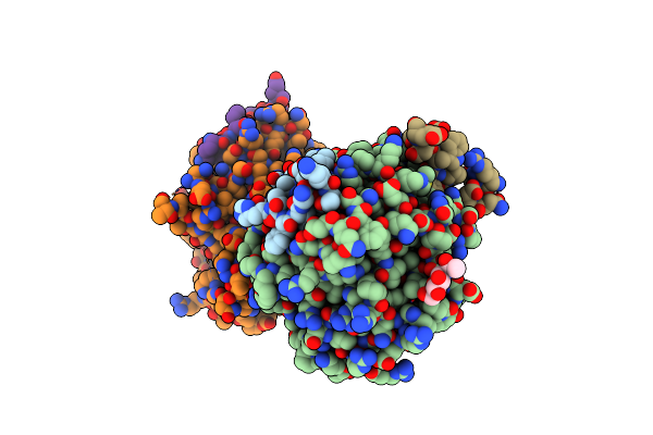
Crystal Structure Of Thrombin Bound To The Extracellular Fragment Of Par1
Organism: Homo sapiens
Method: X-RAY DIFFRACTION Resolution:2.20 Å Release Date: 2008-01-01 Classification: HYDROLASE Ligands: NAG |
Organism: Homo sapiens
Method: X-RAY DIFFRACTION
Release Date: 2008-01-01
Ligands: NAG
 |
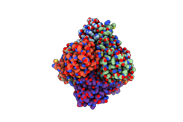
Crystal Structure Of Murine Thrombin Mutant W215A/E217A In Complex With The Extracellular Fragment Of Human Par1
Organism: Mus musculus, Homo sapiens
Method: X-RAY DIFFRACTION Resolution:2.20 Å Release Date: 2009-07-07 Classification: HYDROLASE Ligands: NAG |
Organism: Mus musculus, Homo sapiens
Method: X-RAY DIFFRACTION
Release Date: 2009-07-07
Ligands: NAG
 |
Crystal Structure Of Coagulation Factors Ix/X-Binding Protein (Ix/X-Bp) From Venom Of Habu Snake With A Heterodimer Of C-Type Lectin Domains
Organism: Trimeresurus flavoviridis
Method: X-RAY DIFFRACTION Resolution:2.50 Å Release Date: 1998-05-06 Classification: COAGULATION FACTOR BINDING Ligands: CA |
Organism: Trimeresurus flavoviridis
Method: X-RAY DIFFRACTION
Release Date: 1998-05-06
Ligands: CA