
Search Count: 4
All
Selected
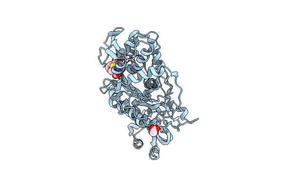 |
Organism: Yersinia pestis
Method: X-RAY DIFFRACTION Resolution:2.63 Å Release Date: 2020-10-14 Classification: CYTOSOLIC PROTEIN Ligands: ACT, CB9 |
 |
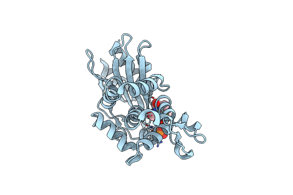
Crystal Structure Of Penicillin-Binding Protein 4 From Listeria Monocytogenes In The Carbenicillin Bound Form
Organism: Listeria monocytogenes
Method: X-RAY DIFFRACTION Resolution:2.01 Å Release Date: 2013-05-29 Classification: PENICILLIN-BINDING PROTEIN Ligands: CB9, GOL |
 |
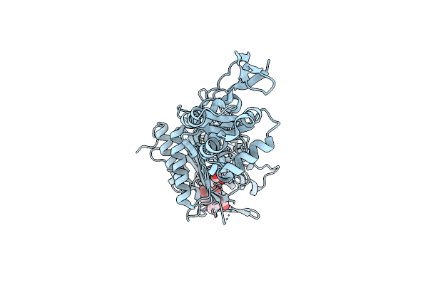
Organism: Mycobacterium tuberculosis
Method: X-RAY DIFFRACTION Resolution:1.41 Å Release Date: 2011-12-14 Classification: HYDROLASE/Antibiotic Ligands: PO4, CB9 |
 |
Crystal Structure Of Penicillin-Binding Protein 3 From Pseudomonas Aeruginosa In Complex With Carbenicillin
Organism: Pseudomonas aeruginosa
Method: X-RAY DIFFRACTION Resolution:2.30 Å Release Date: 2010-11-10 Classification: PENICILLIN-BINDING PROTEIN/ANTIBIOTIC Ligands: CL, CB9, GOL |