

Search Count: 23
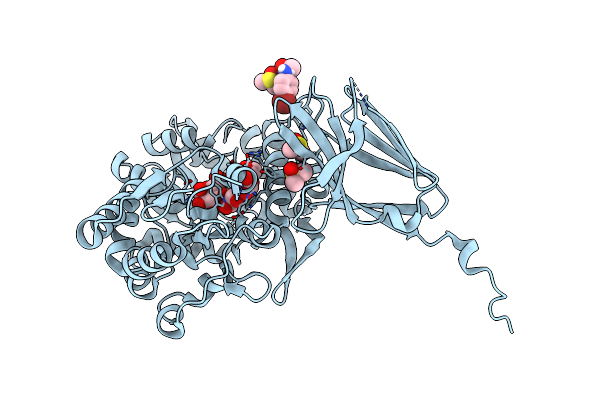 |
Crystal Structure Of Mbp-Trem2 Ig Domain Fusion With Fragment, 2-((4-Bromophenyl)Amino)Ethan-1-Ol
Organism: Methanosarcina mazei, Homo sapiens
Method: X-RAY DIFFRACTION Resolution:1.47 Å Release Date: 2021-02-17 Classification: SIGNALING PROTEIN Ligands: V5S, DMS |
Organism: Methanosarcina mazei, Homo sapiens
Method: X-RAY DIFFRACTION
Release Date: 2021-02-17
Ligands: V5S, DMS
 |
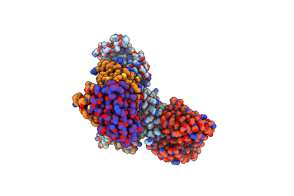
Orexin Receptor 2 (Ox2R) In Complex With G Protein And Natural Peptide-Agonist Orexin B (Oxb)
Organism: Homo sapiens, Mus musculus
Method: ELECTRON MICROSCOPY Release Date: 2021-02-10 Classification: MEMBRANE PROTEIN |
Organism: Homo sapiens, Mus musculus
Method: ELECTRON MICROSCOPY
Release Date: 2021-02-10
 |
Orexin Receptor 2 (Ox2R) In Complex With G Protein And Small-Molecule Agonist Compound 1
Organism: Homo sapiens, Mus musculus, Lama glama
Method: ELECTRON MICROSCOPY Release Date: 2021-02-10 Classification: MEMBRANE PROTEIN Ligands: XGD |
Organism: Homo sapiens, Mus musculus, Lama glama
Method: ELECTRON MICROSCOPY
Release Date: 2021-02-10
Ligands: XGD
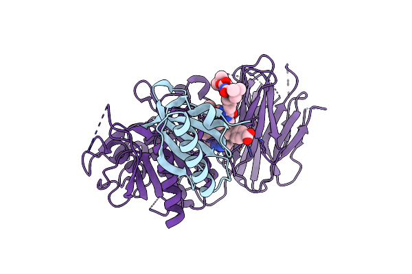 |
Pcsk9 In Complex With Compound 16
Organism: Homo sapiens
Method: X-RAY DIFFRACTION Resolution:1.53 Å Release Date: 2019-11-06 Classification: HYDROLASE/HYDROLASE Inhibitor Ligands: 063 |
Organism: Homo sapiens
Method: X-RAY DIFFRACTION
Release Date: 2019-11-06
Ligands: 063
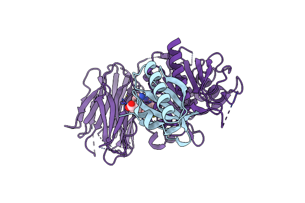 |
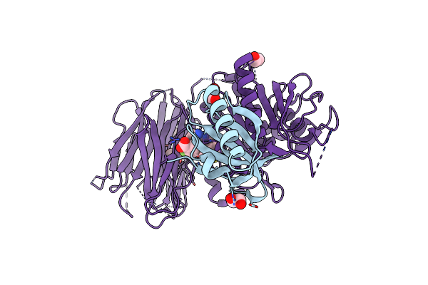
Pcsk9 In Complex With Compound 4
Organism: Homo sapiens
Method: X-RAY DIFFRACTION Resolution:2.15 Å Release Date: 2019-11-06 Classification: HYDROLASE Ligands: PV7 |
Organism: Homo sapiens
Method: X-RAY DIFFRACTION
Release Date: 2019-11-06
Ligands: PV7
 |
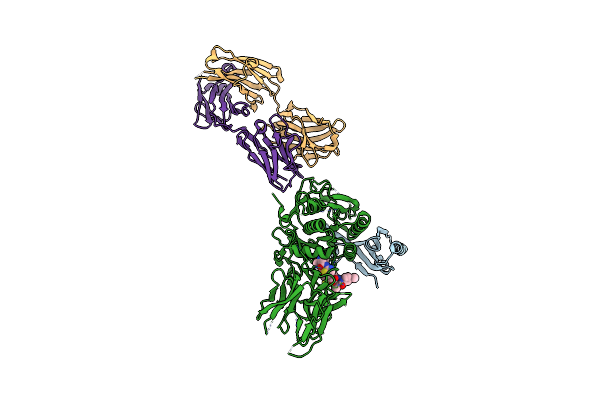
Pcsk9 In Complex With Compound 5
Organism: Homo sapiens
Method: X-RAY DIFFRACTION Resolution:2.04 Å Release Date: 2019-11-06 Classification: HYDROLASE Ligands: EDO, PUV |
Organism: Homo sapiens
Method: X-RAY DIFFRACTION
Release Date: 2019-11-06
Ligands: EDO, PUV
 |
Pcsk9 In Complex With A Fab And Compound 14
Organism: Homo sapiens
Method: X-RAY DIFFRACTION Resolution:2.70 Å Release Date: 2019-11-06 Classification: HYDROLASE/Immune System Ligands: PVM |
Organism: Homo sapiens
Method: X-RAY DIFFRACTION
Release Date: 2019-11-06
Ligands: PVM
 |
Pcsk9 In Complex With A Fab And Compound 8
Organism: Homo sapiens
Method: X-RAY DIFFRACTION Resolution:2.73 Å Release Date: 2019-11-06 Classification: HYDROLASE/Immune System Ligands: PVJ |
Organism: Homo sapiens
Method: X-RAY DIFFRACTION
Release Date: 2019-11-06
Ligands: PVJ
 |
Pcsk9 In Complex With Compound 2
Organism: Homo sapiens
Method: X-RAY DIFFRACTION Resolution:2.64 Å Release Date: 2019-11-06 Classification: HYDROLASE Ligands: PVV |
Organism: Homo sapiens
Method: X-RAY DIFFRACTION
Release Date: 2019-11-06
Ligands: PVV
 |
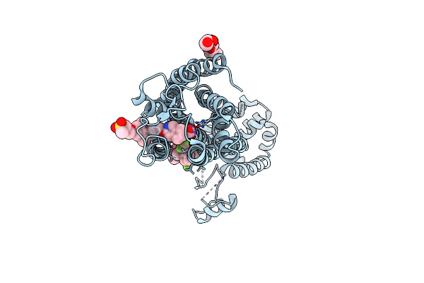
Gpr40 In Complex With Partial Agonist Mk-8666
Organism: Homo sapiens, Enterobacteria phage t4
Method: X-RAY DIFFRACTION Resolution:2.20 Å Release Date: 2017-06-07 Classification: FATTY ACID BINDING PROTEIN/HYDROLASE |
Organism: Homo sapiens, Enterobacteria phage t4
Method: X-RAY DIFFRACTION
Release Date: 2017-06-07
 |
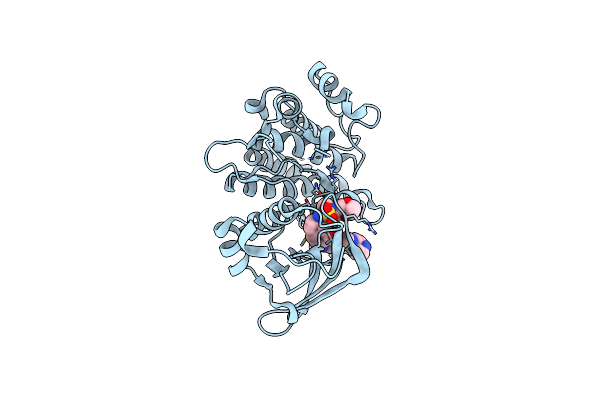
Gpr40 In Complex With Agopam Ap8 And Partial Agonist Mk-8666
Organism: Homo sapiens, Enterobacteria phage t4
Method: X-RAY DIFFRACTION Resolution:3.22 Å Release Date: 2017-06-07 Classification: FATTY ACID BINDING PROTEIN/HYDROLASE Ligands: OLC, MK6, 7OS |
Organism: Homo sapiens, Enterobacteria phage t4
Method: X-RAY DIFFRACTION
Release Date: 2017-06-07
Ligands: OLC, MK6, 7OS
 |
Structure Of Dually Phosphorylated C-Met Receptor Kinase In Complex With An Mk-2461 Analog
Organism: Homo sapiens
Method: X-RAY DIFFRACTION Resolution:2.30 Å Release Date: 2012-02-01 Classification: TRANSFERASE/TRANSFERASE INHIBITOR Ligands: M61 |
Organism: Homo sapiens
Method: X-RAY DIFFRACTION
Release Date: 2012-02-01
Ligands: M61
 |
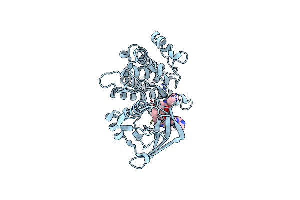
Structure Of The Apo Met Receptor Kinase In The Dually-Phosphorylated, Activated State
Organism: Homo sapiens
Method: X-RAY DIFFRACTION Resolution:1.60 Å Release Date: 2011-01-19 Classification: TRANSFERASE |
Organism: Homo sapiens
Method: X-RAY DIFFRACTION
Release Date: 2011-01-19
 |
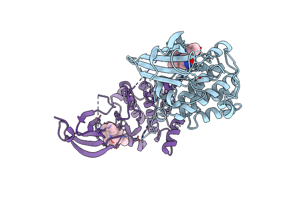
Structure Of Dually-Phosphorylated Met Receptor Kinase In Complex With An Mk-2461 Analog With Specificity For The Activated Receptor
Organism: Homo sapiens
Method: X-RAY DIFFRACTION Resolution:1.75 Å Release Date: 2011-01-19 Classification: transferase/transferase inhibitor Ligands: Q6W |
Organism: Homo sapiens
Method: X-RAY DIFFRACTION
Release Date: 2011-01-19
Ligands: Q6W
 |
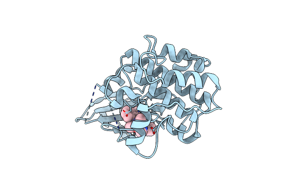
Crystal Structure Of Unphosphorylated P70S6K1 (Form I)
Organism: Homo sapiens
Method: X-RAY DIFFRACTION Resolution:2.80 Å Release Date: 2009-10-27 Classification: TRANSFERASE Ligands: STU |
Organism: Homo sapiens
Method: X-RAY DIFFRACTION
Release Date: 2009-10-27
Ligands: STU
 |
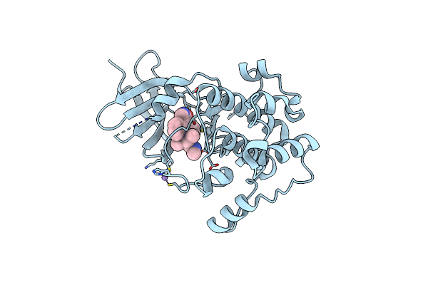
Crystal Structure Of Unphosphorylated P70S6K1 (Form Ii)
Organism: Homo sapiens
Method: X-RAY DIFFRACTION Resolution:3.43 Å Release Date: 2009-10-27 Classification: TRANSFERASE Ligands: STU |
Organism: Homo sapiens
Method: X-RAY DIFFRACTION
Release Date: 2009-10-27
Ligands: STU
 |
Crystal Structure Of Phosphorylated P70S6K1
Organism: Homo sapiens
Method: X-RAY DIFFRACTION Resolution:2.35 Å Release Date: 2009-10-27 Classification: TRANSFERASE Ligands: STU, MN |
Organism: Homo sapiens
Method: X-RAY DIFFRACTION
Release Date: 2009-10-27
Ligands: STU, MN
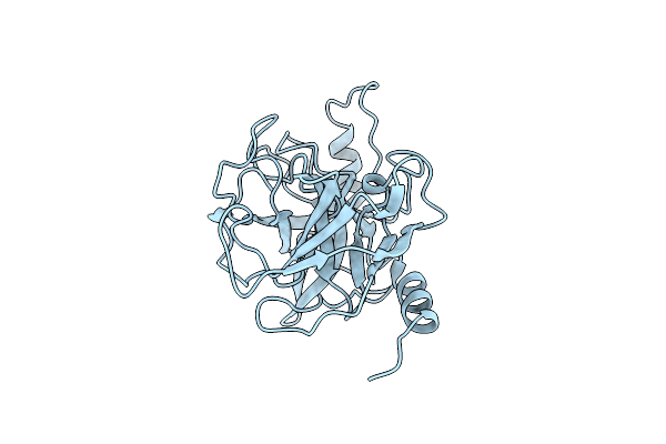 |
Crystal Structure Of Human Prostasin
Organism: Homo sapiens
Method: X-RAY DIFFRACTION Resolution:1.45 Å Release Date: 2008-10-14 Classification: HYDROLASE Ligands: CL |
Organism: Homo sapiens
Method: X-RAY DIFFRACTION
Release Date: 2008-10-14
Ligands: CL
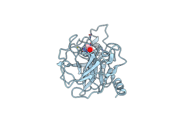 |
Crystal Structure Of Human Prostasin Complexed To 4-Guanidinobenzoic Acid
Organism: Homo sapiens
Method: X-RAY DIFFRACTION Resolution:2.00 Å Release Date: 2008-10-14 Classification: HYDROLASE Ligands: GBS |
Organism: Homo sapiens
Method: X-RAY DIFFRACTION
Release Date: 2008-10-14
Ligands: GBS
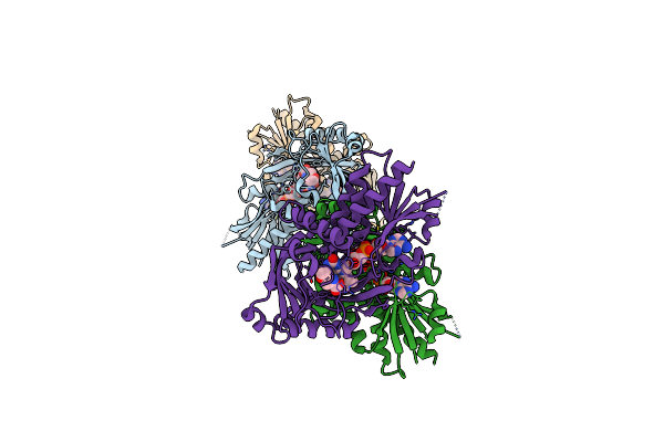 |
Crystal Structure Of Human D-Amino Acid Oxidase: Bound To An Inhibitor
Organism: Homo sapiens
Method: X-RAY DIFFRACTION Resolution:2.49 Å Release Date: 2008-07-22 Classification: OXIDOREDUCTASE Ligands: FAD, 4P5 |
Organism: Homo sapiens
Method: X-RAY DIFFRACTION
Release Date: 2008-07-22
Ligands: FAD, 4P5