

Search Count: 16
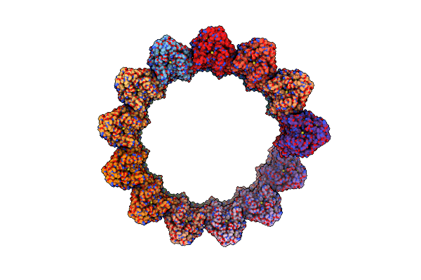 |
Structure Of The Native Microtubule Lattice Nucleated From The Yeast Spindle Pole Body
Organism: Saccharomyces cerevisiae
Method: ELECTRON MICROSCOPY Release Date: 2024-04-24 Classification: CELL CYCLE |
Organism: Saccharomyces cerevisiae
Method: ELECTRON MICROSCOPY
Release Date: 2024-04-24
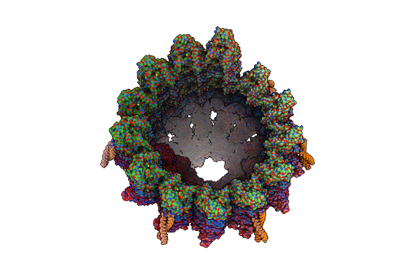 |
Structure Of The Native Y-Tubulin Ring Complex (Yturc) Capping Microtubule Minus Ends At The Spindle Pole Body
Organism: Saccharomyces cerevisiae
Method: ELECTRON MICROSCOPY Release Date: 2024-04-24 Classification: CELL CYCLE Ligands: GTP, GDP |
Organism: Saccharomyces cerevisiae
Method: ELECTRON MICROSCOPY
Release Date: 2024-04-24
Ligands: GTP, GDP
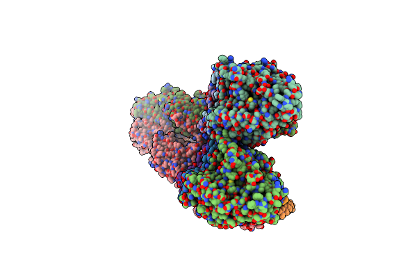 |
Structure Of The Y-Tubulin Small Complex (Ytusc) As Part Of The Native Y-Tubulin Ring Complex (Yturc) Capping Microtubule Minus Ends At The Spindle Pole Body
Organism: Saccharomyces cerevisiae
Method: ELECTRON MICROSCOPY Release Date: 2024-04-24 Classification: CELL CYCLE Ligands: GTP |
Organism: Saccharomyces cerevisiae
Method: ELECTRON MICROSCOPY
Release Date: 2024-04-24
Ligands: GTP
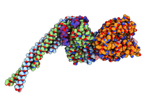 |
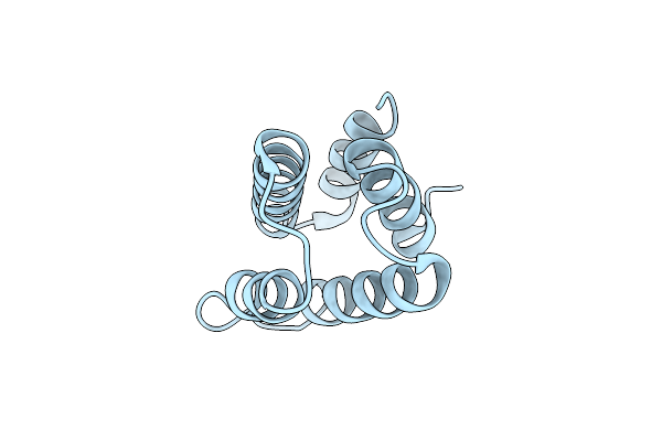
Outer Kinetochore Ndc80-Dam1 Alpha/Beta-Tubulin Complex
Organism: Saccharomyces cerevisiae, Sus scrofa
Method: ELECTRON MICROSCOPY Release Date: 2024-04-03 Classification: CELL CYCLE Ligands: GTP, MG, GDP, TA1 |
Organism: Saccharomyces cerevisiae, Sus scrofa
Method: ELECTRON MICROSCOPY
Release Date: 2024-04-03
Ligands: GTP, MG, GDP, TA1
 |
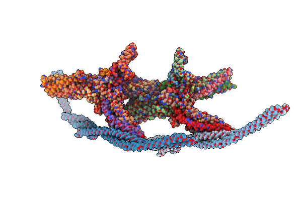
Crystal Structure Of The Yeast Spindle Body Component Spc98
Organism: Saccharomyces cerevisiae
Method: X-RAY DIFFRACTION Resolution:1.87 Å Release Date: 2024-04-03 Classification: CELL CYCLE |
Organism: Saccharomyces cerevisiae
Method: X-RAY DIFFRACTION
Release Date: 2024-04-03
 |
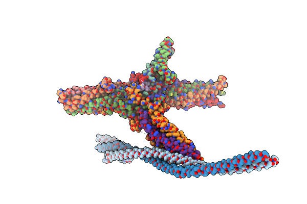
Outer Kinetochore Dam1 Protomer Dimer Ndc80-Nuf2 Coiled-Coil Complex
Organism: Saccharomyces cerevisiae
Method: ELECTRON MICROSCOPY Release Date: 2023-12-06 Classification: CELL CYCLE |
Organism: Saccharomyces cerevisiae
Method: ELECTRON MICROSCOPY
Release Date: 2023-12-06
 |
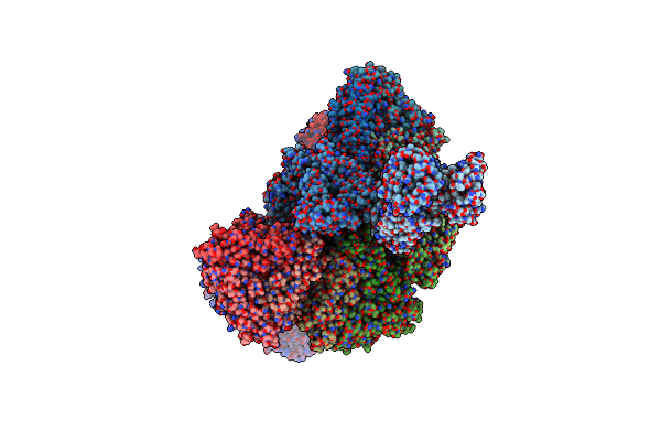
Outer Kinetochore Dam1 Protomer Monomer Ndc80-Nuf2 Coiled-Coil Complex
Organism: Saccharomyces cerevisiae
Method: ELECTRON MICROSCOPY Release Date: 2023-12-06 Classification: CELL CYCLE |
Organism: Saccharomyces cerevisiae
Method: ELECTRON MICROSCOPY
Release Date: 2023-12-06
 |
Subtomogram Average Of Immature Rotavirus Tlp Penton
Organism: Rotavirus a
Method: ELECTRON MICROSCOPY Release Date: 2023-05-10 Classification: VIRAL PROTEIN |
Organism: Rotavirus a
Method: ELECTRON MICROSCOPY
Release Date: 2023-05-10
 |
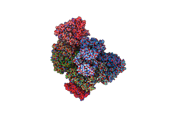
Spa Of Trypsin Untreated Rotavirus Tlp Spike
Organism: Rotavirus a
Method: ELECTRON MICROSCOPY Release Date: 2023-04-05 Classification: VIRAL PROTEIN Ligands: CA, ZN |
Organism: Rotavirus a
Method: ELECTRON MICROSCOPY
Release Date: 2023-04-05
Ligands: CA, ZN
 |
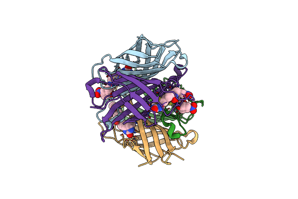
In Situ Subtomogram Average Of Immature Rotavirus Tlp Spike
Organism: Rotavirus a
Method: ELECTRON MICROSCOPY Release Date: 2023-04-05 Classification: VIRAL PROTEIN |
Organism: Rotavirus a
Method: ELECTRON MICROSCOPY
Release Date: 2023-04-05
 |
Avidin-Biotin-Phenol
Organism: Gallus gallus
Method: X-RAY DIFFRACTION Resolution:1.58 Å Release Date: 2021-03-03 Classification: STRUCTURAL PROTEIN Ligands: V8M |
Organism: Gallus gallus
Method: X-RAY DIFFRACTION
Release Date: 2021-03-03
Ligands: V8M
 |
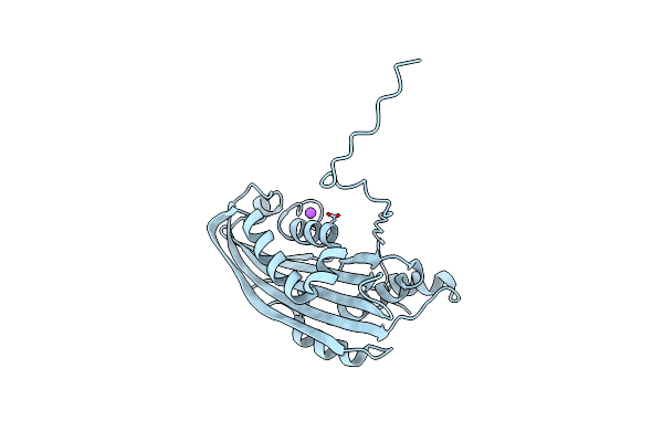
Native Structure Of Mosquitocidal Cyt1A Protoxin Obtained By Serial Femtosecond Crystallography On In Vivo Grown Crystals At Ph 7
Organism: Bacillus thuringiensis subsp. israelensis
Method: X-RAY DIFFRACTION Resolution:1.86 Å Release Date: 2020-10-14 Classification: TOXIN |
Organism: Bacillus thuringiensis subsp. israelensis
Method: X-RAY DIFFRACTION
Release Date: 2020-10-14
 |
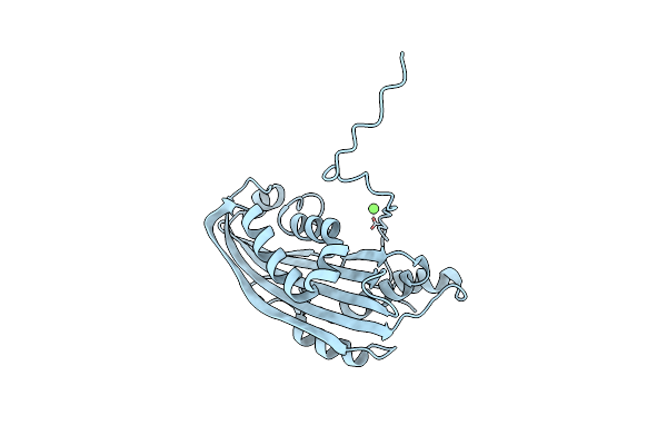
Structure Of Mosquitocidal Cyt1A Protoxin Obtained By Serial Femtosecond Crystallography On In Vivo Grown Crystals Soaked With Dtt At Ph 7
Organism: Bacillus thuringiensis subsp. israelensis
Method: X-RAY DIFFRACTION Resolution:1.85 Å Release Date: 2020-10-14 Classification: TOXIN |
Organism: Bacillus thuringiensis subsp. israelensis
Method: X-RAY DIFFRACTION
Release Date: 2020-10-14
 |
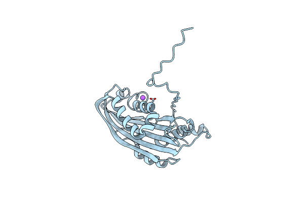
Structure Of Mosquitocidal Cyt1Aa Protoxin Obtained By Serial Femtosecond Crystallography On In Vivo Grown Crystals At Ph 10
Organism: Bacillus thuringiensis subsp. israelensis
Method: X-RAY DIFFRACTION Resolution:1.85 Å Release Date: 2020-10-14 Classification: TOXIN Ligands: CA |
Organism: Bacillus thuringiensis subsp. israelensis
Method: X-RAY DIFFRACTION
Release Date: 2020-10-14
Ligands: CA
 |
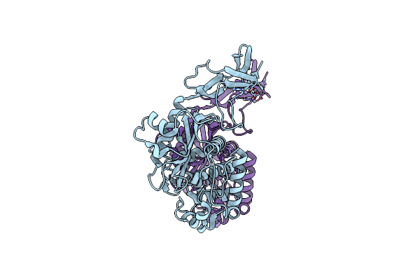
Structure Of The C7S Mutant Of Mosquitocidal Cyt1A Protoxin Obtained By Serial Femtosecond Crystallography On In Vivo Grown Crystals At Ph 7
Organism: Bacillus thuringiensis subsp. israelensis
Method: X-RAY DIFFRACTION Resolution:2.00 Å Release Date: 2020-10-14 Classification: TOXIN |
Organism: Bacillus thuringiensis subsp. israelensis
Method: X-RAY DIFFRACTION
Release Date: 2020-10-14
 |
Crystal Structure Of S. Aureus Penicillin Binding Protein 4 (Pbp4) Mutant (S75C)
Organism: Staphylococcus aureus (strain col)
Method: X-RAY DIFFRACTION Resolution:1.86 Å Release Date: 2018-12-19 Classification: HYDROLASE Ligands: ZN |
Organism: Staphylococcus aureus (strain col)
Method: X-RAY DIFFRACTION
Release Date: 2018-12-19
Ligands: ZN