

Search Count: 50
 |
Non-Helical Dna Triplex Forms A Unique Aptamer Scaffold For High Affinity Recognition Of Nerve Growth Factor
Organism: Homo sapiens, Synthetic construct
Method: X-RAY DIFFRACTION Resolution:2.45 Å Release Date: 2015-06-10 Classification: Immune system/DNA Ligands: EPE |
Organism: Homo sapiens, Synthetic construct
Method: X-RAY DIFFRACTION
Release Date: 2015-06-10
Ligands: EPE
 |
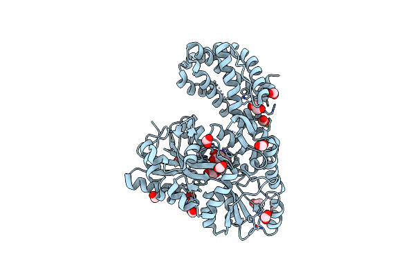
Structure Of Mcl1 Bound To Brd Inhibitor Ligand 1 At 1.7A
Organism: Homo sapiens
Method: X-RAY DIFFRACTION Resolution:1.70 Å Release Date: 2015-05-06 Classification: Apoptosis/Inhibitor Ligands: ZN, 865, POP |
Organism: Homo sapiens
Method: X-RAY DIFFRACTION
Release Date: 2015-05-06
Ligands: ZN, 865, POP
 |
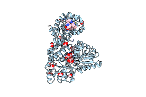
Structure Of Apo Mbp-Mcl1 At 1.9A
Organism: Escherichia coli, Homo sapiens
Method: X-RAY DIFFRACTION Resolution:1.90 Å Release Date: 2015-05-06 Classification: APOPTOSIS Ligands: MG, EDO, FMT |
Organism: Escherichia coli, Homo sapiens
Method: X-RAY DIFFRACTION
Release Date: 2015-05-06
Ligands: MG, EDO, FMT
 |
Structure Of Mbp-Mcl1 Bound To Ligand 1 At 2.35A
Organism: Escherichia coli, Homo sapiens
Method: X-RAY DIFFRACTION Resolution:2.35 Å Release Date: 2015-05-06 Classification: APOPTOSIS Ligands: 865, EDO, FMT |
Organism: Escherichia coli, Homo sapiens
Method: X-RAY DIFFRACTION
Release Date: 2015-05-06
Ligands: 865, EDO, FMT
 |
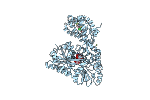
Structure Of Mbp-Mcl1 Bound To Ligand 2 At 1.55A
Organism: Escherichia coli, Homo sapiens
Method: X-RAY DIFFRACTION Resolution:1.55 Å Release Date: 2015-05-06 Classification: APOPTOSIS Ligands: 19H, MG, FMT, NA, EDO |
Organism: Escherichia coli, Homo sapiens
Method: X-RAY DIFFRACTION
Release Date: 2015-05-06
Ligands: 19H, MG, FMT, NA, EDO
 |
Structure Of Mbp-Mcl1 Bound To Ligand 4 At 2.4A
Organism: Escherichia coli, Homo sapiens
Method: X-RAY DIFFRACTION Resolution:2.40 Å Release Date: 2015-05-06 Classification: APOPTOSIS Ligands: CL, MG, 3R4 |
Organism: Escherichia coli, Homo sapiens
Method: X-RAY DIFFRACTION
Release Date: 2015-05-06
Ligands: CL, MG, 3R4
 |
The Structure Of Mbp-Mcl1 Bound To Ligand 5 At 1.9A
Organism: Escherichia coli, Homo sapiens
Method: X-RAY DIFFRACTION Resolution:1.90 Å Release Date: 2015-05-06 Classification: APOPTOSIS Ligands: MG, EDO, FMT, 3R6 |
Organism: Escherichia coli, Homo sapiens
Method: X-RAY DIFFRACTION
Release Date: 2015-05-06
Ligands: MG, EDO, FMT, 3R6
 |
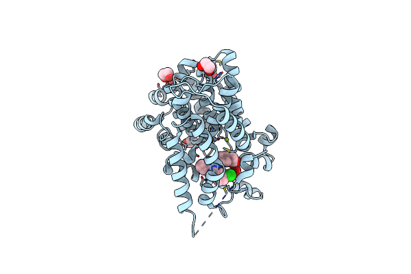
The Structure Of Mbp-Mcl1 Bound To Ligand 6 At 2.0A
Organism: Escherichia coli, Homo sapiens
Method: X-RAY DIFFRACTION Resolution:2.00 Å Release Date: 2015-05-06 Classification: APOPTOSIS Ligands: 3R7, FMT, MG |
Organism: Escherichia coli, Homo sapiens
Method: X-RAY DIFFRACTION
Release Date: 2015-05-06
Ligands: 3R7, FMT, MG
 |
Pde4 Catalytic Domain
Organism: Homo sapiens
Method: X-RAY DIFFRACTION Resolution:2.15 Å Release Date: 2014-10-15 Classification: hydrolase/hydrolase inhibitor Ligands: ZN, MG, NA, 2O5, EDO |
Organism: Homo sapiens
Method: X-RAY DIFFRACTION
Release Date: 2014-10-15
Ligands: ZN, MG, NA, 2O5, EDO
 |
Selective Inhibition Of The Catalytic Domain Of Human Phosphodiesterase 4B With A-33
Organism: Homo sapiens
Method: X-RAY DIFFRACTION Resolution:1.90 Å Release Date: 2014-01-01 Classification: HYDROLASE/HYDROLASE INHIBITOR Ligands: 19T, MG, ZN, NA, EDO |
Organism: Homo sapiens
Method: X-RAY DIFFRACTION
Release Date: 2014-01-01
Ligands: 19T, MG, ZN, NA, EDO
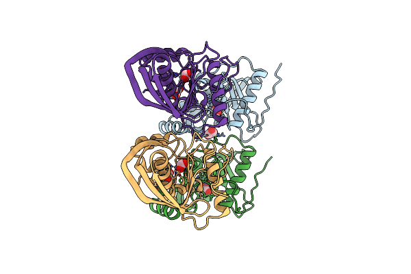 |
Crystal Structure Of A Peptide Deformylase From Synechococcus Elongatus
Organism: Synechococcus elongatus
Method: X-RAY DIFFRACTION Resolution:1.55 Å Release Date: 2013-03-06 Classification: HYDROLASE Ligands: ZN, EDO, FMT, CL |
Organism: Synechococcus elongatus
Method: X-RAY DIFFRACTION
Release Date: 2013-03-06
Ligands: ZN, EDO, FMT, CL
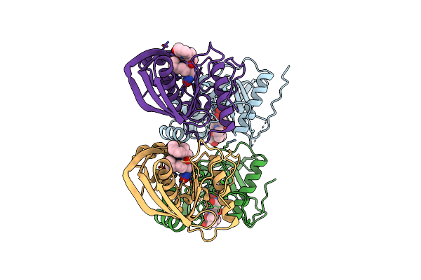 |
Crystal Structure Of A Peptide Deformylase From Synechococcus Elongatus In Complex With Actinonin
Organism: Synechococcus elongatus
Method: X-RAY DIFFRACTION Resolution:1.90 Å Release Date: 2013-01-16 Classification: HYDROLASE/HYDROLASE INHIBITOR Ligands: ZN, BR, BB2 |
Organism: Synechococcus elongatus
Method: X-RAY DIFFRACTION
Release Date: 2013-01-16
Ligands: ZN, BR, BB2
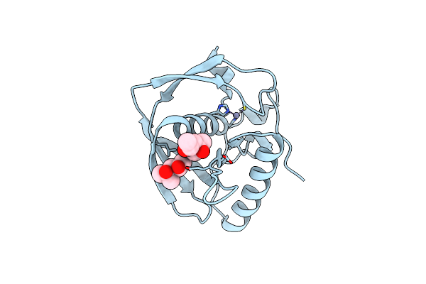 |
Crystal Structure Of A Probable Peptide Deformylase From Synechococcus Phage S-Ssm7
Organism: Synechococcus phage s-ssm7
Method: X-RAY DIFFRACTION Resolution:1.95 Å Release Date: 2013-01-09 Classification: HYDROLASE Ligands: ZN, MRD |
Organism: Synechococcus phage s-ssm7
Method: X-RAY DIFFRACTION
Release Date: 2013-01-09
Ligands: ZN, MRD
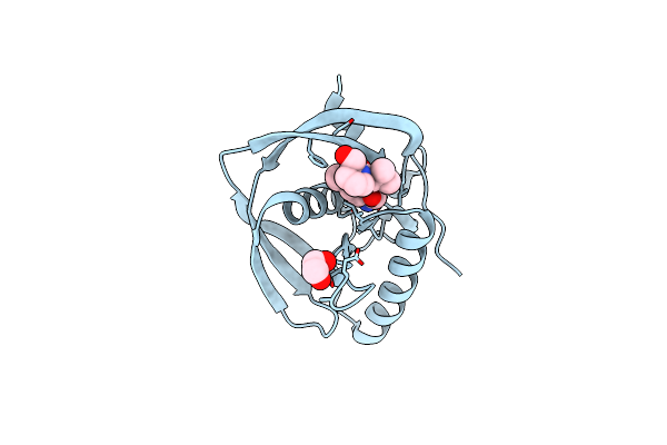 |
Crystal Structure Of A Probable Peptide Deformylase From Strucynechococcus Phage S-Ssm7 In Complex With Actinonin
Organism: Synechococcus phage s-ssm7
Method: X-RAY DIFFRACTION Resolution:1.70 Å Release Date: 2013-01-09 Classification: HYDROLASE/ANTIBIOTIC Ligands: ZN, CL, EDO, BB2 |
Organism: Synechococcus phage s-ssm7
Method: X-RAY DIFFRACTION
Release Date: 2013-01-09
Ligands: ZN, CL, EDO, BB2
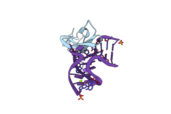 |
Crystal Structure Of Human Pdgf-Bb In Complex With A Modified Nucleotide Aptamer (Somamer Sl5)
Organism: Homo sapiens
Method: X-RAY DIFFRACTION Resolution:2.20 Å Release Date: 2012-11-21 Classification: HORMONE/DNA |
Organism: Homo sapiens
Method: X-RAY DIFFRACTION
Release Date: 2012-11-21
 |
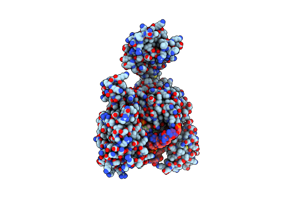
Crystal Structure Of Human Pdgf-Bb In Complex With A Modified Nucleotide Aptamer (Somamer Sl4)
Organism: Homo sapiens
Method: X-RAY DIFFRACTION Resolution:2.30 Å Release Date: 2012-11-21 Classification: HORMONE/DNA |
Organism: Homo sapiens
Method: X-RAY DIFFRACTION
Release Date: 2012-11-21
 |
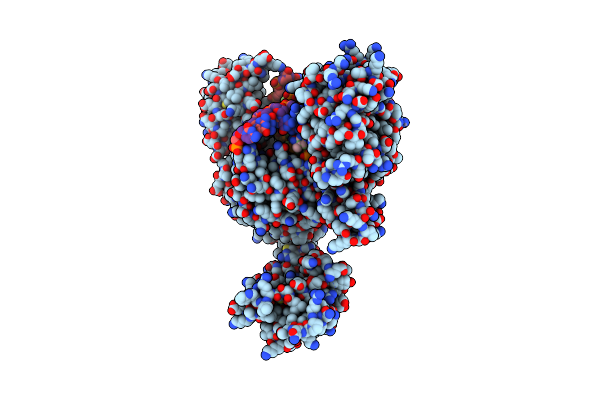
Topoisomerase Ii-Dna Cleavage Complex, Apo
Organism: Saccharomyces cerevisiae
Method: X-RAY DIFFRACTION Resolution:2.48 Å Release Date: 2010-05-26 Classification: Isomerase/DNA Ligands: TSP |
Organism: Saccharomyces cerevisiae
Method: X-RAY DIFFRACTION
Release Date: 2010-05-26
Ligands: TSP
 |
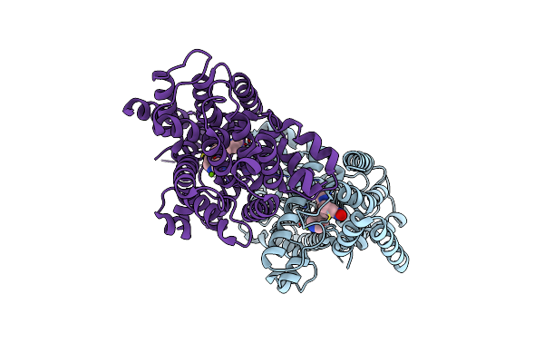
Topoisomerase Ii-Dna Cleavage Complex, Metal-Bound
Organism: Saccharomyces cerevisiae
Method: X-RAY DIFFRACTION Resolution:2.98 Å Release Date: 2010-05-26 Classification: Isomerase/DNA Ligands: ZN, TSP |
Organism: Saccharomyces cerevisiae
Method: X-RAY DIFFRACTION
Release Date: 2010-05-26
Ligands: ZN, TSP
 |
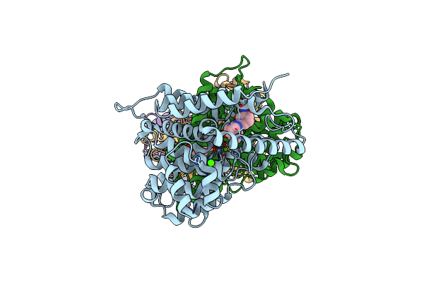
Crystal Structure Of Human Phosphodiesterase 4B With Regulatory Domain And D155988
Organism: Homo sapiens
Method: X-RAY DIFFRACTION Resolution:2.63 Å Release Date: 2010-01-19 Classification: HYDROLASE Ligands: ZN, MG, 988 |
Organism: Homo sapiens
Method: X-RAY DIFFRACTION
Release Date: 2010-01-19
Ligands: ZN, MG, 988
 |
Crystal Structure Of Human Phosphodiesterase 4D With Regulatory Domain And D155871
Organism: Homo sapiens
Method: X-RAY DIFFRACTION Resolution:2.30 Å Release Date: 2010-01-19 Classification: HYDROLASE Ligands: ZN, MG, CA, D71 |
Organism: Homo sapiens
Method: X-RAY DIFFRACTION
Release Date: 2010-01-19
Ligands: ZN, MG, CA, D71