

Search Count: 12
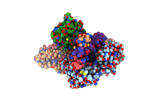 |
The Structure Of The Native Cardiac Thin Filament Troponin Core In Ca2+-Free State From The Upper Strand
Organism: Sus scrofa
Method: ELECTRON MICROSCOPY Release Date: 2024-03-06 Classification: MOTOR PROTEIN |
Organism: Sus scrofa
Method: ELECTRON MICROSCOPY
Release Date: 2024-03-06
 |
The Structure Of The Native Cardiac Thin Filament Troponin Core In Ca2+-Free Tilted State From The Upper Strand
Organism: Sus scrofa
Method: ELECTRON MICROSCOPY Release Date: 2024-03-06 Classification: MOTOR PROTEIN |
Organism: Sus scrofa
Method: ELECTRON MICROSCOPY
Release Date: 2024-03-06
 |
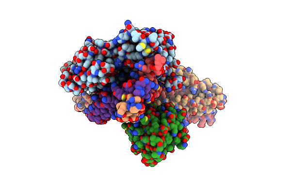
The Structure Of The Native Cardiac Thin Filament Troponin Core In Ca2+-Free Rotated State From The Upper Strand
Organism: Sus scrofa
Method: ELECTRON MICROSCOPY Release Date: 2024-03-06 Classification: MOTOR PROTEIN |
Organism: Sus scrofa
Method: ELECTRON MICROSCOPY
Release Date: 2024-03-06
 |
The Structure Of The Native Cardiac Thin Filament Troponin Core In Ca2+-Free State From The Lower Strand
Organism: Sus scrofa
Method: ELECTRON MICROSCOPY Release Date: 2024-03-06 Classification: MOTOR PROTEIN Ligands: ADP, MG |
Organism: Sus scrofa
Method: ELECTRON MICROSCOPY
Release Date: 2024-03-06
Ligands: ADP, MG
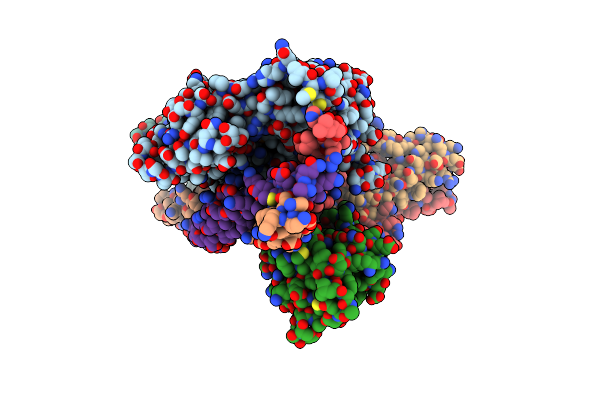 |
The Structure Of The Native Cardiac Thin Filament Troponin Core In Ca2+-Free Rotated State From The Lower Strand
Organism: Sus scrofa
Method: ELECTRON MICROSCOPY Release Date: 2024-03-06 Classification: MOTOR PROTEIN Ligands: ADP, MG |
Organism: Sus scrofa
Method: ELECTRON MICROSCOPY
Release Date: 2024-03-06
Ligands: ADP, MG
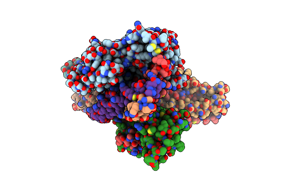 |
The Structure Of The Native Cardiac Thin Filament Troponin Core In Ca2+-Free Tilted State From The Lower Strand
Organism: Sus scrofa
Method: ELECTRON MICROSCOPY Release Date: 2024-03-06 Classification: MOTOR PROTEIN Ligands: ADP, MG |
Organism: Sus scrofa
Method: ELECTRON MICROSCOPY
Release Date: 2024-03-06
Ligands: ADP, MG
 |
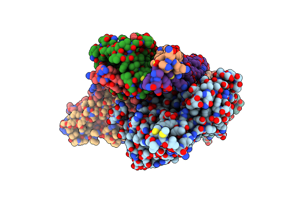
The Structure Of The Native Cardiac Thin Filament Troponin Core In Ca2+-Bound Fully Activated State From The Upper Strand
Organism: Sus scrofa
Method: ELECTRON MICROSCOPY Release Date: 2024-03-06 Classification: MOTOR PROTEIN Ligands: ADP, MG, CA |
Organism: Sus scrofa
Method: ELECTRON MICROSCOPY
Release Date: 2024-03-06
Ligands: ADP, MG, CA
 |
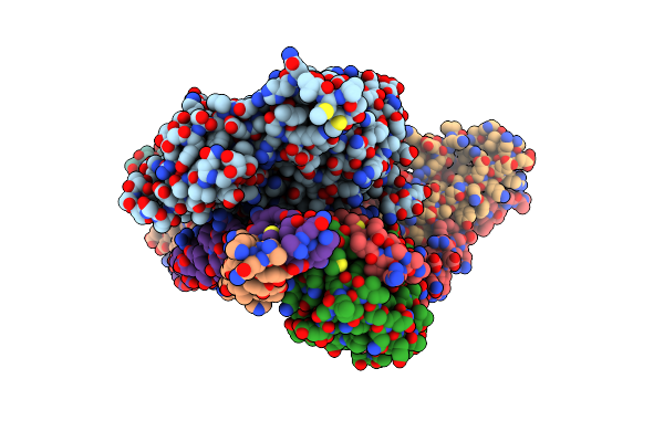
The Structure Of The Native Cardiac Thin Filament Troponin Core In Ca2+-Bound Partially Activated State From The Upper Strand
Organism: Sus scrofa
Method: ELECTRON MICROSCOPY Release Date: 2024-03-06 Classification: MOTOR PROTEIN Ligands: ADP, MG, CA |
Organism: Sus scrofa
Method: ELECTRON MICROSCOPY
Release Date: 2024-03-06
Ligands: ADP, MG, CA
 |
The Structure Of The Native Cardiac Thin Filament Troponin Core In Ca2+-Bound Fully Activated State 1 From The Lower Strand
Organism: Sus scrofa
Method: ELECTRON MICROSCOPY Release Date: 2024-03-06 Classification: MOTOR PROTEIN Ligands: ADP, MG, CA |
Organism: Sus scrofa
Method: ELECTRON MICROSCOPY
Release Date: 2024-03-06
Ligands: ADP, MG, CA
 |
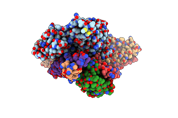
The Structure Of The Native Cardiac Thin Filament Troponin Core In Ca2+-Bound Fully Activated State 2 From The Lower Strand
Organism: Sus scrofa
Method: ELECTRON MICROSCOPY Release Date: 2024-03-06 Classification: MOTOR PROTEIN Ligands: ADP, MG, CA |
Organism: Sus scrofa
Method: ELECTRON MICROSCOPY
Release Date: 2024-03-06
Ligands: ADP, MG, CA
 |
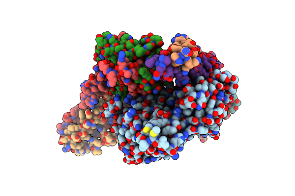
The Structure Of The Native Cardiac Thin Filament Troponin Core In Ca2+-Bound Partially Activated State From The Lower Strand
Organism: Sus scrofa
Method: ELECTRON MICROSCOPY Release Date: 2024-03-06 Classification: MOTOR PROTEIN Ligands: ADP, MG, CA |
Organism: Sus scrofa
Method: ELECTRON MICROSCOPY
Release Date: 2024-03-06
Ligands: ADP, MG, CA
 |
The Structure Of The Native Cardiac Thin Filament Troponin Core In Ca2+-Free State From The Upper Strand Activated By The C1-Domain Of Cardiac Myosin Binding Protein C
Organism: Sus scrofa
Method: ELECTRON MICROSCOPY Release Date: 2024-03-06 Classification: MOTOR PROTEIN Ligands: ADP, MG |
Organism: Sus scrofa
Method: ELECTRON MICROSCOPY
Release Date: 2024-03-06
Ligands: ADP, MG