

Search Count: 22
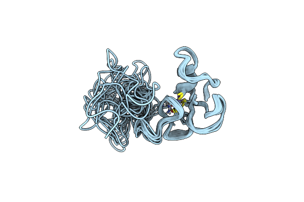 |
Solution Nmr Structure Of Human Gata2 N-Terminal Zinc Finger Domain
Organism: Homo sapiens
Method: SOLUTION NMR Release Date: 2021-06-30 Classification: TRANSCRIPTION Ligands: ZN |
Organism: Homo sapiens
Method: SOLUTION NMR
Release Date: 2021-06-30
Ligands: ZN
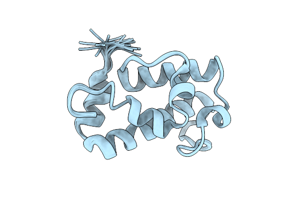 |
A1-Type Acp Domain From Module 5 Of Mlsa1
Organism: Mycobacterium ulcerans (strain agy99)
Method: SOLUTION NMR Release Date: 2019-03-06 Classification: PROTEIN BINDING |
Organism: Mycobacterium ulcerans (strain agy99)
Method: SOLUTION NMR
Release Date: 2019-03-06
 |
B1-Type Acp Domain From Module 7 Of Mlsb
Organism: Mycobacterium ulcerans (strain agy99)
Method: SOLUTION NMR Release Date: 2019-03-06 Classification: PROTEIN BINDING |
Organism: Mycobacterium ulcerans (strain agy99)
Method: SOLUTION NMR
Release Date: 2019-03-06
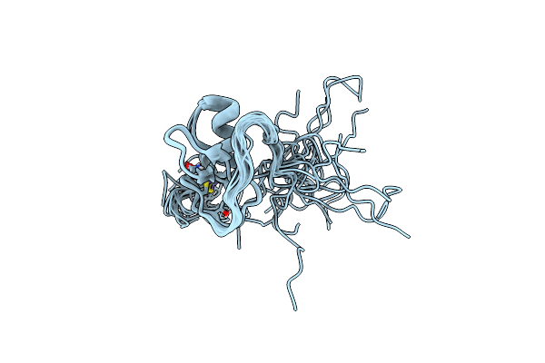 |
Solution Nmr Structure Of Human Gata2 C-Terminal Zinc Finger Domain
Organism: Homo sapiens
Method: SOLUTION NMR Release Date: 2019-02-06 Classification: TRANSCRIPTION Ligands: ZN |
Organism: Homo sapiens
Method: SOLUTION NMR
Release Date: 2019-02-06
Ligands: ZN
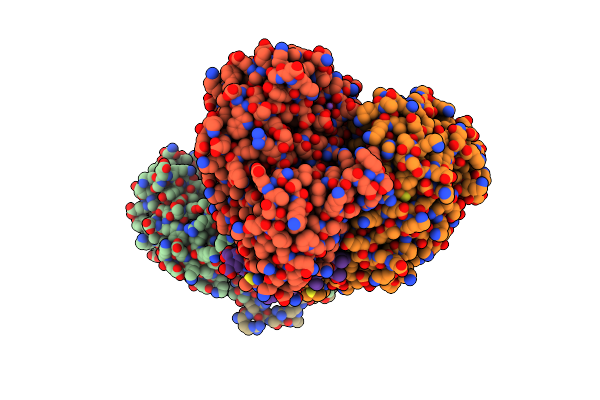 |
Enolase In Complex With Rnase E
Organism: Escherichia coli (strain k12)
Method: X-RAY DIFFRACTION Resolution:2.00 Å Release Date: 2017-07-26 Classification: LYASE Ligands: MG, NA, PO4 |
Organism: Escherichia coli (strain k12)
Method: X-RAY DIFFRACTION
Release Date: 2017-07-26
Ligands: MG, NA, PO4
 |
Structure Of The N-Terminal Domain Of The Escherichia Coli Proq Rna Binding Protein
Organism: Escherichia coli o45:k1 (strain s88 / expec)
Method: SOLUTION NMR Release Date: 2017-05-03 Classification: RNA |
Organism: Escherichia coli o45:k1 (strain s88 / expec)
Method: SOLUTION NMR
Release Date: 2017-05-03
 |
Structure Of The C-Terminal Domain Of The Escherichia Coli Proq Rna Binding Protein
Organism: Escherichia coli (strain k12)
Method: SOLUTION NMR Release Date: 2017-05-03 Classification: CHAPERONE |
Organism: Escherichia coli (strain k12)
Method: SOLUTION NMR
Release Date: 2017-05-03
 |
Solution Structure Of The Qua1 Dimerization Domain Of Pxqua, The Xenopus Ortholog Of Quaking.
|
Organism: Xenopus laevis
Method: SOLUTION NMR
Release Date: 2013-10-30
 |
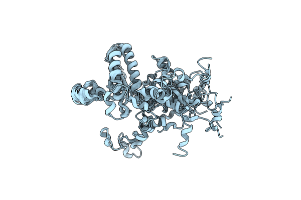
Multienzyme Docking In Hybrid Megasynthetases
Organism: Angiococcus disciformis
Method: SOLUTION NMR Release Date: 2007-12-11 Classification: BIOSYNTHETIC PROTEIN |
Organism: Angiococcus disciformis
Method: SOLUTION NMR
Release Date: 2007-12-11
 |
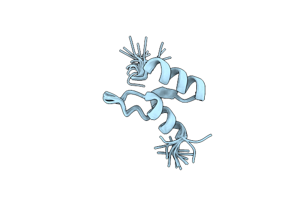
Solution Structure Of The Kh-Qua2 Region Of The Xenopus Star-Gsg Quaking Protein.
|
Organism: Xenopus laevis
Method: SOLUTION NMR
Release Date: 2005-04-14
 |
Nmr Structure Of The Peripheral-Subunit Binding Domain Of Bacillus Stearothermophilus E2P
Organism: Geobacillus stearothermophilus
Method: SOLUTION NMR Release Date: 2004-07-20 Classification: TRANSFERASE |
Organism: Geobacillus stearothermophilus
Method: SOLUTION NMR
Release Date: 2004-07-20
 |
Structure Of Fused Docking Domains From The Erythromycin Polyketide Synthase (Debs), A Model For The Interaction Between Debs 2 And Debs 3: The A Domain
Organism: Saccharopolyspora erythraea
Method: SOLUTION NMR Release Date: 2004-02-24 Classification: TRANSFERASE |
Organism: Saccharopolyspora erythraea
Method: SOLUTION NMR
Release Date: 2004-02-24
 |
Structure Of Fused Docking Domains From The Erythromycin Polyketide Synthase (Debs), A Model For The Interaction Between Debs2 And Debs3: The B Domain
Organism: Saccharopolyspora erythraea
Method: SOLUTION NMR Release Date: 2004-02-24 Classification: TRANSFERASE |
Organism: Saccharopolyspora erythraea
Method: SOLUTION NMR
Release Date: 2004-02-24
 |
Solution Structure Of A Dimeric N-Terminal Fragment Of Human Ubiquitin
|
Organism: Homo sapiens
Method: SOLUTION NMR
Release Date: 2001-12-13
 |
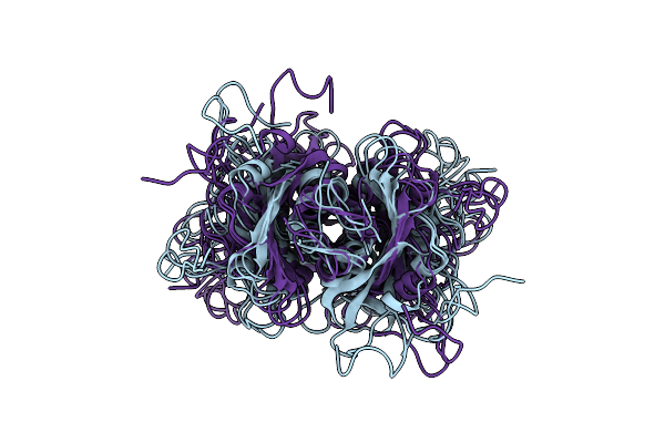
Solution Structure Of The Lipoyl Domain Of The Chimeric Dihydrolipoyl Dehydrogenase P64K From Neisseria Meningitidis
Organism: Neisseria meningitidis
Method: SOLUTION NMR Release Date: 2001-11-28 Classification: OXIDOREDUCTASE |
Organism: Neisseria meningitidis
Method: SOLUTION NMR
Release Date: 2001-11-28
 |
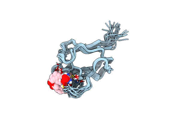
Mouse Hp1 (M31) C Terminal (Shadow Chromo) Domain
Organism: Mus musculus
Method: SOLUTION NMR Release Date: 2000-04-09 Classification: CHROMATIN STRUCTURE |
Organism: Mus musculus
Method: SOLUTION NMR
Release Date: 2000-04-09
 |
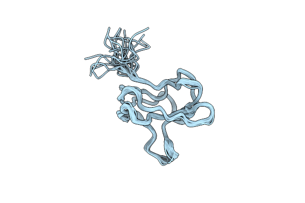
Solution Structure Of Holo-Biotinyl Domain From Acetyl Coenzyme A Carboxylase Of Escherichia Coli Determined By Triple-Resonance Nmr Spectroscopy
Organism: Escherichia coli
Method: SOLUTION NMR Release Date: 1999-04-27 Classification: BIOTIN Ligands: BTN |
Organism: Escherichia coli
Method: SOLUTION NMR
Release Date: 1999-04-27
Ligands: BTN
 |
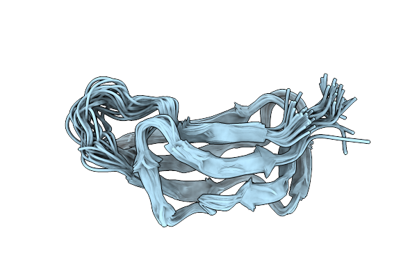
Solution Structure Of Apo-Biotinyl Domain From Acetyl Coenzyme A Carboxylase Of Escherichia Coli Determined By Triple-Resonance Nmr Spectroscopy
|
Organism: Escherichia coli
Method: SOLUTION NMR
Release Date: 1999-04-26
 |
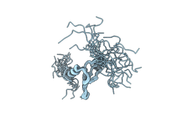
Lipoyl Domain From The Dihydrolipoyl Succinyltransferase Component Of The 2-Oxoglutarate Dehydrogenase Multienzyme Complex Of Escherichia Coli, Nmr, 25 Structures
Organism: Escherichia coli
Method: SOLUTION NMR Release Date: 1998-07-29 Classification: TRANSFERASE |
Organism: Escherichia coli
Method: SOLUTION NMR
Release Date: 1998-07-29
 |
Structure Of The Chromatin Binding (Chromo) Domain From Mouse Modifier Protein 1, Nmr, 26 Structures
Organism: Mus musculus
Method: SOLUTION NMR Release Date: 1998-07-22 Classification: CHROMATIN-BINDING |
Organism: Mus musculus
Method: SOLUTION NMR
Release Date: 1998-07-22