

Search Count: 31
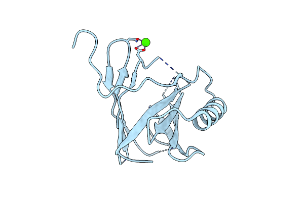 |
X-Ray Structure Of Canis Familiaris Odorant Binding Protein 1
Organism: Canis lupus familiaris
Method: X-RAY DIFFRACTION Resolution:3.00 Å Release Date: 2023-08-16 Classification: TRANSPORT PROTEIN Ligands: CA |
Organism: Canis lupus familiaris
Method: X-RAY DIFFRACTION
Release Date: 2023-08-16
Ligands: CA
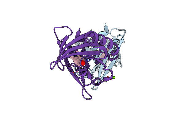 |
X-Ray Structure Of Canis Familiaris Odorant Binding Protein 2 Bound To Citronellal
Organism: Canis lupus familiaris
Method: X-RAY DIFFRACTION Resolution:1.74 Å Release Date: 2023-08-16 Classification: TRANSPORT PROTEIN Ligands: ODM, MG |
Organism: Canis lupus familiaris
Method: X-RAY DIFFRACTION
Release Date: 2023-08-16
Ligands: ODM, MG
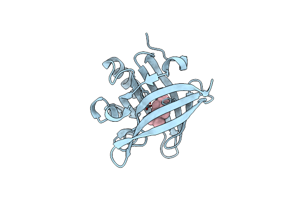 |
X-Ray Structure Of Canis Familiaris Odorant Binding Protein 3 Bound To Menthone
Organism: Canis lupus familiaris
Method: X-RAY DIFFRACTION Resolution:1.84 Å Release Date: 2023-08-16 Classification: TRANSPORT PROTEIN Ligands: LWE |
Organism: Canis lupus familiaris
Method: X-RAY DIFFRACTION
Release Date: 2023-08-16
Ligands: LWE
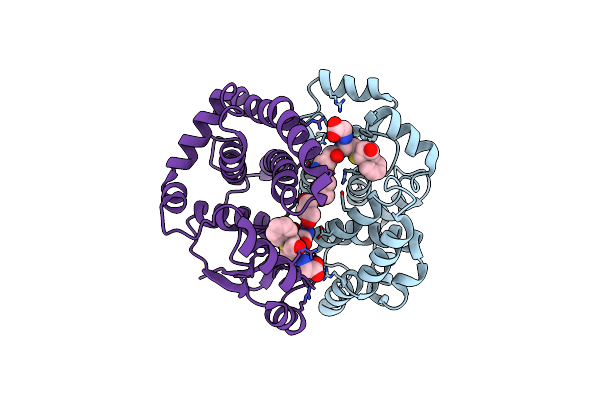 |
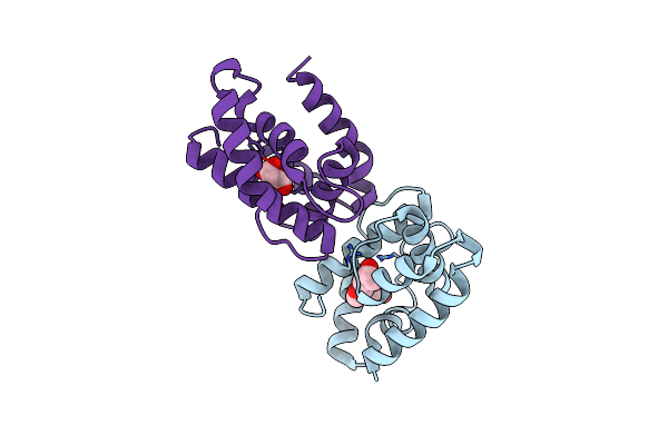
Crystal Structure Of Human Gsta1-1 Bound To The Glutathione Adduct Of Cinnamaldehyde
Organism: Homo sapiens
Method: X-RAY DIFFRACTION Resolution:2.19 Å Release Date: 2020-08-26 Classification: TRANSFERASE Ligands: P9H, GOL |
Organism: Homo sapiens
Method: X-RAY DIFFRACTION
Release Date: 2020-08-26
Ligands: P9H, GOL
 |
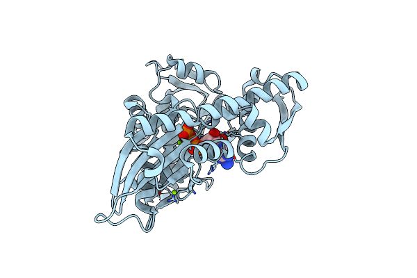
Odorant-Binding Protein Dmelobp28A From Drosophila Melanogaster
Organism: Drosophila melanogaster
Method: X-RAY DIFFRACTION Resolution:2.00 Å Release Date: 2020-03-18 Classification: TRANSPORT PROTEIN Ligands: 1PE, MLA |
Organism: Drosophila melanogaster
Method: X-RAY DIFFRACTION
Release Date: 2020-03-18
Ligands: 1PE, MLA
 |
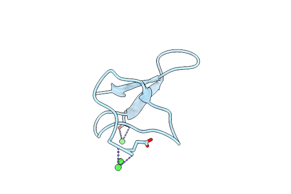
Structure Of Nucleotide Binding Domain Of Hsp110, Atp And Mg2+ Complexed
Organism: Homo sapiens
Method: X-RAY DIFFRACTION Resolution:2.00 Å Release Date: 2019-05-08 Classification: CHAPERONE Ligands: MG, ATP |
Organism: Homo sapiens
Method: X-RAY DIFFRACTION
Release Date: 2019-05-08
Ligands: MG, ATP
 |
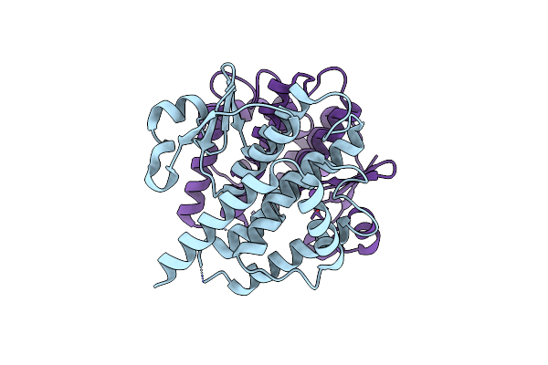
Crystal Structure Of Gurmarin, A Sweet Taste Suppressing Polypeptide
Organism: Gymnema sylvestre
Method: X-RAY DIFFRACTION Resolution:1.45 Å Release Date: 2018-08-08 Classification: PLANT PROTEIN Ligands: NI |
Organism: Gymnema sylvestre
Method: X-RAY DIFFRACTION
Release Date: 2018-08-08
Ligands: NI
 |
Structure Of The Glutathione Transferase Delta 2 From Drosophila Melanogaster
Organism: Drosophila melanogaster
Method: X-RAY DIFFRACTION Resolution:1.60 Å Release Date: 2017-04-05 Classification: TRANSFERASE |
Organism: Drosophila melanogaster
Method: X-RAY DIFFRACTION
Release Date: 2017-04-05
 |
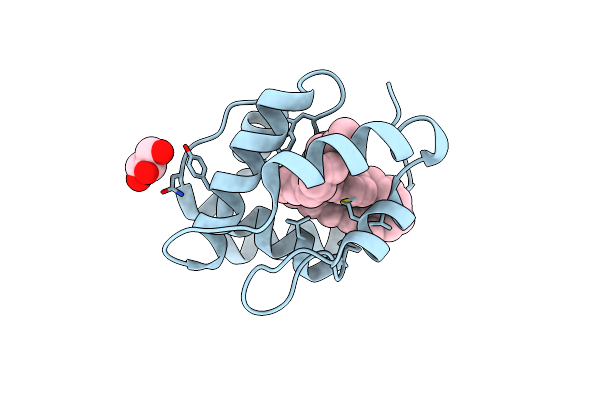
Crystal Structure Of Rat Odorant Binding Protein 3 (Obp3)
Organism: Rattus norvegicus
Method: X-RAY DIFFRACTION Resolution:2.80 Å Release Date: 2014-03-12 Classification: ODORANT BINDING PROTEIN |
Organism: Rattus norvegicus
Method: X-RAY DIFFRACTION
Release Date: 2014-03-12
 |
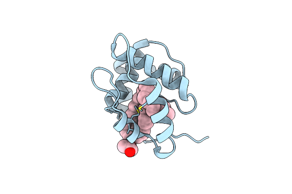
Crystal Structure Of A Pheromone Binding Protein From Apis Mellifera With A Serendipitous Ligand At Ph 5.5
Organism: Apis mellifera
Method: X-RAY DIFFRACTION Resolution:1.80 Å Release Date: 2009-12-01 Classification: Pheromone binding protein Ligands: CMJ, GOL, CL |
Organism: Apis mellifera
Method: X-RAY DIFFRACTION
Release Date: 2009-12-01
Ligands: CMJ, GOL, CL
 |
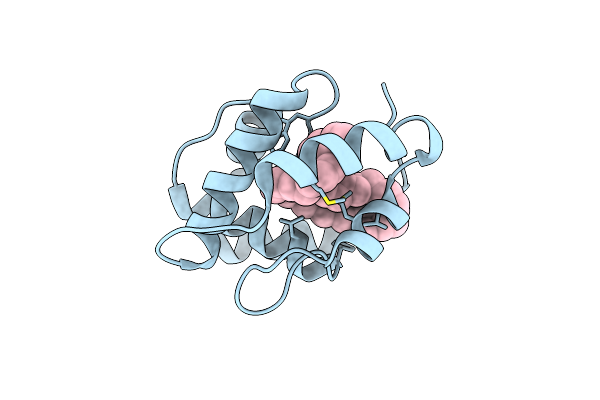
Crystal Structure Of A Pheromone Binding Protein From Apis Mellifera With A Serendipitous Ligand Soaked At Ph 4.0
Organism: Apis mellifera
Method: X-RAY DIFFRACTION Resolution:1.90 Å Release Date: 2009-12-01 Classification: Pheromone binding protein Ligands: CMJ, GOL, CL |
Organism: Apis mellifera
Method: X-RAY DIFFRACTION
Release Date: 2009-12-01
Ligands: CMJ, GOL, CL
 |
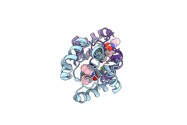
Crystal Structure Of A Pheromone Binding Protein From Apis Mellifera With A Serendipitous Ligand Soaked At Ph 7.0
Organism: Apis mellifera
Method: X-RAY DIFFRACTION Resolution:1.75 Å Release Date: 2009-12-01 Classification: Pheromone binding protein Ligands: CMJ, CL |
Organism: Apis mellifera
Method: X-RAY DIFFRACTION
Release Date: 2009-12-01
Ligands: CMJ, CL
 |
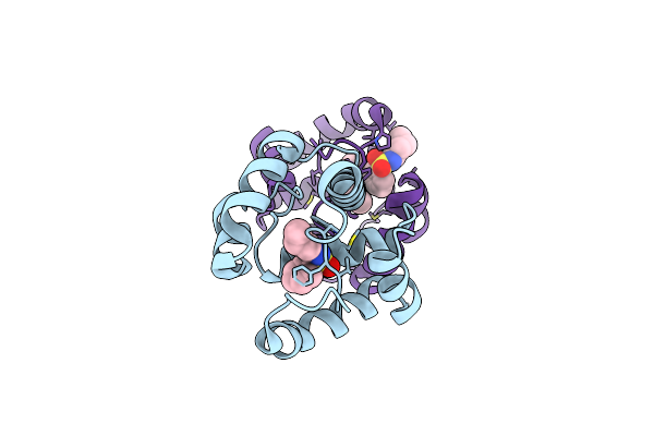
Crystal Structure Of A Pheromone Binding Protein Mutant D35A, From Apis Mellifera, At Ph 7.0
Organism: Apis mellifera
Method: X-RAY DIFFRACTION Resolution:2.03 Å Release Date: 2009-05-26 Classification: Pheromone Binding Protein Ligands: NBB |
Organism: Apis mellifera
Method: X-RAY DIFFRACTION
Release Date: 2009-05-26
Ligands: NBB
 |
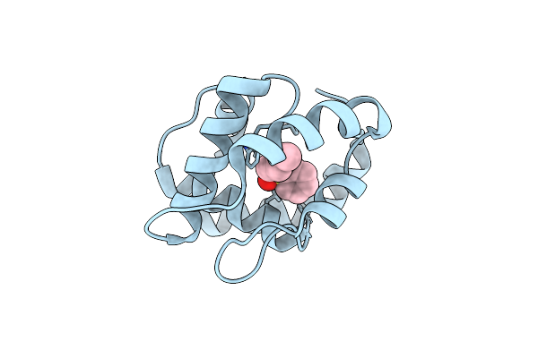
Crystal Structure Of A Pheromone Binding Protein Mutant D35A, From Apis Mellifera, Soaked At Ph 5.5
Organism: Apis mellifera
Method: X-RAY DIFFRACTION Resolution:2.10 Å Release Date: 2009-05-26 Classification: Pheromone Binding Protein Ligands: NBB |
Organism: Apis mellifera
Method: X-RAY DIFFRACTION
Release Date: 2009-05-26
Ligands: NBB
 |
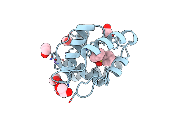
Crystal Structure Of A Pheromone Binding Protein Mutant D35N, From Apis Mellifera, At Ph 5.5
Organism: Apis mellifera
Method: X-RAY DIFFRACTION Resolution:2.30 Å Release Date: 2009-05-26 Classification: Pheromone Binding Protein Ligands: NBB |
Organism: Apis mellifera
Method: X-RAY DIFFRACTION
Release Date: 2009-05-26
Ligands: NBB
 |
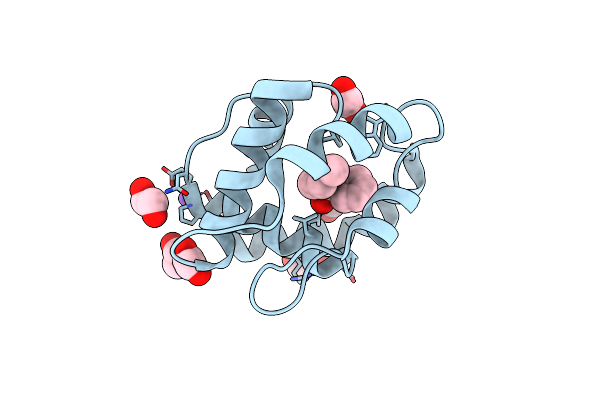
Crystal Structure Of A Pheromone Binding Protein Mutant D35N, From Apis Mellifera, Soaked At Ph 7.0
Organism: Apis mellifera
Method: X-RAY DIFFRACTION Resolution:1.90 Å Release Date: 2009-05-26 Classification: Pheromone binding protein |
Organism: Apis mellifera
Method: X-RAY DIFFRACTION
Release Date: 2009-05-26
 |
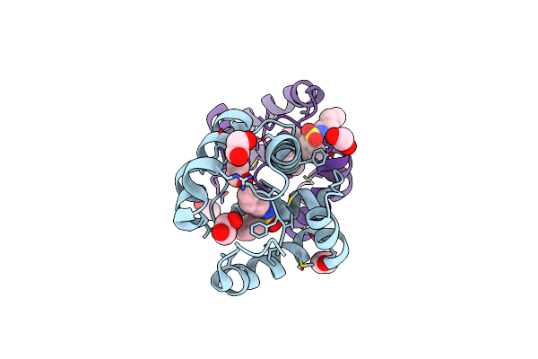
Crystal Structure Of A Pheromone Binding Protein Mutant D35N, From Apis Mellifera, Soaked At Ph 4.0
Organism: Apis mellifera
Method: X-RAY DIFFRACTION Resolution:1.70 Å Release Date: 2009-05-26 Classification: Pheromone binding protein |
Organism: Apis mellifera
Method: X-RAY DIFFRACTION
Release Date: 2009-05-26
 |
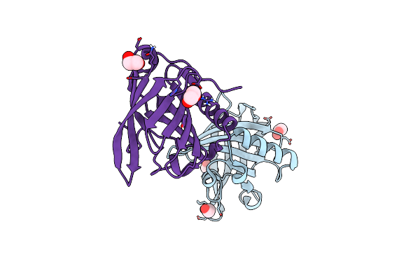
Dimeric Crystal Structure Of A Pheromone Binding Protein Mutant D35N, From Apis Mellifera, At Ph 7.0
Organism: Apis mellifera
Method: X-RAY DIFFRACTION Resolution:1.60 Å Release Date: 2009-05-26 Classification: Pheromone binding protein Ligands: NBB, EDO |
Organism: Apis mellifera
Method: X-RAY DIFFRACTION
Release Date: 2009-05-26
Ligands: NBB, EDO
 |
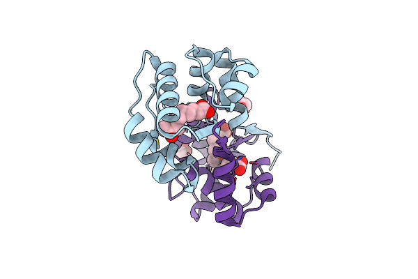
Odorant Binding Protein Obp1
Organism: Rattus norvegicus
Method: X-RAY DIFFRACTION Resolution:1.60 Å Release Date: 2009-05-19 Classification: TRANSPORT PROTEIN Ligands: EDO |
Organism: Rattus norvegicus
Method: X-RAY DIFFRACTION
Release Date: 2009-05-19
Ligands: EDO
 |
Dimeric Crystal Structure Of A Pheromone Binding Protein From Apis Mellifera In Complex With 9-Keto-2(E)-Decenoic Acid At Ph 7.0
Organism: Apis mellifera
Method: X-RAY DIFFRACTION Resolution:1.80 Å Release Date: 2009-04-28 Classification: Pheromone Binding Protein Ligands: 9OD, CL, MG, GOL |
Organism: Apis mellifera
Method: X-RAY DIFFRACTION
Release Date: 2009-04-28
Ligands: 9OD, CL, MG, GOL