

Search Count: 24
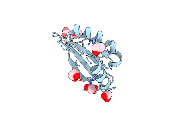 |
An Enhanced Repressor Of Human Papillomavirus E2 Protein
Organism: Human papillomavirus type 16
Method: X-RAY DIFFRACTION Resolution:2.20 Å Release Date: 2011-04-06 Classification: VIRAL PROTEIN Ligands: EDO |
Organism: Human papillomavirus type 16
Method: X-RAY DIFFRACTION
Release Date: 2011-04-06
Ligands: EDO
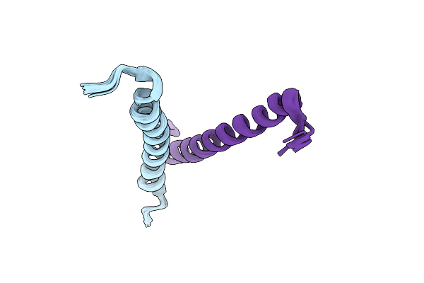 |
Structure Of Rab11-Fip2 C-Terminal Coiled-Coil Domain
Organism: Homo sapiens
Method: SOLUTION NMR Release Date: 2009-06-16 Classification: PROTEIN TRANSPORT |
Organism: Homo sapiens
Method: SOLUTION NMR
Release Date: 2009-06-16
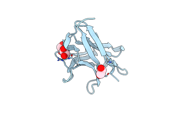 |
Crystal Structure Of The Surrogate Light Chain Variable Domain Vprebj
Organism: Homo sapiens
Method: X-RAY DIFFRACTION Resolution:2.00 Å Release Date: 2008-03-04 Classification: IMMUNE SYSTEM Ligands: EDO |
Organism: Homo sapiens
Method: X-RAY DIFFRACTION
Release Date: 2008-03-04
Ligands: EDO
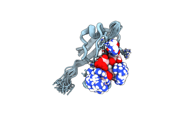 |
Structure Of The Hdlg/Sap97 Pdz2 In Complex With Hpv-18 Papillomavirus E6 Peptide
Organism: Homo sapiens
Method: SOLUTION NMR Release Date: 2007-09-04 Classification: PEPTIDE BINDING PROTEIN |
Organism: Homo sapiens
Method: SOLUTION NMR
Release Date: 2007-09-04
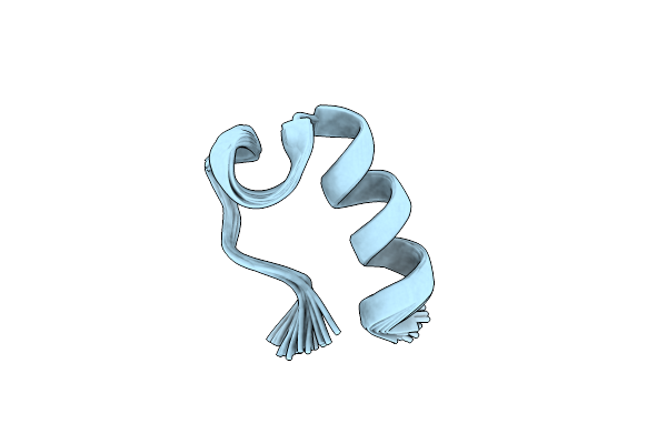 |
E6-Bind Trp-Cage (E6Apn1)
|
Method: SOLUTION NMR
Release Date: 2004-08-03
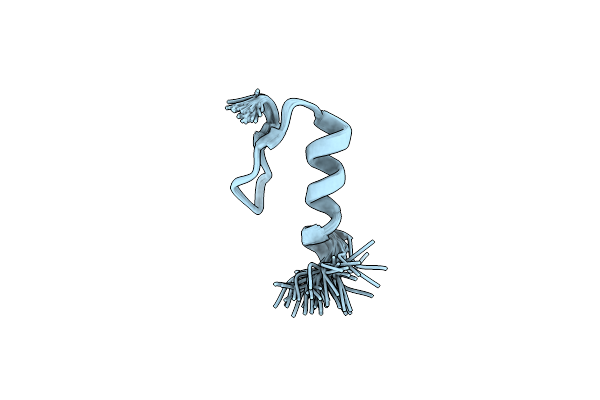 |
E6-Binding Zinc Finger (E6Apc1)
|
Method: SOLUTION NMR
Release Date: 2004-08-03
 |
E6-Binding Zinc Finger (E6Apc2)
|
Method: SOLUTION NMR
Release Date: 2004-08-03
 |
Solution Structure Of The Reps1 Eh Domain
Organism: Mus musculus
Method: SOLUTION NMR Release Date: 2001-07-18 Classification: endocytosis/exocytosis Ligands: CA |
Organism: Mus musculus
Method: SOLUTION NMR
Release Date: 2001-07-18
Ligands: CA
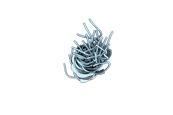 |
Solution Structure Determination And Mutational Analysis Of The Papillomavirus E6-Interacting Peptide Of E6Ap
|
Organism: Homo sapiens
Method: SOLUTION NMR
Release Date: 2001-02-28
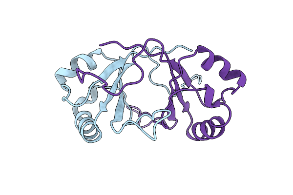 |
E2 Dna-Binding Domain From Papillomavirus Bpv-1
Organism: Bovine papillomavirus type 1
Method: SOLUTION NMR Release Date: 2000-01-01 Classification: GENE REGULATION |
Organism: Bovine papillomavirus type 1
Method: SOLUTION NMR
Release Date: 2000-01-01
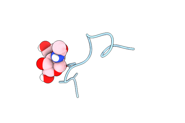 |
A Novel Conotoxin From Conus Textile With Unusual Post-Translational Modifications Reduces Presynaptic Calcium Influx, Nmr, 1 Structure, Glycosylated Protein
Organism: Conus textile
Method: SOLUTION NMR Release Date: 1999-06-08 Classification: GAMMA-CARBOXY GLUTAMIC ACID |
Organism: Conus textile
Method: SOLUTION NMR
Release Date: 1999-06-08
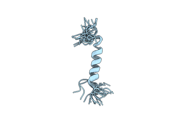 |
Microcin Leader Peptide From E. Coli, Nmr, 25 Structures
Organism: Escherichia coli
Method: SOLUTION NMR Release Date: 1998-07-22 Classification: LEADER PEPTIDE |
Organism: Escherichia coli
Method: SOLUTION NMR
Release Date: 1998-07-22
 |
Gal4 (Cd), Nmr, 24 Structures
Organism: Saccharomyces cerevisiae
Method: SOLUTION NMR Release Date: 1998-04-15 Classification: TRANSCRIPTION REGULATION Ligands: CD |
Organism: Saccharomyces cerevisiae
Method: SOLUTION NMR
Release Date: 1998-04-15
Ligands: CD
 |
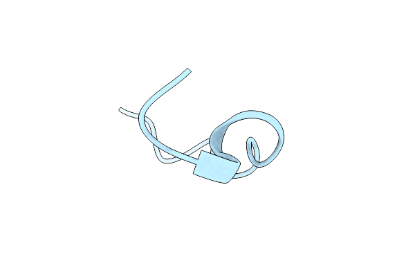
Nmr Structure Of Calcium Bound Conformer Of Conantokin G, Minimized Average Structure
|
Organism: Conus geographus
Method: SOLUTION NMR
Release Date: 1998-04-08
 |
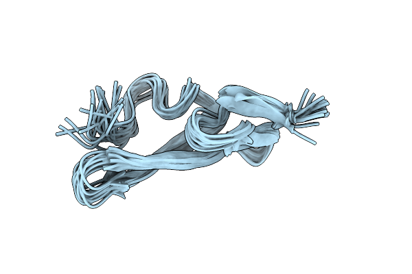
Nmr Structure Of Metal-Free Conantokin G, 1 Structure
Organism: Conus geographus
Method: SOLUTION NMR Release Date: 1997-08-20 Classification: GAMMA-CARBOXYGLUTAMIC ACID |
Organism: Conus geographus
Method: SOLUTION NMR
Release Date: 1997-08-20
 |
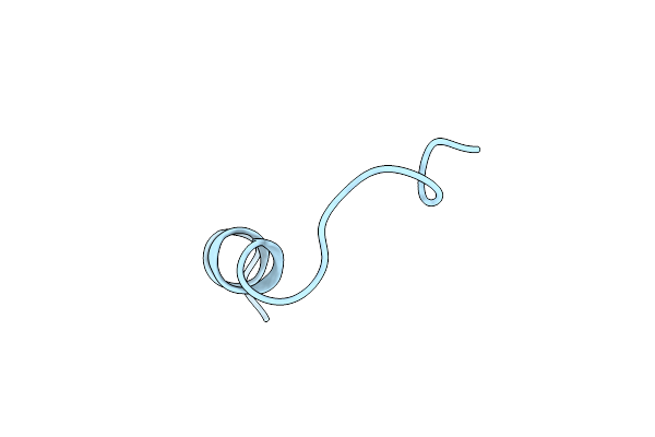
Structure Of The Egf Domain Of P-Selectin, Nmr, 19 Structures
Organism: Homo sapiens
Method: SOLUTION NMR Release Date: 1997-04-01 Classification: CELL ADHESION PROTEIN |
Organism: Homo sapiens
Method: SOLUTION NMR
Release Date: 1997-04-01
 |
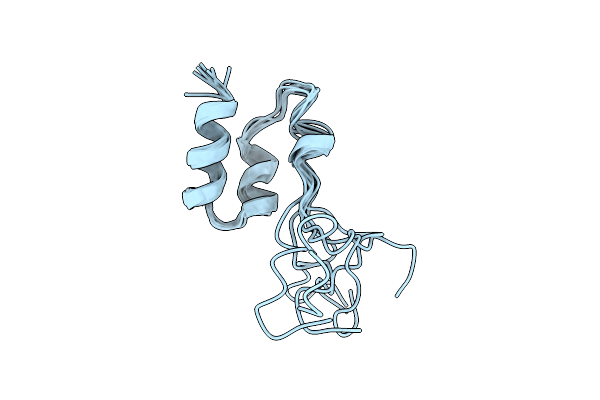
Coagulation Factor Viii, Nmr, 1 Structure
Organism: Homo sapiens
Method: SOLUTION NMR Release Date: 1997-01-11 Classification: COAGULATION FACTOR |
Organism: Homo sapiens
Method: SOLUTION NMR
Release Date: 1997-01-11
 |
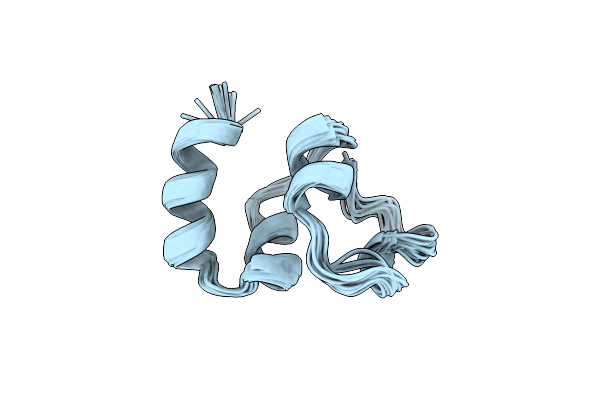
Coagulation Factor, Mg(Ii), Nmr, 7 Structures (Backbone Atoms Only)
Organism: Homo sapiens
Method: SOLUTION NMR Release Date: 1996-11-08 Classification: COAGULATION FACTOR |
Organism: Homo sapiens
Method: SOLUTION NMR
Release Date: 1996-11-08
 |
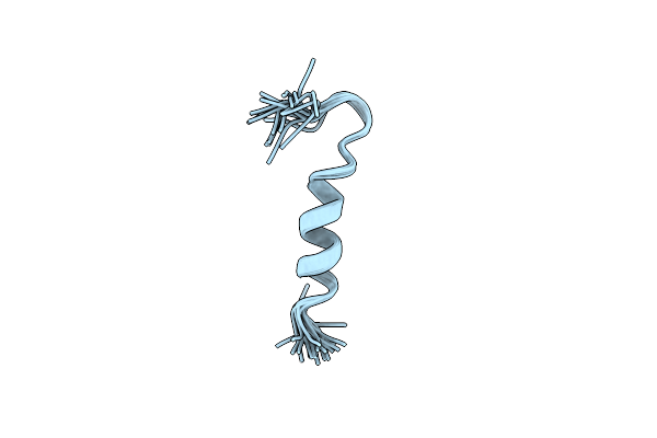
Nmr Structure Of Calcium Ion-Bound Gamma-Carboxy-Glutamic Acid-Rich Domain Of Factor Ix
Organism: Homo sapiens
Method: SOLUTION NMR Release Date: 1996-06-20 Classification: COAGULATION FACTOR |
Organism: Homo sapiens
Method: SOLUTION NMR
Release Date: 1996-06-20
 |
Membrane-Binding Peptide From The C2 Domain Of Factor Viii Forms An Amphipathic Structure As Determined By Nmr Spectroscopy
Organism: Homo sapiens
Method: SOLUTION NMR Release Date: 1995-11-02 Classification: COAGULATION FACTOR |
Organism: Homo sapiens
Method: SOLUTION NMR
Release Date: 1995-11-02