

Search Count: 19
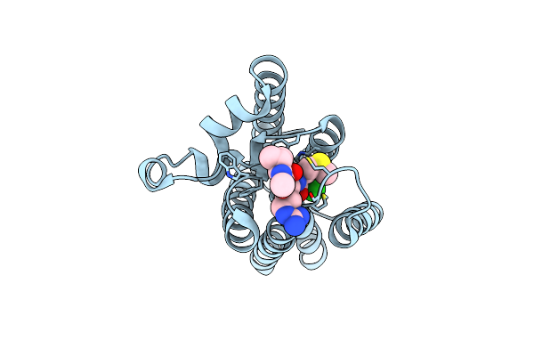 |
Crystal Structure Of Glpg In Complex With Peptide Chloromethylketone Inhibitor
Organism: Escherichia coli, Drosophila melanogaster
Method: X-RAY DIFFRACTION Resolution:2.30 Å Release Date: 2020-09-16 Classification: HYDROLASE/HYDROLASE INHIBITOR |
Organism: Escherichia coli, Drosophila melanogaster
Method: X-RAY DIFFRACTION
Release Date: 2020-09-16
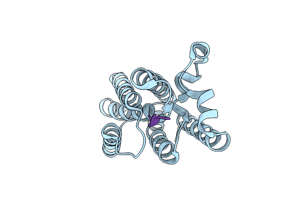 |
Crystal Structure Of Glpg In Complex With Peptide Boronate Inhibitor
Organism: Escherichia coli, Drosophila melanogaster
Method: X-RAY DIFFRACTION Resolution:2.30 Å Release Date: 2020-09-16 Classification: HYDROLASE/HYDROLASE INHIBITOR |
Organism: Escherichia coli, Drosophila melanogaster
Method: X-RAY DIFFRACTION
Release Date: 2020-09-16
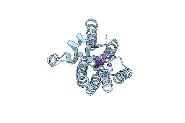 |
Crystal Structure Of Glpg In Complex With Peptide Boronate Inhibitor, Ac-Krfrsmqysa-B(Oh)2
Organism: Escherichia coli, Drosophila melanogaster
Method: X-RAY DIFFRACTION Resolution:2.30 Å Release Date: 2020-09-16 Classification: HYDROLASE/HYDROLASE INHIBITOR Ligands: CL, NA |
Organism: Escherichia coli, Drosophila melanogaster
Method: X-RAY DIFFRACTION
Release Date: 2020-09-16
Ligands: CL, NA
 |
Crystal Structure Of Glpg In Complex With Peptide Ketoamide Inhibitor, Ac-Rvwha-Ketoamide-Phenylbutyl
Organism: Escherichia coli, Drosophila melanogaster
Method: X-RAY DIFFRACTION Resolution:2.40 Å Release Date: 2020-09-16 Classification: HYDROLASE/HYDROLASE INHIBITOR Ligands: V87 |
Organism: Escherichia coli, Drosophila melanogaster
Method: X-RAY DIFFRACTION
Release Date: 2020-09-16
Ligands: V87
 |
Time-Resolved Structural Snapshot Of Proteolysis By Glpg Inside The Membrane
Organism: Escherichia coli, Drosophila melanogaster
Method: X-RAY DIFFRACTION Resolution:2.50 Å Release Date: 2019-11-27 Classification: MEMBRANE PROTEIN |
Organism: Escherichia coli, Drosophila melanogaster
Method: X-RAY DIFFRACTION
Release Date: 2019-11-27
 |
Time-Resolved Structural Snapshot Of Proteolysis By Glpg Inside The Membrane
Organism: Escherichia coli, Drosophila melanogaster
Method: X-RAY DIFFRACTION Resolution:2.50 Å Release Date: 2019-11-27 Classification: MEMBRANE PROTEIN/INHIBITOR |
Organism: Escherichia coli, Drosophila melanogaster
Method: X-RAY DIFFRACTION
Release Date: 2019-11-27
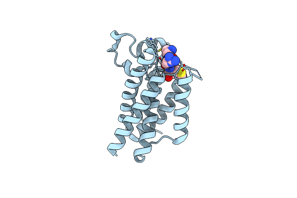 |
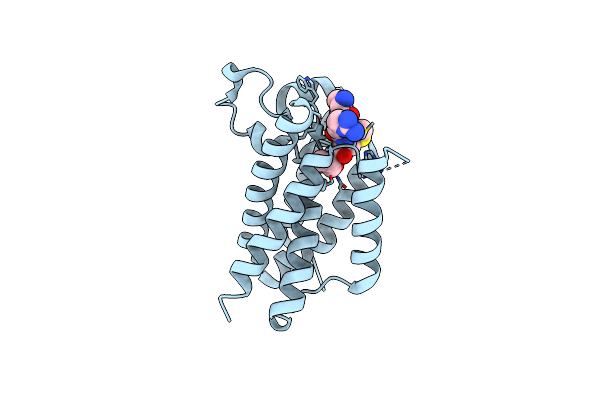
Time-Resolved Structural Snapshot Of Proteolysis By Glpg Inside The Membrane
Organism: Escherichia coli, Drosophila melanogaster
Method: X-RAY DIFFRACTION Resolution:2.30 Å Release Date: 2019-10-02 Classification: MEMBRANE PROTEIN/INHIBITOR |
Organism: Escherichia coli, Drosophila melanogaster
Method: X-RAY DIFFRACTION
Release Date: 2019-10-02
 |
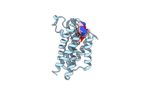
Time-Resolved Structural Snapshot Of Proteolysis By Glpg Inside The Membrane
Organism: Escherichia coli, Drosophila melanogaster
Method: X-RAY DIFFRACTION Resolution:2.40 Å Release Date: 2019-10-02 Classification: MEMBRANE PROTEIN/INHIBITOR |
Organism: Escherichia coli, Drosophila melanogaster
Method: X-RAY DIFFRACTION
Release Date: 2019-10-02
 |
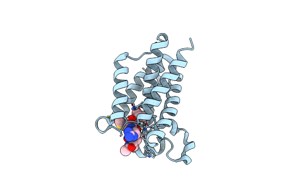
Time-Resolved Structural Snapshot Of Proteolysis By Glpg Inside The Membrane
Organism: Escherichia coli, Drosophila melanogaster
Method: X-RAY DIFFRACTION Resolution:2.30 Å Release Date: 2019-10-02 Classification: MEMBRANE PROTEIN/INHIBITOR |
Organism: Escherichia coli, Drosophila melanogaster
Method: X-RAY DIFFRACTION
Release Date: 2019-10-02
 |
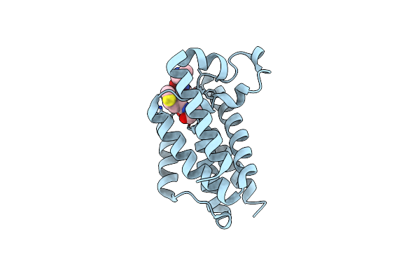
Time-Resolved Structural Snapshot Of Proteolysis By Glpg Inside The Membrane
Organism: Escherichia coli, Drosophila melanogaster
Method: X-RAY DIFFRACTION Resolution:2.40 Å Release Date: 2019-10-02 Classification: MEMBRANE PROTEIN/INHIBITOR |
Organism: Escherichia coli, Drosophila melanogaster
Method: X-RAY DIFFRACTION
Release Date: 2019-10-02
 |
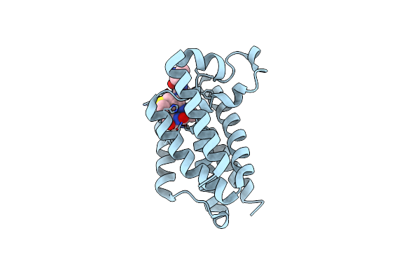
Time-Resolved Structural Snapshot Of Proteolysis By Glpg Inside The Membrane
Organism: Escherichia coli, Drosophila melanogaster
Method: X-RAY DIFFRACTION Resolution:2.50 Å Release Date: 2019-10-02 Classification: MEMBRANE PROTEIN |
Organism: Escherichia coli, Drosophila melanogaster
Method: X-RAY DIFFRACTION
Release Date: 2019-10-02
 |
Time-Resolved Structural Snapshot Of Proteolysis By Glpg Inside The Membrane
Organism: Escherichia coli, Drosophila melanogaster
Method: X-RAY DIFFRACTION Resolution:2.60 Å Release Date: 2019-10-02 Classification: MEMBRANE PROTEIN |
Organism: Escherichia coli, Drosophila melanogaster
Method: X-RAY DIFFRACTION
Release Date: 2019-10-02
 |
Time-Resolved Structural Snapshot Of Proteolysis By Glpg Inside The Membrane
Organism: Escherichia coli, Drosophila melanogaster
Method: X-RAY DIFFRACTION Resolution:2.45 Å Release Date: 2019-10-02 Classification: MEMBRANE PROTEIN |
Organism: Escherichia coli, Drosophila melanogaster
Method: X-RAY DIFFRACTION
Release Date: 2019-10-02
 |
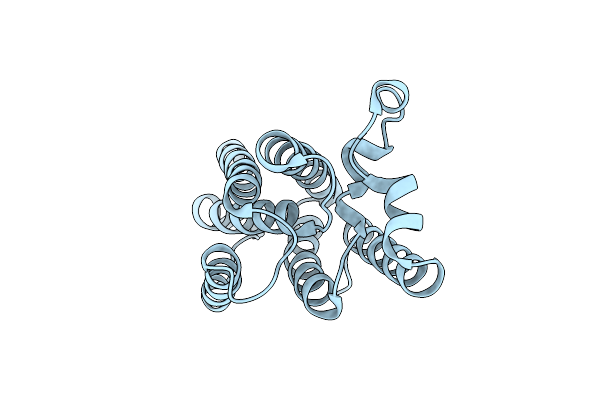
Time-Resolved Structural Snapshot Of Proteolysis By Glpg Inside The Membrane
Organism: Escherichia coli, Drosophila melanogaster
Method: X-RAY DIFFRACTION Resolution:2.40 Å Release Date: 2019-10-02 Classification: MEMBRANE PROTEIN/INHIBITOR Ligands: CL |
Organism: Escherichia coli, Drosophila melanogaster
Method: X-RAY DIFFRACTION
Release Date: 2019-10-02
Ligands: CL
 |
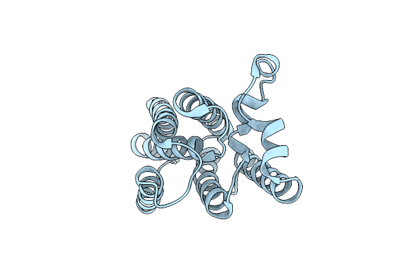
Crystal Structure Of E.Coli Glpg At Ph 4.5
Organism: Escherichia coli
Method: X-RAY DIFFRACTION Resolution:2.40 Å Release Date: 2013-12-25 Classification: HYDROLASE/MEMBRANE PROTEIN |
Organism: Escherichia coli
Method: X-RAY DIFFRACTION
Release Date: 2013-12-25
 |
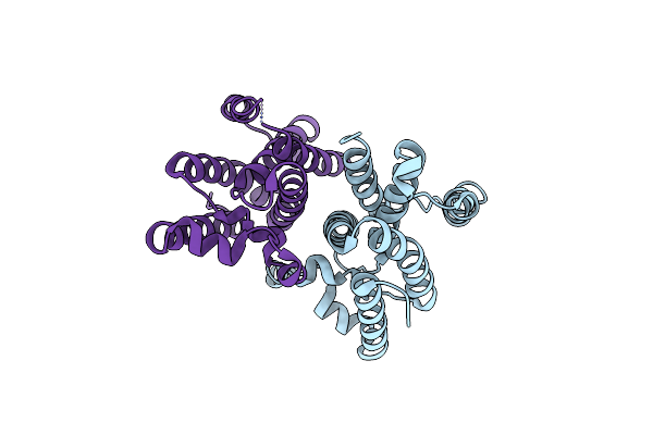
Proteolysis Inside The Membrane Is A Rate-Governed Reaction Not Driven By Substrate Affinity
Organism: Escherichia coli
Method: X-RAY DIFFRACTION Resolution:2.40 Å Release Date: 2013-12-25 Classification: HYDROLASE/MEMBRANE PROTEIN |
Organism: Escherichia coli
Method: X-RAY DIFFRACTION
Release Date: 2013-12-25
 |
Crystal Structure Of Glpg, A Rhomboid Family Intramembrane Protease
Organism: Escherichia coli
Method: X-RAY DIFFRACTION Resolution:2.60 Å Release Date: 2006-11-14 Classification: MEMBRANE PROTEIN |
Organism: Escherichia coli
Method: X-RAY DIFFRACTION
Release Date: 2006-11-14
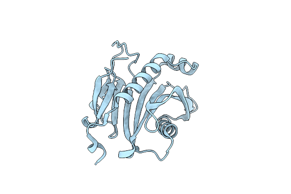 |
Protein Mog1 E65A Mutant
Organism: Saccharomyces cerevisiae
Method: X-RAY DIFFRACTION Resolution:1.90 Å Release Date: 2003-06-03 Classification: GENE REGULATION |
Organism: Saccharomyces cerevisiae
Method: X-RAY DIFFRACTION
Release Date: 2003-06-03
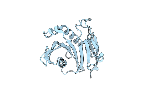 |
1.9 Angstrom Resolution Crystal Structure Of The Saccharomyces Cerevisiae Ran-Binding Protein Mog1P
Organism: Saccharomyces cerevisiae
Method: X-RAY DIFFRACTION Resolution:1.90 Å Release Date: 2000-05-27 Classification: PROTEIN TRANSPORT |
Organism: Saccharomyces cerevisiae
Method: X-RAY DIFFRACTION
Release Date: 2000-05-27