

Search Count: 15
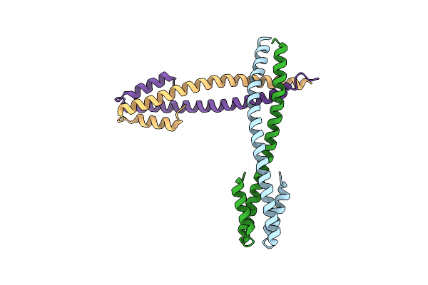 |
Crystal Structure Of The Pre-Assembly Scaffolding Protein Gp7 From The Double-Stranded Dna Bacteriophage Phi29
Organism: Bacillus phage phi29
Method: X-RAY DIFFRACTION Resolution:2.20 Å Release Date: 2003-07-01 Classification: VIRAL PROTEIN |
Organism: Bacillus phage phi29
Method: X-RAY DIFFRACTION
Release Date: 2003-07-01
 |
The Structure Of Bacteriophage Phi29 Scaffolding Protein Gp7 After Prohead Assembly
Organism: Bacillus phage phi29
Method: X-RAY DIFFRACTION Resolution:2.80 Å Release Date: 2003-07-01 Classification: VIRAL PROTEIN |
Organism: Bacillus phage phi29
Method: X-RAY DIFFRACTION
Release Date: 2003-07-01
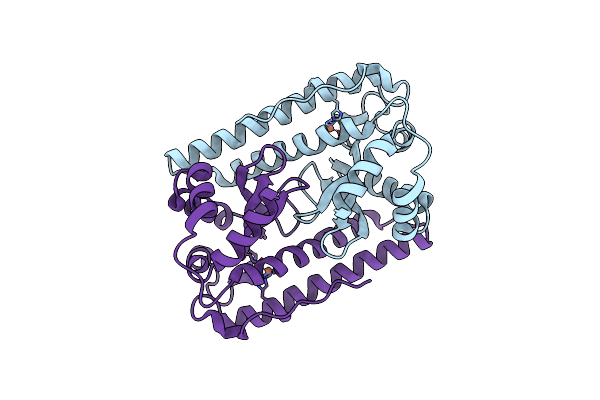 |
H145Q Mutant Of Mycobacterium Tuberculosis Iron-Superoxide Dismutase.
Organism: Mycobacterium tuberculosis
Method: X-RAY DIFFRACTION Resolution:4.00 Å Release Date: 2001-10-05 Classification: OXIDOREDUCTASE Ligands: FE |
Organism: Mycobacterium tuberculosis
Method: X-RAY DIFFRACTION
Release Date: 2001-10-05
Ligands: FE
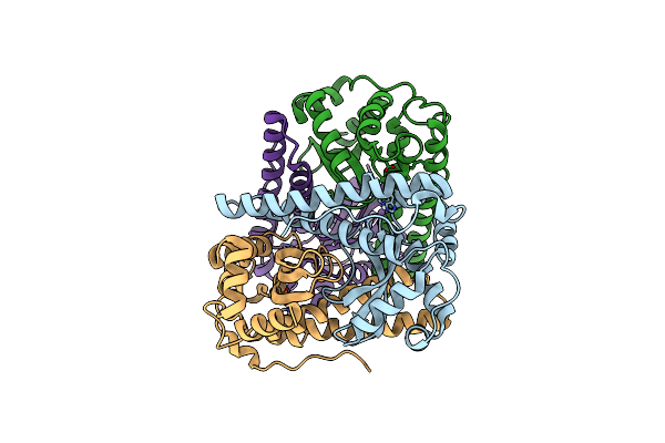 |
H145E Mutant Of Mycobacterium Tuberculosis Iron-Superoxide Dismutase.
Organism: Mycobacterium tuberculosis
Method: X-RAY DIFFRACTION Resolution:2.50 Å Release Date: 2001-10-05 Classification: OXIDOREDUCTASE Ligands: MN |
Organism: Mycobacterium tuberculosis
Method: X-RAY DIFFRACTION
Release Date: 2001-10-05
Ligands: MN
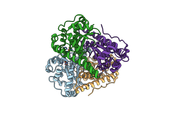 |
G152A Mutant Of Mycobacterium Tuberculosis Iron-Superoxide Dismutase.
Organism: Mycobacterium tuberculosis
Method: X-RAY DIFFRACTION Resolution:2.90 Å Release Date: 2001-10-05 Classification: OXIDOREDUCTASE Ligands: FE |
Organism: Mycobacterium tuberculosis
Method: X-RAY DIFFRACTION
Release Date: 2001-10-05
Ligands: FE
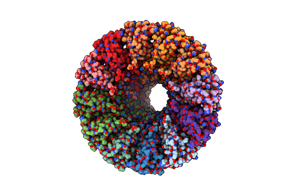 |
Connector Protein From Bacteriophage Phi29
Organism: Bacillus phage phi29
Method: X-RAY DIFFRACTION Resolution:3.20 Å Release Date: 2001-08-15 Classification: VIRAL PROTEIN |
Organism: Bacillus phage phi29
Method: X-RAY DIFFRACTION
Release Date: 2001-08-15
 |
Structure Of The Bacteriophage Phi29 Head-Tail Connector Protein
Organism: Bacillus phage phi29
Method: X-RAY DIFFRACTION Resolution:2.90 Å Release Date: 2001-05-09 Classification: VIRAL PROTEIN |
Organism: Bacillus phage phi29
Method: X-RAY DIFFRACTION
Release Date: 2001-05-09
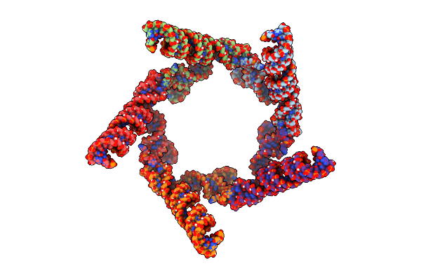 |
Pentameric Model Of The Bacteriophage Phi29 Prohead Rna
Organism: Bacillus phage phi29
Method: ELECTRON MICROSCOPY Resolution:20.00 Å Release Date: 2000-12-22 Classification: RNA |
Organism: Bacillus phage phi29
Method: ELECTRON MICROSCOPY
Release Date: 2000-12-22
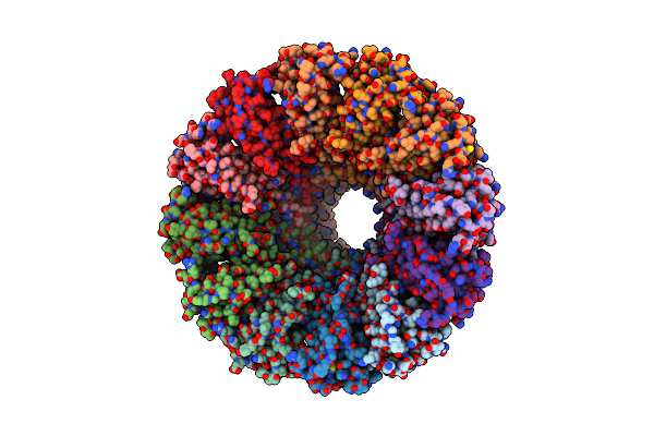 |
Connector Protein From Bacteriophage Phi29
Organism: Bacillus phage phi29
Method: X-RAY DIFFRACTION Resolution:3.20 Å Release Date: 2000-12-22 Classification: VIRAL PROTEIN |
Organism: Bacillus phage phi29
Method: X-RAY DIFFRACTION
Release Date: 2000-12-22
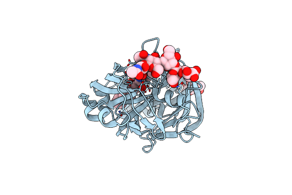 |
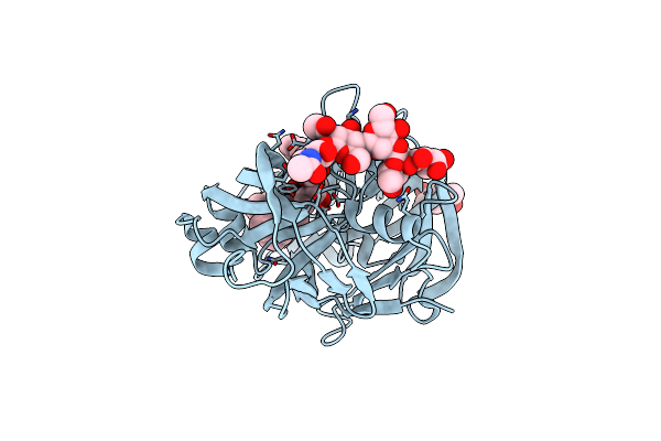
Crystal Structure Of A Complex Between Hydroxyethylene Inhibitor Cp-108,420 And Yeast Aspartic Proteinase A
Organism: Saccharomyces cerevisiae
Method: X-RAY DIFFRACTION Resolution:2.70 Å Release Date: 2000-09-20 Classification: HYDROLASE/HYDROLASE INHIBITOR Ligands: 2Y2, NAG |
Organism: Saccharomyces cerevisiae
Method: X-RAY DIFFRACTION
Release Date: 2000-09-20
Ligands: 2Y2, NAG
 |
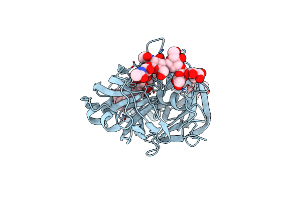
X-Ray Structure Of A Cyclic Statine Inhibitor Pd-129,541 Bound To Yeast Proteinase A
Organism: Saccharomyces cerevisiae
Method: X-RAY DIFFRACTION Resolution:2.40 Å Release Date: 2000-09-20 Classification: HYDROLASE/HYDROLASE INHIBITOR Ligands: NAG, 0GM |
Organism: Saccharomyces cerevisiae
Method: X-RAY DIFFRACTION
Release Date: 2000-09-20
Ligands: NAG, 0GM
 |
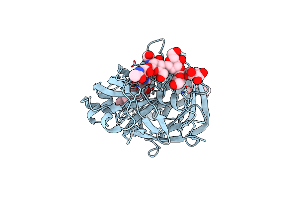
X-Ray Structure Of Glycol Inhibitor Pd-133,450 Bound To Saccharopepsin
Organism: Saccharomyces cerevisiae
Method: X-RAY DIFFRACTION Resolution:2.70 Å Release Date: 2000-09-20 Classification: HYDROLASE/HYDROLASE INHIBITOR Ligands: 0QF, NAG |
Organism: Saccharomyces cerevisiae
Method: X-RAY DIFFRACTION
Release Date: 2000-09-20
Ligands: 0QF, NAG
 |
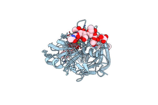
X-Ray Structure Of Inhibitor Cp-72,647 Bound To Saccharopepsin
Organism: Saccharomyces cerevisiae
Method: X-RAY DIFFRACTION Resolution:2.80 Å Release Date: 2000-09-20 Classification: HYDROLASE/HYDROLASE INHIBITOR Ligands: 2Y3, NAG |
Organism: Saccharomyces cerevisiae
Method: X-RAY DIFFRACTION
Release Date: 2000-09-20
Ligands: 2Y3, NAG
 |
X-Ray Structure Of Difluorostatine Inhibitor Cp81,198 Bound To Saccharopepsin
Organism: Saccharomyces cerevisiae
Method: X-RAY DIFFRACTION Resolution:2.80 Å Release Date: 2000-09-20 Classification: HYDROLASE/HYDROLASE INHIBITOR Ligands: 2Y4, NAG |
Organism: Saccharomyces cerevisiae
Method: X-RAY DIFFRACTION
Release Date: 2000-09-20
Ligands: 2Y4, NAG
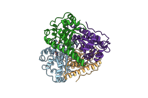 |
X-Ray Structure Analysis Of The Iron-Dependent Superoxide Dismutase From Mycobacterium Tuberculosis At 2.0 Angstroms Resolutions Reveals Novel Dimer-Dimer Interactions
Organism: Mycobacterium tuberculosis
Method: X-RAY DIFFRACTION Resolution:2.00 Å Release Date: 1994-12-20 Classification: SUPEROXIDE DISMUTASE Ligands: FE |
Organism: Mycobacterium tuberculosis
Method: X-RAY DIFFRACTION
Release Date: 1994-12-20
Ligands: FE