

Search Count: 28
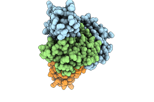 |
Structure Of The Cross-Hla Supertype Antibody R302 Bound To A Class I Mhc Presenting A Divarasib-Modified Kras-G12C Peptide On Hla-A*02
Organism: Homo sapiens
Method: ELECTRON MICROSCOPY Release Date: 2025-10-08 Classification: PROTEIN BINDING Ligands: A1AWR |
Organism: Homo sapiens
Method: ELECTRON MICROSCOPY
Release Date: 2025-10-08
Ligands: A1AWR
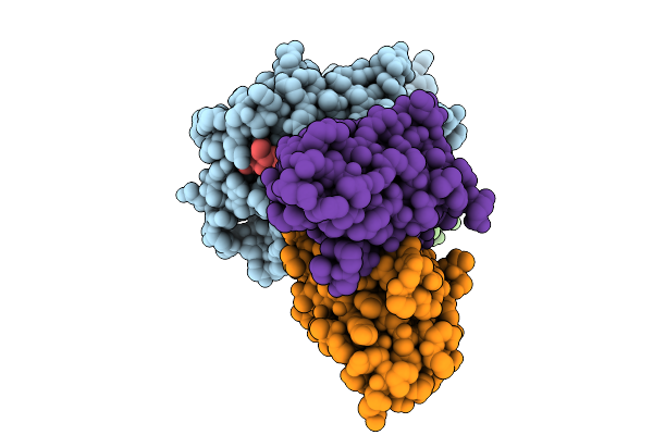 |
Structure Of The Cross-Hla Supertype Antibody R302 Bound To A Class I Mhc Presenting A Divarasib-Modified Kras-G12C Peptide On Hla-A*03
Organism: Homo sapiens
Method: ELECTRON MICROSCOPY Release Date: 2025-10-08 Classification: PROTEIN BINDING Ligands: A1AWR |
Organism: Homo sapiens
Method: ELECTRON MICROSCOPY
Release Date: 2025-10-08
Ligands: A1AWR
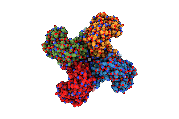 |
Composite Map Of Human Kv1.3 Channel In Apo State With Beta Subunits
Organism: Homo sapiens
Method: ELECTRON MICROSCOPY Release Date: 2022-02-09 Classification: MEMBRANE PROTEIN Ligands: K, NAP |
Organism: Homo sapiens
Method: ELECTRON MICROSCOPY
Release Date: 2022-02-09
Ligands: K, NAP
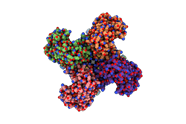 |
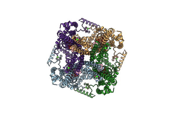
Composite Map Of Human Kv1.3 Channel In Dalazatide-Bound State With Beta Subunits
Organism: Homo sapiens
Method: ELECTRON MICROSCOPY Release Date: 2022-02-09 Classification: MEMBRANE PROTEIN Ligands: NAP, K |
Organism: Homo sapiens
Method: ELECTRON MICROSCOPY
Release Date: 2022-02-09
Ligands: NAP, K
 |
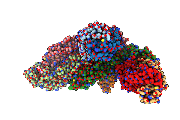
Pi3P And Calcium Bound Full-Length Trpy1 In Detergent
Organism: Saccharomyces cerevisiae
Method: ELECTRON MICROSCOPY Release Date: 2021-04-21 Classification: MEMBRANE PROTEIN Ligands: CA, PWE |
Organism: Saccharomyces cerevisiae
Method: ELECTRON MICROSCOPY
Release Date: 2021-04-21
Ligands: CA, PWE
 |
Cryoem Structure Of Zika Virus With Fab At 4.1 Angstrom
Organism: Homo sapiens, Zika virus
Method: ELECTRON MICROSCOPY Release Date: 2020-07-08 Classification: VIRUS |
Organism: Homo sapiens, Zika virus
Method: ELECTRON MICROSCOPY
Release Date: 2020-07-08
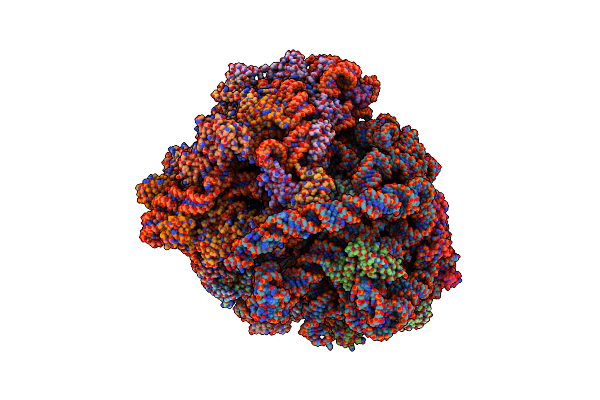 |
M. Smegmatis P/P State 70S Ribosome Structure
Organism: Mycobacterium smegmatis str. mc2 155, Escherichia coli
Method: ELECTRON MICROSCOPY Resolution:3.40 Å Release Date: 2018-09-26 Classification: RIBOSOME |
Organism: Mycobacterium smegmatis str. mc2 155, Escherichia coli
Method: ELECTRON MICROSCOPY
Release Date: 2018-09-26
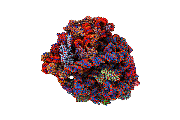 |
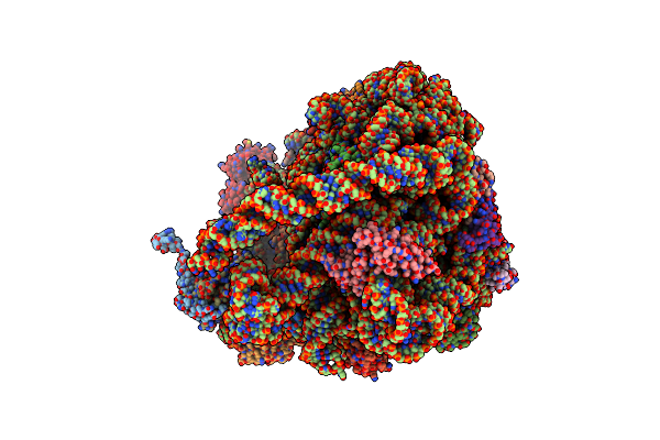
M. Smegmatis Hibernating State 70S Ribosome Structure
Organism: Mycobacterium smegmatis str. mc2 155, Escherichia coli
Method: ELECTRON MICROSCOPY Resolution:3.40 Å Release Date: 2018-09-26 Classification: RIBOSOME |
Organism: Mycobacterium smegmatis str. mc2 155, Escherichia coli
Method: ELECTRON MICROSCOPY
Release Date: 2018-09-26
 |
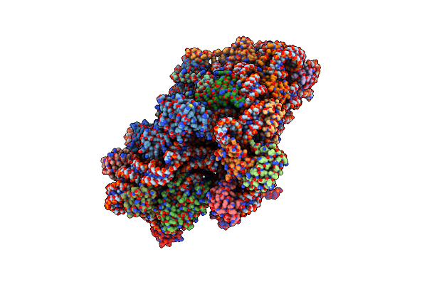
M. Smegmatis P/P State 50S Ribosomal Subunit
Organism: Mycobacterium smegmatis str. mc2 155
Method: ELECTRON MICROSCOPY Resolution:3.20 Å Release Date: 2018-09-26 Classification: RIBOSOME |
Organism: Mycobacterium smegmatis str. mc2 155
Method: ELECTRON MICROSCOPY
Release Date: 2018-09-26
 |
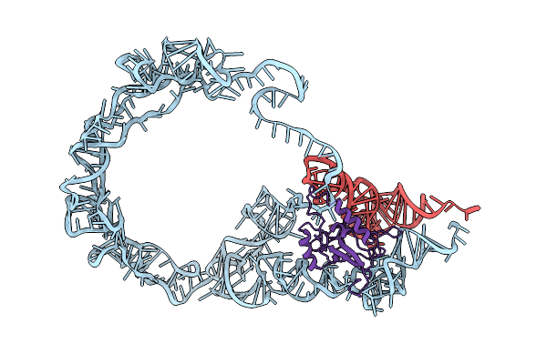
M. Smegmatis P/P State 30S Ribosomal Subunit
Organism: Mycobacterium smegmatis (strain atcc 700084 / mc(2)155), Escherichia coli
Method: ELECTRON MICROSCOPY Resolution:3.70 Å Release Date: 2018-09-26 Classification: RIBOSOME |
 |
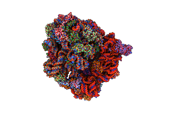
M. Smegmatis Trans-Translation State 70S Ribosome
Organism: Mycobacterium smegmatis (strain atcc 700084 / mc(2)155), Escherichia coli
Method: ELECTRON MICROSCOPY Release Date: 2018-09-26 Classification: RNA BINDING PROTEIN/RNA |
 |
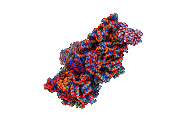
Structure Of The 70S Chloroplast Ribosome From Spinach
Organism: Spinacia oleracea
Method: ELECTRON MICROSCOPY Release Date: 2017-06-14 Classification: RIBOSOME |
Organism: Spinacia oleracea
Method: ELECTRON MICROSCOPY
Release Date: 2017-06-14
 |
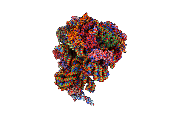
Structure Of The 30S Small Subunit Of Chloroplast Ribosome From Spinach
Organism: Spinacia oleracea
Method: ELECTRON MICROSCOPY Release Date: 2017-06-07 Classification: RIBOSOME |
Organism: Spinacia oleracea
Method: ELECTRON MICROSCOPY
Release Date: 2017-06-07
 |
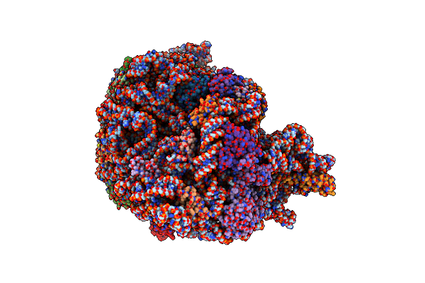
Structure Of The 50S Large Subunit Of Chloroplast Ribosome From Spinach
Organism: Spinacia oleracea
Method: ELECTRON MICROSCOPY Release Date: 2017-06-07 Classification: RIBOSOME |
Organism: Spinacia oleracea
Method: ELECTRON MICROSCOPY
Release Date: 2017-06-07
 |
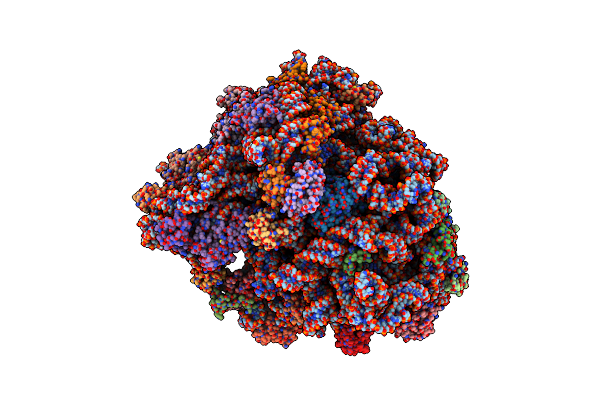
Methicillin Resistant, Linezolid Resistant Staphylococcus Aureus 70S Ribosome (Delta S145 Ul3)
Organism: Staphylococcus aureus
Method: ELECTRON MICROSCOPY Release Date: 2017-05-31 Classification: RIBOSOME Ligands: MG |
Organism: Staphylococcus aureus
Method: ELECTRON MICROSCOPY
Release Date: 2017-05-31
Ligands: MG
 |
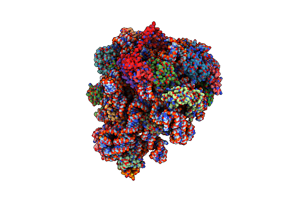
Methicillin Sensitive Staphylococcus Aureus 70S Ribosome
Organism: Staphylococcus aureus subsp. aureus nctc 8325, Staphylococcus aureus (strain nctc 8325)
Method: ELECTRON MICROSCOPY Release Date: 2017-05-24 Classification: ribosome/antibiotic Ligands: PAR, MG |
Organism: Staphylococcus aureus subsp. aureus nctc 8325, Staphylococcus aureus (strain nctc 8325)
Method: ELECTRON MICROSCOPY
Release Date: 2017-05-24
Ligands: PAR, MG
 |
Structure Of The Large Subunit Of The Chloro-Ribosome
Organism: Spinacia oleracea
Method: ELECTRON MICROSCOPY Release Date: 2017-02-01 Classification: RIBOSOME |
Organism: Spinacia oleracea
Method: ELECTRON MICROSCOPY
Release Date: 2017-02-01
 |
Crystal Structure Of The Braf (R509H) Kinase Domain Monomer Bound To Vemurafenib
Organism: Homo sapiens
Method: X-RAY DIFFRACTION Resolution:2.99 Å Release Date: 2016-08-10 Classification: Transferase/Transferase Inhibitor Ligands: 032 |
Organism: Homo sapiens
Method: X-RAY DIFFRACTION
Release Date: 2016-08-10
Ligands: 032
 |
Crystal Structure Of Braf (R509H) Kinase Domain Bound To Az628
Organism: Homo sapiens
Method: X-RAY DIFFRACTION Resolution:3.49 Å Release Date: 2016-08-10 Classification: Transferase/Transferase Inhibitor Ligands: B1E |
Organism: Homo sapiens
Method: X-RAY DIFFRACTION
Release Date: 2016-08-10
Ligands: B1E
 |
Crystal Structure Of Braf Kinase Domain Bound To Az-Vem
Organism: Homo sapiens
Method: X-RAY DIFFRACTION Resolution:1.95 Å Release Date: 2016-08-10 Classification: TRANSFERASE/TRANSFERASE INHIBITOR Ligands: 6DC |
Organism: Homo sapiens
Method: X-RAY DIFFRACTION
Release Date: 2016-08-10
Ligands: 6DC