

Search Count: 25
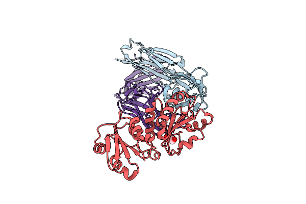 |
Oxidoreductase Fragment Of Human Qsox1 In Complex With A Fab Fragment Of A Humanized Anti-Qsox1 Antibody
Organism: Homo sapiens
Method: X-RAY DIFFRACTION Resolution:2.10 Å Release Date: 2023-08-02 Classification: IMMUNE SYSTEM Ligands: GOL |
Organism: Homo sapiens
Method: X-RAY DIFFRACTION
Release Date: 2023-08-02
Ligands: GOL
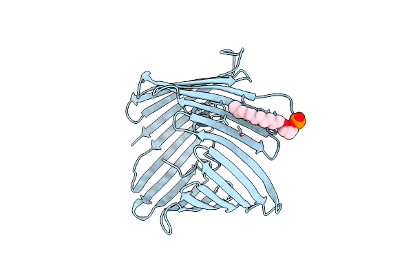 |
Vdac K12E Mutant
Organism: Mus musculus
Method: X-RAY DIFFRACTION Resolution:2.60 Å Release Date: 2023-01-11 Classification: MEMBRANE PROTEIN Ligands: MC3 |
Organism: Mus musculus
Method: X-RAY DIFFRACTION
Release Date: 2023-01-11
Ligands: MC3
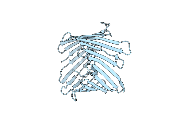 |
Microed Structure Of Mvdac
Organism: Mus musculus
Method: ELECTRON CRYSTALLOGRAPHY Resolution:3.12 Å Release Date: 2020-12-23 Classification: TRANSPORT PROTEIN |
Organism: Mus musculus
Method: ELECTRON CRYSTALLOGRAPHY
Release Date: 2020-12-23
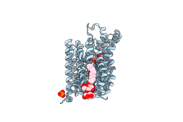 |
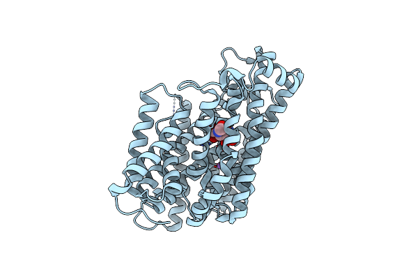
Substrate-Bound Outward-Open State Of A Na+-Coupled Sialic Acid Symporter Reveals A Novel Na+-Site
Organism: Proteus mirabilis (strain hi4320)
Method: X-RAY DIFFRACTION Resolution:1.95 Å Release Date: 2018-04-04 Classification: MEMBRANE PROTEIN |
Organism: Proteus mirabilis (strain hi4320)
Method: X-RAY DIFFRACTION
Release Date: 2018-04-04
 |
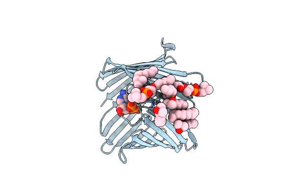
Substrate-Bound Outward-Open State Of A Na+-Coupled Sialic Acid Symporter Reveals A Novel Na+-Site
Organism: Proteus mirabilis (strain hi4320)
Method: X-RAY DIFFRACTION Resolution:2.26 Å Release Date: 2018-04-04 Classification: MEMBRANE PROTEIN |
Organism: Proteus mirabilis (strain hi4320)
Method: X-RAY DIFFRACTION
Release Date: 2018-04-04
 |
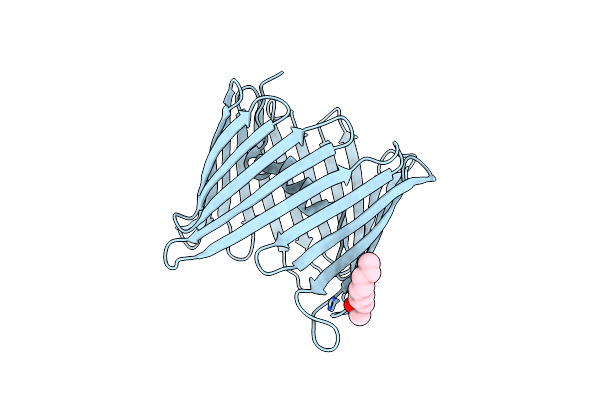
Atp Binding To Murine Voltage-Dependent Anion Channel 1 (Mvdac1).
Organism: Mus musculus
Method: X-RAY DIFFRACTION Resolution:2.28 Å Release Date: 2014-06-04 Classification: TRANSPORT PROTEIN Ligands: LDA, MC3, ATP |
Organism: Mus musculus
Method: X-RAY DIFFRACTION
Release Date: 2014-06-04
Ligands: LDA, MC3, ATP
 |
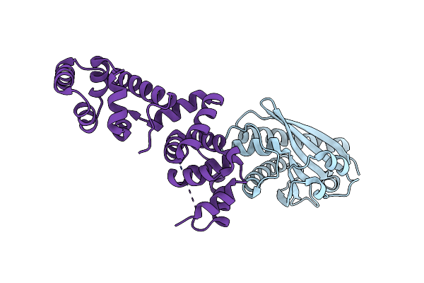
Crystal Structure Of The Voltage Dependant Anion Channel 2 From Zebrafish.
Organism: Danio rerio
Method: X-RAY DIFFRACTION Resolution:2.80 Å Release Date: 2014-03-26 Classification: MEMBRANE PROTEIN Ligands: LDA |
Organism: Danio rerio
Method: X-RAY DIFFRACTION
Release Date: 2014-03-26
Ligands: LDA
 |
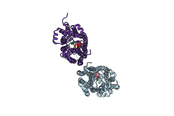
Crystal Structure Of The Complex Between The Flagellar Motor Proteins Flig And Flim.
Organism: Thermotoga maritima
Method: X-RAY DIFFRACTION Resolution:1.93 Å Release Date: 2012-08-22 Classification: MOTOR PROTEIN |
Organism: Thermotoga maritima
Method: X-RAY DIFFRACTION
Release Date: 2012-08-22
 |
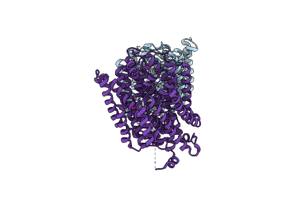
Crystal Structure Of Lacy In Complex With An Affinity Inactivator
Organism: Escherichia coli
Method: X-RAY DIFFRACTION Resolution:3.38 Å Release Date: 2011-06-15 Classification: TRANSPORT PROTEIN Ligands: BA, TGA |
Organism: Escherichia coli
Method: X-RAY DIFFRACTION
Release Date: 2011-06-15
Ligands: BA, TGA
 |
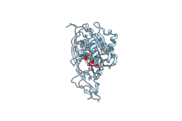
Structure Of The K294A Mutant Of Vsglt
Organism: Vibrio parahaemolyticus
Method: X-RAY DIFFRACTION Resolution:2.73 Å Release Date: 2010-12-08 Classification: TRANSPORT PROTEIN Ligands: PEG |
Organism: Vibrio parahaemolyticus
Method: X-RAY DIFFRACTION
Release Date: 2010-12-08
Ligands: PEG
 |
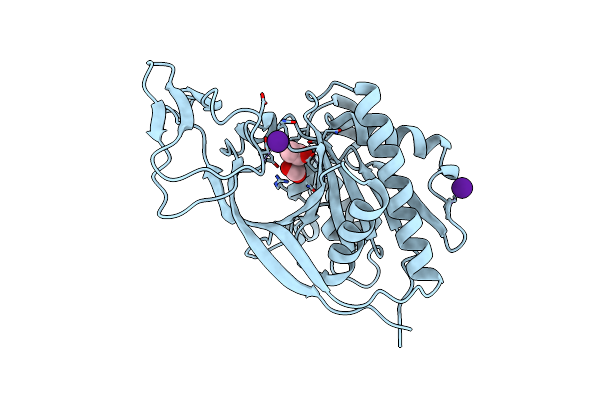
Crystal Structure Of Murine Aminoacylase 3 In Complex With N-Acetyl-L-Tyrosine
Organism: Mus musculus
Method: X-RAY DIFFRACTION Resolution:2.15 Å Release Date: 2010-10-20 Classification: HYDROLASE Ligands: ZN, CL, 3NF |
Organism: Mus musculus
Method: X-RAY DIFFRACTION
Release Date: 2010-10-20
Ligands: ZN, CL, 3NF
 |
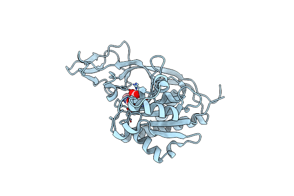
Crystal Structure Of Murine Aminoacylase 3
Organism: Mus musculus
Method: X-RAY DIFFRACTION Resolution:2.00 Å Release Date: 2010-10-20 Classification: HYDROLASE Ligands: ZN, CS, CL, ACT, FMT |
Organism: Mus musculus
Method: X-RAY DIFFRACTION
Release Date: 2010-10-20
Ligands: ZN, CS, CL, ACT, FMT
 |
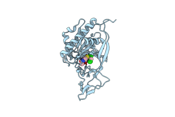
Crystal Structure Of E177A-Mutant Murine Aminoacylase 3
Organism: Mus musculus
Method: X-RAY DIFFRACTION Resolution:2.09 Å Release Date: 2010-10-20 Classification: HYDROLASE Ligands: ZN, CL, ACT, FMT |
Organism: Mus musculus
Method: X-RAY DIFFRACTION
Release Date: 2010-10-20
Ligands: ZN, CL, ACT, FMT
 |
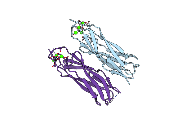
Crystal Structure Of Murine Aminoacylase 3 In Complex With N-Acetyl-S-1,2-Dichlorovinyl-L-Cysteine
Organism: Mus musculus
Method: X-RAY DIFFRACTION Resolution:2.80 Å Release Date: 2010-10-20 Classification: HYDROLASE Ligands: ZN, CL, DC2 |
Organism: Mus musculus
Method: X-RAY DIFFRACTION
Release Date: 2010-10-20
Ligands: ZN, CL, DC2
 |
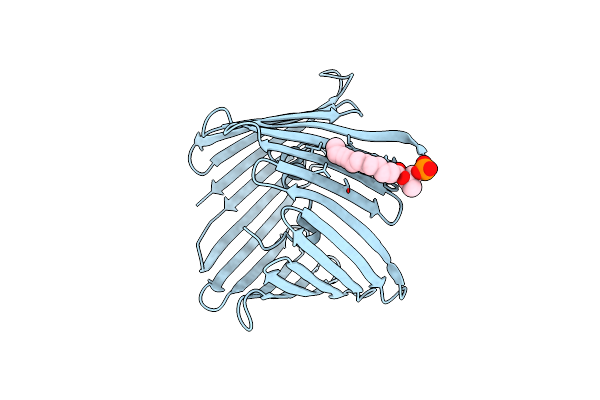
Crystal Structure Of E454K-Cbd1
Organism: Canis lupus familiaris
Method: X-RAY DIFFRACTION Resolution:2.40 Å Release Date: 2009-04-21 Classification: METAL BINDING PROTEIN Ligands: CA |
Organism: Canis lupus familiaris
Method: X-RAY DIFFRACTION
Release Date: 2009-04-21
Ligands: CA
 |
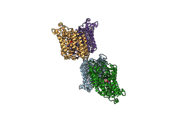
The Crystal Structure Of Mouse Vdac1 At 2.3 A Resolution
Organism: Mus musculus
Method: X-RAY DIFFRACTION Resolution:2.30 Å Release Date: 2008-12-16 Classification: MEMBRANE PROTEIN Ligands: MC3 |
Organism: Mus musculus
Method: X-RAY DIFFRACTION
Release Date: 2008-12-16
Ligands: MC3
 |
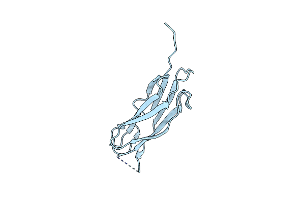
Crystal Structure Of Sodium/Sugar Symporter With Bound Galactose From Vibrio Parahaemolyticus
Organism: Vibrio parahaemolyticus
Method: X-RAY DIFFRACTION Resolution:2.70 Å Release Date: 2008-08-05 Classification: TRANSPORT PROTEIN Ligands: GAL, ER3, NA |
Organism: Vibrio parahaemolyticus
Method: X-RAY DIFFRACTION
Release Date: 2008-08-05
Ligands: GAL, ER3, NA
 |
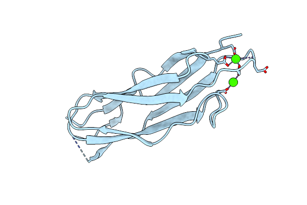
The Second Ca2+-Binding Domain Of The Na+-Ca2+ Exchanger Is Essential For Regulation: Crystal Structures And Mutational Analysis
Organism: Canis lupus familiaris
Method: X-RAY DIFFRACTION Resolution:1.45 Å Release Date: 2007-11-13 Classification: METAL BINDING PROTEIN |
Organism: Canis lupus familiaris
Method: X-RAY DIFFRACTION
Release Date: 2007-11-13
 |
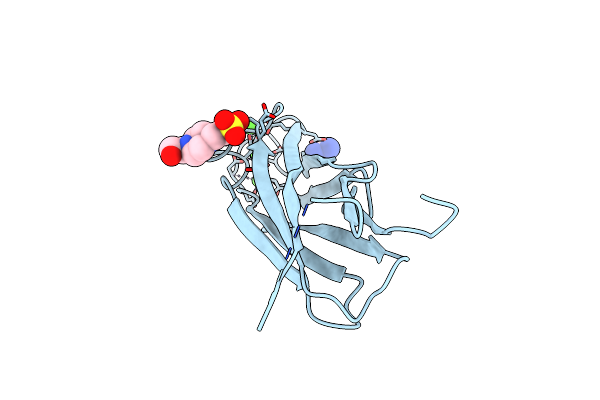
The Second Ca2+-Binding Domain Of The Na+-Ca2+ Exchanger Is Essential For Regulation: Crystal Structures And Mutational Analysis
Organism: Canis lupus familiaris
Method: X-RAY DIFFRACTION Resolution:1.70 Å Release Date: 2007-11-13 Classification: METAL BINDING PROTEIN Ligands: CA |
Organism: Canis lupus familiaris
Method: X-RAY DIFFRACTION
Release Date: 2007-11-13
Ligands: CA
 |
The Crystal Structure Of The Primary Ca2+ Sensor Of The Na+/Ca2+ Exchanger
Organism: Canis lupus familiaris
Method: X-RAY DIFFRACTION Resolution:2.50 Å Release Date: 2006-06-13 Classification: METAL TRANSPORT Ligands: CA, GAI, EPE |
Organism: Canis lupus familiaris
Method: X-RAY DIFFRACTION
Release Date: 2006-06-13
Ligands: CA, GAI, EPE