
Search Count: 2,014
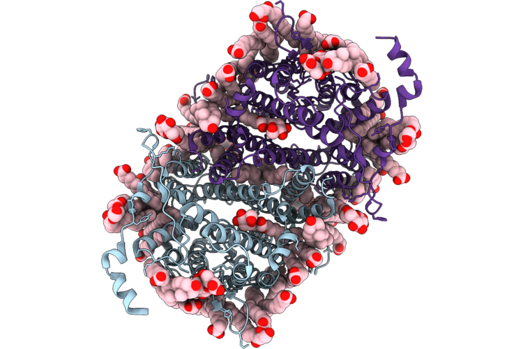 |
Uapa (Wt) In Ddm, Apo State
Organism: Aspergillus nidulans fgsc a4
Method: ELECTRON MICROSCOPY Resolution:2.06 Å Release Date: 2026-07-01 Classification: MEMBRANE PROTEIN Ligands: LMT, MYR, PLM, ERG, DAO, DGA |
Organism: Aspergillus nidulans fgsc a4
Method: ELECTRON MICROSCOPY
Release Date: 2026-07-01
Ligands: LMT, MYR, PLM, ERG, DAO, DGA
 |
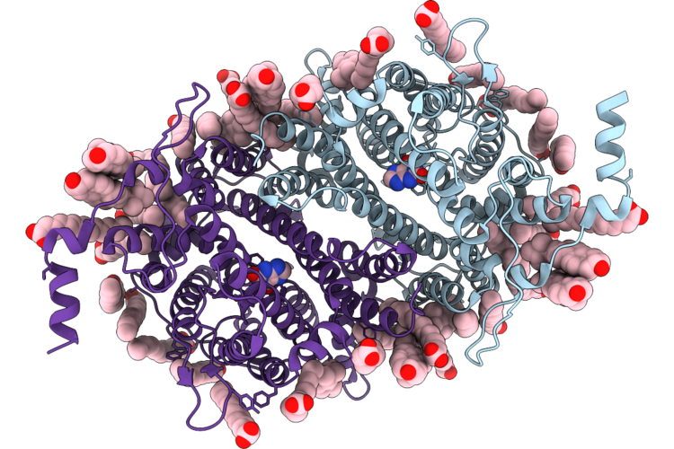
Uapa (Q408E) In Ddm, Apo State
Organism: Aspergillus nidulans fgsc a4
Method: ELECTRON MICROSCOPY Resolution:2.59 Å Release Date: 2026-07-01 Classification: MEMBRANE PROTEIN Ligands: LMT, MYR, PLM, ERG, DAO, FAW |
Organism: Aspergillus nidulans fgsc a4
Method: ELECTRON MICROSCOPY
Release Date: 2026-07-01
Ligands: LMT, MYR, PLM, ERG, DAO, FAW
 |
Uapa (Q408E) In Msp1E3D1 Nanodiscs, Xanthine-Bound State
Organism: Aspergillus nidulans fgsc a4
Method: ELECTRON MICROSCOPY Resolution:3.48 Å Release Date: 2026-07-01 Classification: MEMBRANE PROTEIN Ligands: MYR, ERG, XAN |
Organism: Aspergillus nidulans fgsc a4
Method: ELECTRON MICROSCOPY
Release Date: 2026-07-01
Ligands: MYR, ERG, XAN
 |
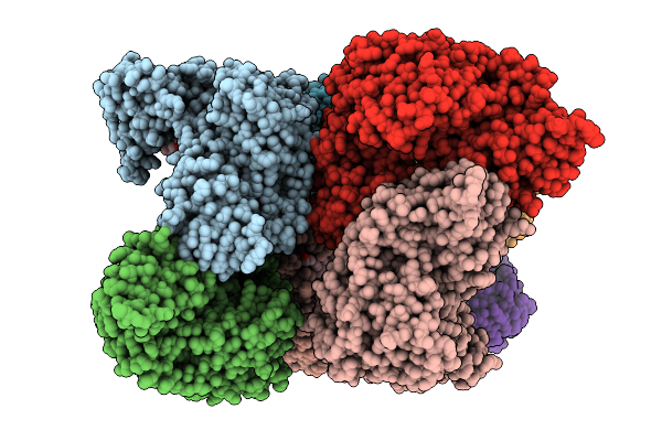
N. Brasiliensis Glft2 In A Styrene Maleic Acid Liponanoparticle
Organism: Nocardia brasiliensis
Method: ELECTRON MICROSCOPY Release Date: 2026-05-20 Classification: TRANSFERASE Ligands: MG |
Organism: Nocardia brasiliensis
Method: ELECTRON MICROSCOPY
Release Date: 2026-05-20
Ligands: MG
 |
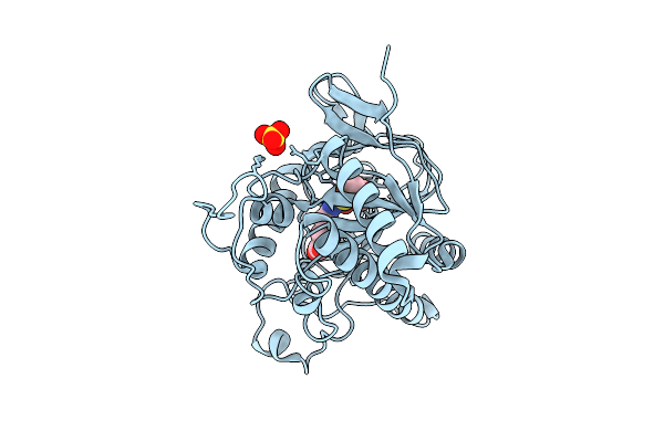
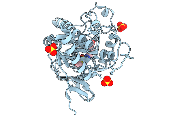
Isopenicillin N Synthase In Complex With Fe And Ipn
Organism: Aspergillus nidulans fgsc a4
Method: X-RAY DIFFRACTION Resolution:1.79 Å Release Date: 2026-04-08 Classification: OXIDOREDUCTASE Ligands: FE, SO4, IP1 |
Organism: Aspergillus nidulans fgsc a4
Method: X-RAY DIFFRACTION
Release Date: 2026-04-08
Ligands: FE, SO4, IP1
 |
Isopenicillin N Synthase In Complex With Fe And Ipn Using Tr-Sfx
Organism: Aspergillus nidulans fgsc a4
Method: X-RAY DIFFRACTION Resolution:1.70 Å Release Date: 2026-04-08 Classification: OXIDOREDUCTASE Ligands: FE, SO4, IP1 |
Organism: Aspergillus nidulans fgsc a4
Method: X-RAY DIFFRACTION
Release Date: 2026-04-08
Ligands: FE, SO4, IP1
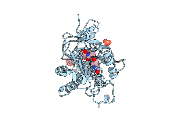 |
Isopenicillin N Synthase In Complex With Fe And Acv
Organism: Aspergillus nidulans fgsc a4
Method: X-RAY DIFFRACTION Resolution:1.45 Å Release Date: 2026-04-08 Classification: OXIDOREDUCTASE Ligands: SO4, FE, ACV |
Organism: Aspergillus nidulans fgsc a4
Method: X-RAY DIFFRACTION
Release Date: 2026-04-08
Ligands: SO4, FE, ACV
 |
Isopenicillin N Synthase In Complex With Fe And Ipn Using Tr-Ssx
Organism: Aspergillus nidulans fgsc a4
Method: X-RAY DIFFRACTION Resolution:1.90 Å Release Date: 2026-04-08 Classification: OXIDOREDUCTASE Ligands: FE, SO4, IP1 |
Organism: Aspergillus nidulans fgsc a4
Method: X-RAY DIFFRACTION
Release Date: 2026-04-08
Ligands: FE, SO4, IP1
 |
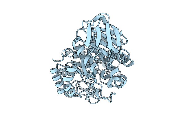
Glft2 From Nocardia Brasiliensis Bound To Galf Tetrasaccharide
Organism: Nocardia brasiliensis
Method: X-RAY DIFFRACTION Resolution:3.15 Å Release Date: 2026-03-11 Classification: TRANSFERASE Ligands: MG, TON, A1BYH, TRT |
Organism: Nocardia brasiliensis
Method: X-RAY DIFFRACTION
Release Date: 2026-03-11
Ligands: MG, TON, A1BYH, TRT
 |
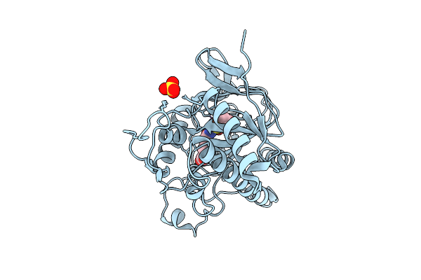
Isopenicillin N Synthase N252E Variant In Complex With Fe And Ipn After O2 Exposure
Organism: Aspergillus nidulans fgsc a4
Method: X-RAY DIFFRACTION Resolution:1.40 Å Release Date: 2026-01-14 Classification: OXIDOREDUCTASE Ligands: FE, SO4, IP1 |
Organism: Aspergillus nidulans fgsc a4
Method: X-RAY DIFFRACTION
Release Date: 2026-01-14
Ligands: FE, SO4, IP1
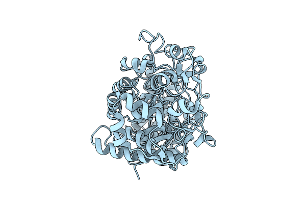 |
Structure Of The Gh13 Domain Of Ruminococcus Bromii Amy4
Organism: Ruminococcus bromii l2-63
Method: ELECTRON MICROSCOPY Release Date: 2025-10-22 Classification: HYDROLASE |
Organism: Ruminococcus bromii l2-63
Method: ELECTRON MICROSCOPY
Release Date: 2025-10-22
 |
Structure Of The Gh13 And Mucbp Domains Of Ruminococcus Bromii Amy10
Organism: Ruminococcus bromii l2-63
Method: ELECTRON MICROSCOPY Release Date: 2025-10-22 Classification: HYDROLASE |
Organism: Ruminococcus bromii l2-63
Method: ELECTRON MICROSCOPY
Release Date: 2025-10-22
 |
Structure Of The Gh13 Domain Of Ruminococcus Bromii Amy16
Organism: Ruminococcus bromii l2-63
Method: ELECTRON MICROSCOPY Release Date: 2025-10-22 Classification: HYDROLASE |
Organism: Ruminococcus bromii l2-63
Method: ELECTRON MICROSCOPY
Release Date: 2025-10-22
 |
Structure Of The Gh13 And Mucbp Domains Of Ruminococcus Bromii Amy12
Organism: Ruminococcus bromii l2-63
Method: ELECTRON MICROSCOPY Release Date: 2025-10-22 Classification: HYDROLASE |
Organism: Ruminococcus bromii l2-63
Method: ELECTRON MICROSCOPY
Release Date: 2025-10-22
 |
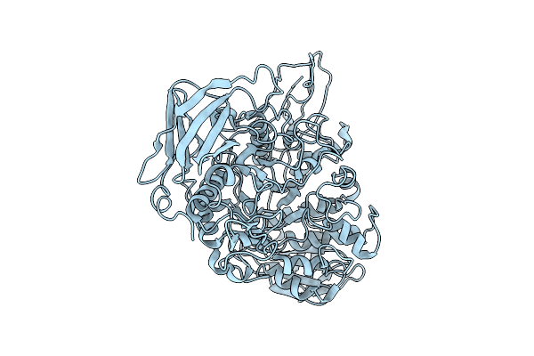
Structural Basis For The Polymer-Protein Binding Mechanism Of Polyvinyl Alcohol Esterase
Organism: Comamonas sp. nyz500
Method: X-RAY DIFFRACTION Resolution:1.92 Å Release Date: 2025-10-08 Classification: HYDROLASE Ligands: SO4, ACT, GOL, CL, NA |
Organism: Comamonas sp. nyz500
Method: X-RAY DIFFRACTION
Release Date: 2025-10-08
Ligands: SO4, ACT, GOL, CL, NA
 |
Structural Basis For The Polymer-Protein Binding Mechanism Of Polyvinyl Alcohol Esterase
Organism: Comamonas sp. nyz500
Method: X-RAY DIFFRACTION Resolution:1.80 Å Release Date: 2025-10-08 Classification: HYDROLASE Ligands: GOL |
Organism: Comamonas sp. nyz500
Method: X-RAY DIFFRACTION
Release Date: 2025-10-08
Ligands: GOL
 |
Structural Basis For The Polymer-Protein Binding Mechanism Of Polyvinyl Alcohol Esterase
Organism: Comamonas sp. nyz500
Method: X-RAY DIFFRACTION Resolution:2.84 Å Release Date: 2025-10-08 Classification: HYDROLASE Ligands: CL, GOL, ACT |
Organism: Comamonas sp. nyz500
Method: X-RAY DIFFRACTION
Release Date: 2025-10-08
Ligands: CL, GOL, ACT
 |
Structural Basis For The Polymer-Protein Binding Mechanism Of Polyvinyl Alcohol Esterase
Organism: Comamonas sp. nyz500
Method: X-RAY DIFFRACTION Resolution:2.13 Å Release Date: 2025-10-08 Classification: HYDROLASE Ligands: CL, A1EGH, ACT, PO4 |
Organism: Comamonas sp. nyz500
Method: X-RAY DIFFRACTION
Release Date: 2025-10-08
Ligands: CL, A1EGH, ACT, PO4
 |
Structural Basis For The Polymer-Protein Binding Mechanism Of Polyvinyl Alcohol Esterase
Organism: Comamonas sp. nyz500
Method: X-RAY DIFFRACTION Resolution:1.78 Å Release Date: 2025-10-08 Classification: HYDROLASE Ligands: A1EGL, CL |
Organism: Comamonas sp. nyz500
Method: X-RAY DIFFRACTION
Release Date: 2025-10-08
Ligands: A1EGL, CL
 |
Ensemble Refined Structure Of Cotb2 In Complex With 2-Fluoro-3,7,18-Dolabellatriene
Organism: Streptomyces melanosporofaciens
Method: X-RAY DIFFRACTION Resolution:1.80 Å Release Date: 2025-09-17 Classification: HYDROLASE Ligands: MG, PPV, EXW, MPD, CL, CCN, 2PE, K |
Organism: Streptomyces melanosporofaciens
Method: X-RAY DIFFRACTION
Release Date: 2025-09-17
Ligands: MG, PPV, EXW, MPD, CL, CCN, 2PE, K