

Search Count: 19
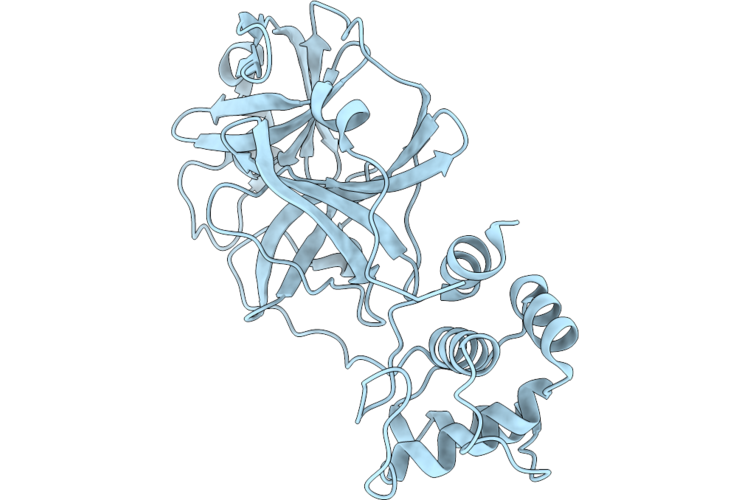 |
Crystal Structure Of The Apo Form Of Main Protease (Mpro) From Hku5-Cov-1
Organism: Pipistrellus bat coronavirus hku5
Method: X-RAY DIFFRACTION Resolution:1.75 Å Release Date: 2026-07-15 Classification: VIRAL PROTEIN |
Organism: Pipistrellus bat coronavirus hku5
Method: X-RAY DIFFRACTION
Release Date: 2026-07-15
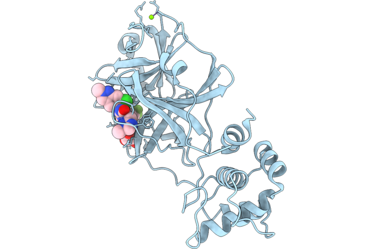 |
Crystal Structure Of Hku5-Cov-1 Main Protease(Mpro) In Complex With Ensitrelvir
Organism: Pipistrellus bat coronavirus hku5
Method: X-RAY DIFFRACTION Resolution:1.55 Å Release Date: 2026-07-15 Classification: VIRAL PROTEIN Ligands: 7YY, GOL, MG |
Organism: Pipistrellus bat coronavirus hku5
Method: X-RAY DIFFRACTION
Release Date: 2026-07-15
Ligands: 7YY, GOL, MG
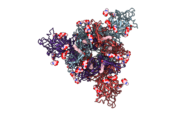 |
Overall Structure Of Hku5 S Protein In Closed Conformation
Organism: Bat coronavirus hku5
Method: ELECTRON MICROSCOPY Release Date: 2025-11-05 Classification: VIRAL PROTEIN Ligands: PLM, NAG, OLA |
Organism: Bat coronavirus hku5
Method: ELECTRON MICROSCOPY
Release Date: 2025-11-05
Ligands: PLM, NAG, OLA
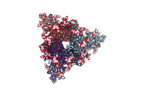 |
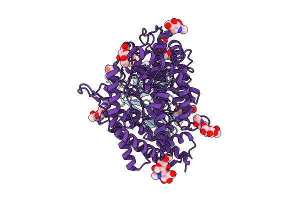
Overall Structure Of Hku5 S Protein With S468A/Y463A Mutation
Organism: Bat coronavirus hku5
Method: ELECTRON MICROSCOPY Release Date: 2025-11-05 Classification: VIRAL PROTEIN Ligands: PLM, NAG |
Organism: Bat coronavirus hku5
Method: ELECTRON MICROSCOPY
Release Date: 2025-11-05
Ligands: PLM, NAG
 |
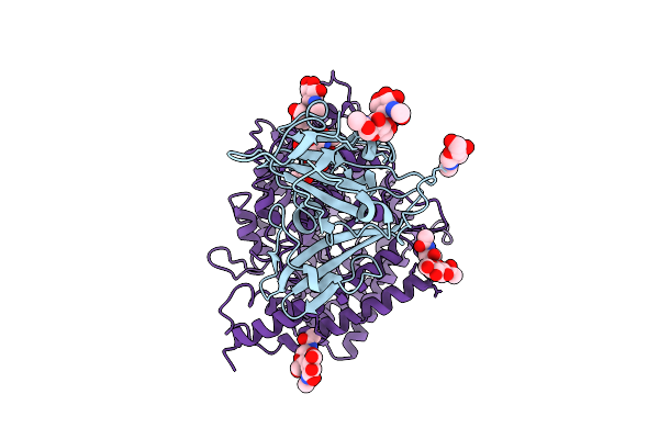
The Rbd Of Hku5 Complex With Pipistrellus Abramus Ace2
Organism: Bat coronavirus hku5, Pipistrellus abramus
Method: ELECTRON MICROSCOPY Release Date: 2025-11-05 Classification: VIRAL PROTEIN Ligands: NAG |
Organism: Bat coronavirus hku5, Pipistrellus abramus
Method: ELECTRON MICROSCOPY
Release Date: 2025-11-05
Ligands: NAG
 |
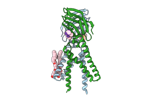
The Rbd Of Hku5 Complex With Pitta Sordida Ace2
Organism: Bat coronavirus hku5, Pitta sordida
Method: ELECTRON MICROSCOPY Release Date: 2025-11-05 Classification: VIRAL PROTEIN Ligands: NAG |
Organism: Bat coronavirus hku5, Pitta sordida
Method: ELECTRON MICROSCOPY
Release Date: 2025-11-05
Ligands: NAG
 |
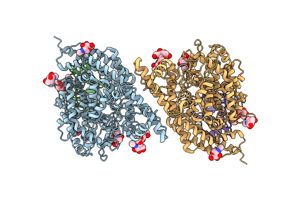
Crystal Structure Of A Coronaviral M Protein In Complex With A C-Terminal Peptide Of The N Protein
Organism: Pipistrellus bat coronavirus hku5
Method: X-RAY DIFFRACTION Resolution:3.14 Å Release Date: 2025-11-05 Classification: MEMBRANE PROTEIN Ligands: N8E |
Organism: Pipistrellus bat coronavirus hku5
Method: X-RAY DIFFRACTION
Release Date: 2025-11-05
Ligands: N8E
 |
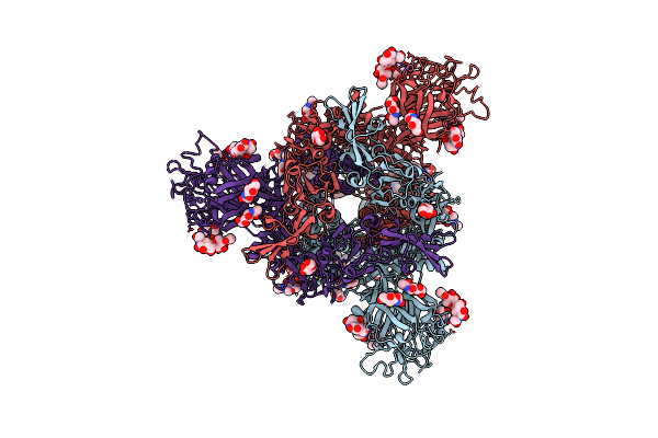
Structure Of Hku5 Spike C-Terminal Domain In Complex With Ace2 From Pipistrellus Abramus
Organism: Pipistrellus abramus, Pipistrellus bat coronavirus hku5
Method: ELECTRON MICROSCOPY Resolution:4.20 Å Release Date: 2025-07-30 Classification: VIRAL PROTEIN Ligands: NAG |
Organism: Pipistrellus abramus, Pipistrellus bat coronavirus hku5
Method: ELECTRON MICROSCOPY
Release Date: 2025-07-30
Ligands: NAG
 |
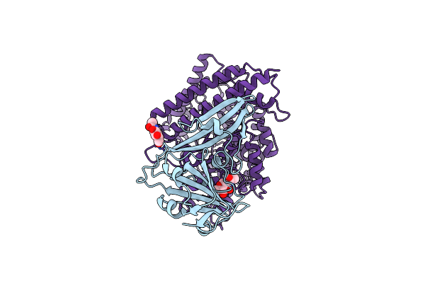
Structure Of Hku5-20S Spike Glycoprotein
Organism: Pipistrellus bat coronavirus hku5
Method: ELECTRON MICROSCOPY Release Date: 2025-06-04 Classification: VIRAL PROTEIN Ligands: NAG |
Organism: Pipistrellus bat coronavirus hku5
Method: ELECTRON MICROSCOPY
Release Date: 2025-06-04
Ligands: NAG
 |
Cryo-Em Structure Of Mrcov Rbd In Complex With Mink Ace2
Organism: Bat coronavirus hku5, Neogale vison
Method: ELECTRON MICROSCOPY Release Date: 2025-03-12 Classification: VIRAL PROTEIN/HYDROLASE Ligands: NAG |
Organism: Bat coronavirus hku5, Neogale vison
Method: ELECTRON MICROSCOPY
Release Date: 2025-03-12
Ligands: NAG
 |
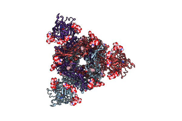
Bthku5-Cov-2-441 Spike Rbd Domain Binding To Hace2
Organism: Pipistrellus bat coronavirus hku5, Homo sapiens
Method: ELECTRON MICROSCOPY Release Date: 2025-03-12 Classification: VIRAL PROTEIN/HYDROLASE Ligands: NAG |
Organism: Pipistrellus bat coronavirus hku5, Homo sapiens
Method: ELECTRON MICROSCOPY
Release Date: 2025-03-12
Ligands: NAG
 |
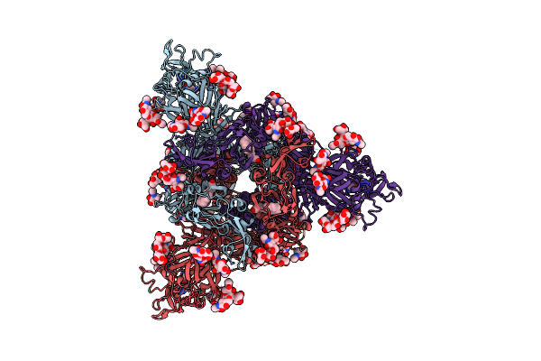
Structure Of The Prefusion Hku5-19S Spike Trimer (Conformation 1)
Organism: Pipistrellus bat coronavirus hku5
Method: ELECTRON MICROSCOPY Resolution:2.00 Å Release Date: 2025-02-26 Classification: VIRAL PROTEIN Ligands: NAG, ZN, FOL, EIC |
Organism: Pipistrellus bat coronavirus hku5
Method: ELECTRON MICROSCOPY
Release Date: 2025-02-26
Ligands: NAG, ZN, FOL, EIC
 |
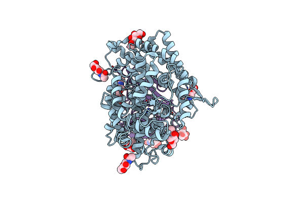
Structure Of The Prefusion Hku5-19S Spike Trimer (Conformation 2)
Organism: Pipistrellus bat coronavirus hku5
Method: ELECTRON MICROSCOPY Resolution:2.00 Å Release Date: 2025-02-26 Classification: VIRAL PROTEIN/IMMUNE SYSTEM Ligands: NAG, FOL, EIC |
Organism: Pipistrellus bat coronavirus hku5
Method: ELECTRON MICROSCOPY
Release Date: 2025-02-26
Ligands: NAG, FOL, EIC
 |
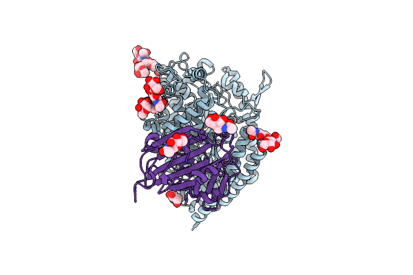
Structure Of The Hku5 Rbd Bound To The P. Abramus Ace2 Receptor
Organism: Pipistrellus abramus, Pipistrellus bat coronavirus hku5
Method: ELECTRON MICROSCOPY Resolution:3.10 Å Release Date: 2025-02-19 Classification: VIRAL PROTEIN/HYDROLASE Ligands: NAG, ZN |
Organism: Pipistrellus abramus, Pipistrellus bat coronavirus hku5
Method: ELECTRON MICROSCOPY
Release Date: 2025-02-19
Ligands: NAG, ZN
 |
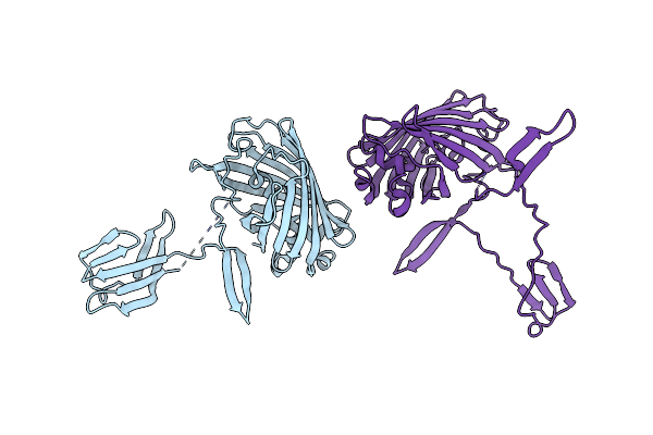
Structure Of The Hku5-19S Rbd Bound To The Bos Taurus Ace2 Receptor
Organism: Bos taurus, Pipistrellus bat coronavirus hku5
Method: ELECTRON MICROSCOPY Release Date: 2025-02-19 Classification: VIRAL PROTEIN/HYDROLASE Ligands: NAG, ZN |
Organism: Bos taurus, Pipistrellus bat coronavirus hku5
Method: ELECTRON MICROSCOPY
Release Date: 2025-02-19
Ligands: NAG, ZN
 |
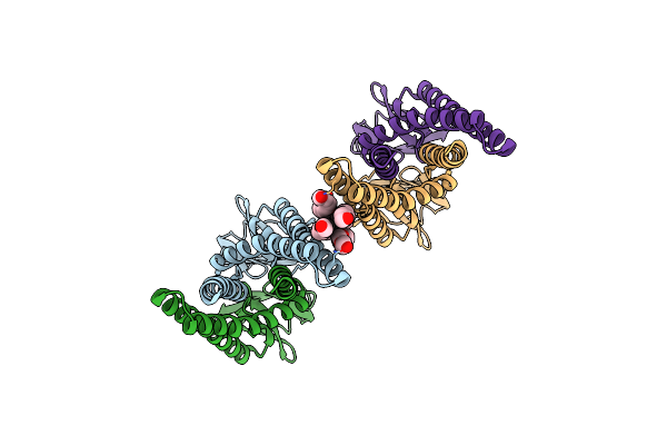
Crystal Structure Of The Carboxy-Terminal Domain Of A Coronavirus M Protein Fused With A Split Gfp
Organism: Aequorea victoria, Pipistrellus bat coronavirus hku5
Method: X-RAY DIFFRACTION Resolution:3.42 Å Release Date: 2022-08-17 Classification: MEMBRANE PROTEIN |
Organism: Aequorea victoria, Pipistrellus bat coronavirus hku5
Method: X-RAY DIFFRACTION
Release Date: 2022-08-17
 |
Crystal Structure Of The Membrane (M) Protein Of A Sars-Cov-2-Related Coronavirus
Organism: Pipistrellus bat coronavirus hku5
Method: X-RAY DIFFRACTION Resolution:3.21 Å Release Date: 2022-08-17 Classification: MEMBRANE PROTEIN Ligands: N8E |
Organism: Pipistrellus bat coronavirus hku5
Method: X-RAY DIFFRACTION
Release Date: 2022-08-17
Ligands: N8E
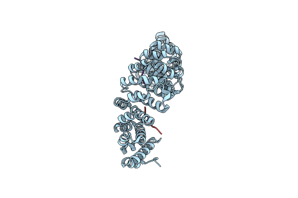 |
Importin Alpha2 In Complex With Orf4B Bat Coronavirus Hku5
Organism: Mus musculus, Bat coronavirus hku5
Method: X-RAY DIFFRACTION Resolution:2.10 Å Release Date: 2021-08-25 Classification: TRANSPORT PROTEIN |
Organism: Mus musculus, Bat coronavirus hku5
Method: X-RAY DIFFRACTION
Release Date: 2021-08-25
 |
Structure Of The S1 Subunit C-Terminal Domain From Bat-Derived Coronavirus Hku5 Spike Protein
Organism: Bat coronavirus hku5
Method: X-RAY DIFFRACTION Resolution:2.10 Å Release Date: 2017-05-10 Classification: VIRAL PROTEIN Ligands: NAG |
Organism: Bat coronavirus hku5
Method: X-RAY DIFFRACTION
Release Date: 2017-05-10
Ligands: NAG