
Search Count: 25,583
All
Selected
 |
Organism: Streptomyces fagopyri
Method: X-RAY DIFFRACTION Resolution:1.87 Å Release Date: 2026-03-18 Classification: BIOSYNTHETIC PROTEIN |
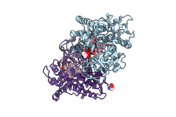 |
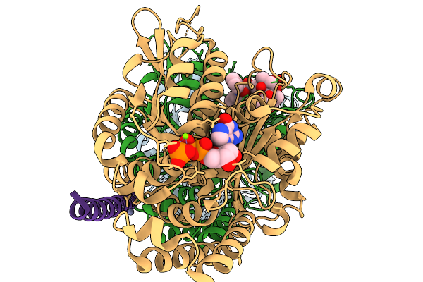
Organism: Priestia megaterium (strain nbrc 15308)
Method: X-RAY DIFFRACTION Resolution:1.57 Å Release Date: 2026-03-18 Classification: OXIDOREDUCTASE Ligands: HEM, IMD, PEG, TRS, CL |
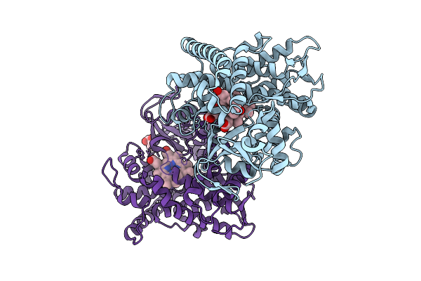 |
Crystal Structure Of A P450 Bm3 Heme Domain Mutant In Complex With Zearalenone
Organism: Priestia megaterium (strain nbrc 15308)
Method: X-RAY DIFFRACTION Resolution:1.80 Å Release Date: 2026-03-18 Classification: OXIDOREDUCTASE Ligands: HEM, ZER, TRS |
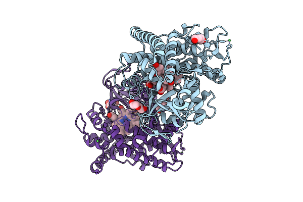 |
Crystal Structure Of A P450 Bm3 Heme Domain Mutant In Complex With Alpha-Zearalanol
Organism: Priestia megaterium (strain nbrc 15308)
Method: X-RAY DIFFRACTION Resolution:2.09 Å Release Date: 2026-03-18 Classification: OXIDOREDUCTASE Ligands: HEM, 36J, PEG, NI, TRS |
 |
Organism: Azotobacter beijerinckii
Method: X-RAY DIFFRACTION Resolution:3.41 Å Release Date: 2026-03-11 Classification: ELECTRON TRANSPORT Ligands: FES |
 |
Organism: Azotobacter beijerinckii
Method: X-RAY DIFFRACTION Resolution:1.70 Å Release Date: 2026-03-04 Classification: ELECTRON TRANSPORT Ligands: FES |
 |
Organism: Azotobacter beijerinckii
Method: X-RAY DIFFRACTION Resolution:1.45 Å Release Date: 2026-03-04 Classification: ELECTRON TRANSPORT Ligands: FES |
 |
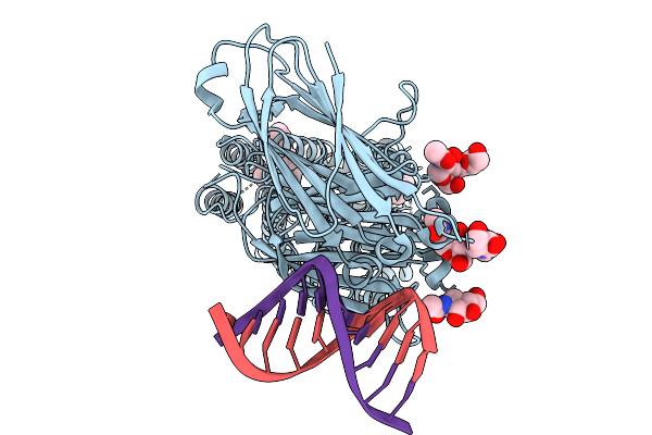
Organism: Caenorhabditis elegans
Method: X-RAY DIFFRACTION Resolution:2.99 Å Release Date: 2026-03-04 Classification: DNA BINDING PROTEIN Ligands: STL, MG |
 |
Organism: Shewanella baltica
Method: X-RAY DIFFRACTION Resolution:2.90 Å Release Date: 2026-02-25 Classification: LIPID BINDING PROTEIN |
 |
Cryo-Em Structure Of The Sah Domain Of Caenorhabditis Elegans Icp-1 Bound To The Paclitaxel-Stabilized Microtubule
Organism: Caenorhabditis elegans, Sus scrofa
Method: ELECTRON MICROSCOPY Release Date: 2026-02-04 Classification: CELL CYCLE Ligands: GTP, ZN, MG, GDP, TA1 |
 |
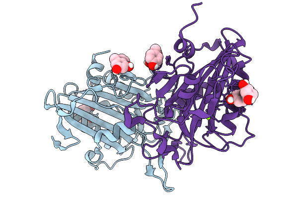
Structure Of Human Fab 245 In Complex With Influenza H1N1 A/Solomon Island/3/2006 Hemagglutinin
Organism: Influenza a virus, Homo sapiens
Method: ELECTRON MICROSCOPY Resolution:2.80 Å Release Date: 2026-02-04 Classification: IMMUNE SYSTEM/VIRAL PROTEIN |
 |
Structure Of Human Fab 604 In Complex With Influenza H1N1 A/Solomon Island/3/2006 Hemagglutinin
Organism: Influenza a virus, Homo sapiens
Method: ELECTRON MICROSCOPY Release Date: 2026-02-04 Classification: IMMUNE SYSTEM/VIRAL PROTEIN |
 |
Organism: Caenorhabditis elegans, Synthetic construct
Method: ELECTRON MICROSCOPY Resolution:3.99 Å Release Date: 2026-01-28 Classification: MEMBRANE PROTEIN/RNA Ligands: NAG, ZN |
 |
Organism: Caenorhabditis elegans, Synthetic construct
Method: ELECTRON MICROSCOPY Release Date: 2026-01-21 Classification: MEMBRANE PROTEIN/RNA Ligands: NAG, ZN, Y01, LBN |
 |
Organism: Caenorhabditis elegans
Method: ELECTRON MICROSCOPY Release Date: 2026-01-21 Classification: MEMBRANE PROTEIN Ligands: NAG, ZN, Y01, LBN |
 |
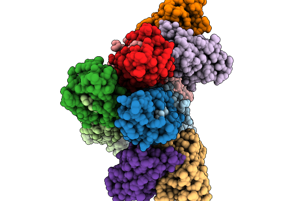
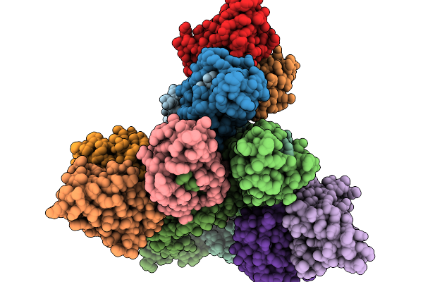
Octameric C. Elegans Borc, Containing Borcs5, Borcs6, Borcs7, Borcs8, Kxd1 And The Shared Borc And Bloc-1 Subunits, Bloc1S1, Bloc1S2 And Snapin
Organism: Caenorhabditis elegans, Caenorhabditis
Method: ELECTRON MICROSCOPY Release Date: 2026-01-21 Classification: TRANSPORT PROTEIN |
 |
Organism: Caenorhabditis elegans
Method: ELECTRON MICROSCOPY Release Date: 2025-12-24 Classification: MEMBRANE PROTEIN Ligands: NAG, CLR |
 |
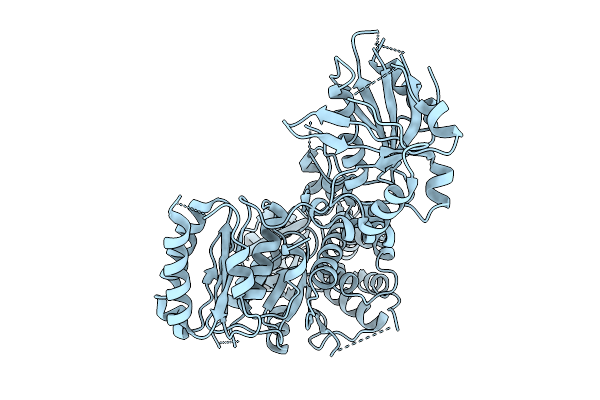
Organism: Caenorhabditis elegans
Method: X-RAY DIFFRACTION Resolution:2.80 Å Release Date: 2025-12-24 Classification: CHAPERONE |
 |
Organism: Caenorhabditis elegans
Method: ELECTRON MICROSCOPY Release Date: 2025-12-24 Classification: MEMBRANE PROTEIN |
 |
Organism: Caenorhabditis elegans
Method: ELECTRON MICROSCOPY Release Date: 2025-12-24 Classification: MEMBRANE PROTEIN |