
Search Count: 3,109
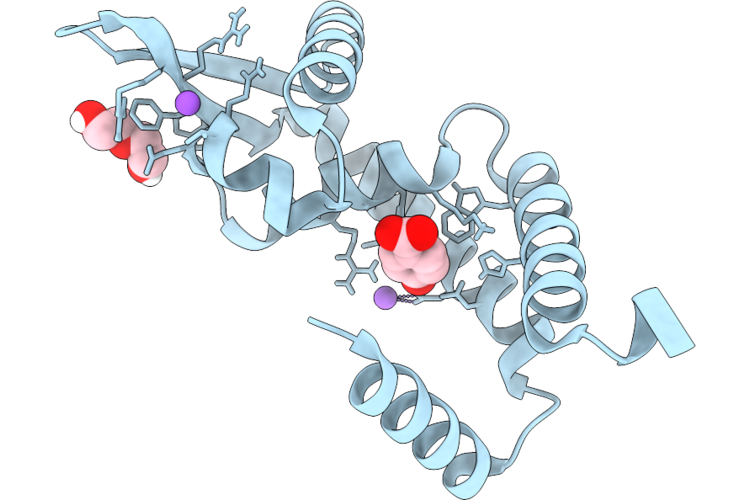 |
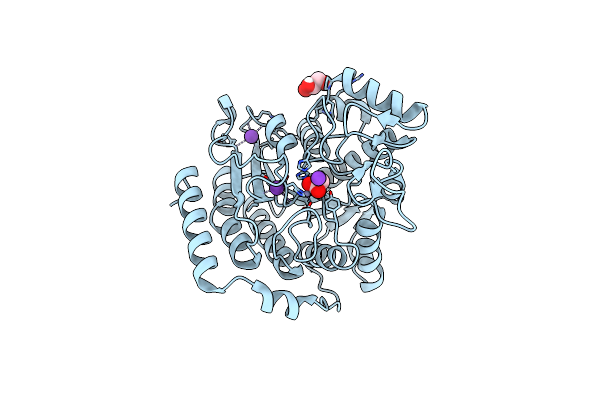
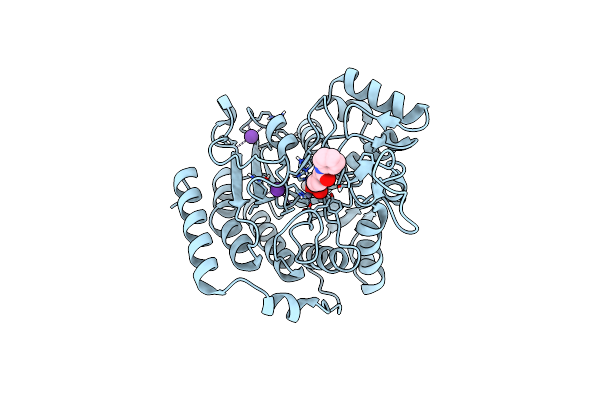
Hosa Transcriptional Regulator From Enteropathogenic Escherichia Coli O127:H6 (Strain E2348/69) Bound With 4-Hydroxy Benzoic Acid - L2' Conformation At 1.41 Angstrom Resolution
Organism: Escherichia coli o127:h6 str. e2348/69
Method: X-RAY DIFFRACTION Resolution:1.41 Å Release Date: 2026-06-17 Classification: DNA BINDING PROTEIN Ligands: PHB, PEG, CL, NA |
 |
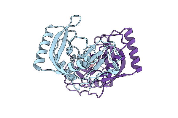
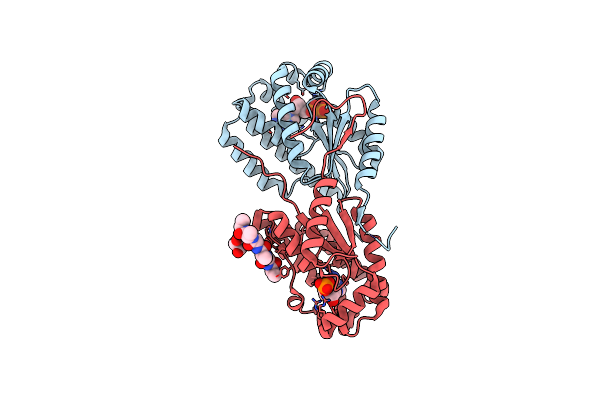
Organism: Streptomyces lividans tk24
Method: X-RAY DIFFRACTION Resolution:2.06 Å Release Date: 2026-04-22 Classification: HYDROLASE |
 |
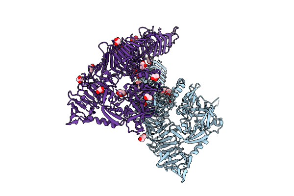
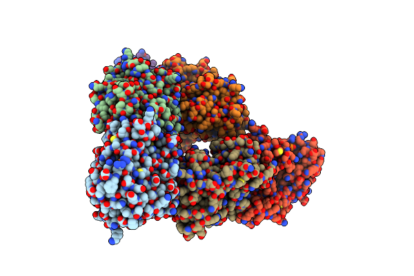
Organism: Cervus canadensis, Severe acute respiratory syndrome coronavirus 2
Method: ELECTRON MICROSCOPY Release Date: 2026-02-25 Classification: VIRAL PROTEIN Ligands: NAG |
 |
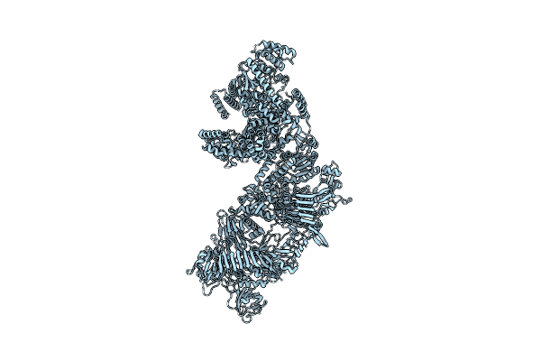
Organism: Severe acute respiratory syndrome coronavirus 2, Cervus canadensis
Method: ELECTRON MICROSCOPY Release Date: 2026-02-25 Classification: VIRAL PROTEIN Ligands: NAG |
 |
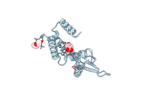
Solution Structure Of The De Novo Designed Monoheme Protein M4D2 With Bound Iron(Iii) 2,4-Dimethyldeuteroporphyrin Ix
Organism: Test organism
Method: SOLUTION NMR Release Date: 2026-02-04 Classification: DE NOVO PROTEIN Ligands: FDD |
 |
Phosphodiesterase From Burkholderia Phage Bcsr5 In The Closed Lid Conformation
Organism: Burkholderia phage bcsr5
Method: X-RAY DIFFRACTION Resolution:2.00 Å Release Date: 2025-11-05 Classification: VIRAL PROTEIN Ligands: FLC |
 |
Phosphodiesterase From Burkholderia Phage Bcsr5 In The Open Lid Conformation
Organism: Burkholderia phage bcsr5
Method: X-RAY DIFFRACTION Resolution:2.03 Å Release Date: 2025-11-05 Classification: VIRAL PROTEIN Ligands: CAC |
 |
Organism: Escherichia coli o127:h6 str. e2348/69
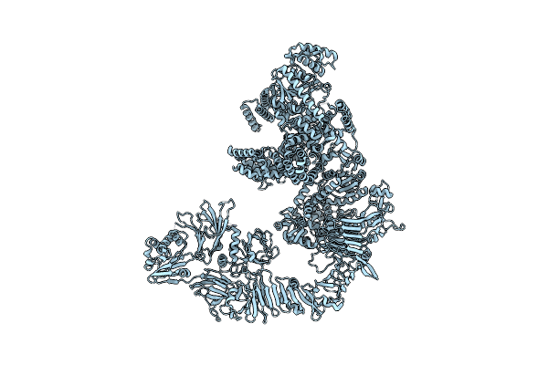
Method: ELECTRON MICROSCOPY Resolution:2.56 Å Release Date: 2025-11-05 Classification: PROTEIN TRANSPORT |
 |
Organism: Escherichia coli o127:h6
Method: X-RAY DIFFRACTION Resolution:2.94 Å Release Date: 2025-10-29 Classification: HYDROLASE Ligands: P6G, PE8, GOL, PEG |
 |
Organism: Escherichia coli o127:h6 str. e2348/69
Method: X-RAY DIFFRACTION Resolution:1.30 Å Release Date: 2025-07-23 Classification: DNA BINDING PROTEIN |
 |
Organism: Escherichia coli o127:h6 str. e2348/69
Method: ELECTRON MICROSCOPY Release Date: 2025-04-09 Classification: TOXIN |
 |
Organism: Escherichia coli o127:h6 str. e2348/69
Method: ELECTRON MICROSCOPY Release Date: 2025-04-09 Classification: TOXIN |
 |
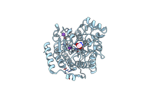
Hosa Transcriptional Regulator From Enteropathogenic Escherichia Coli O127:H6 (Strain E2348/69) Bound With 4-Hydroxy Benzoic Acid - Conformation Ii At 2.16 Angstrom Resolution (Staraniso Processed)
Organism: Escherichia coli o127:h6 str. e2348/69
Method: X-RAY DIFFRACTION Resolution:2.16 Å Release Date: 2024-12-04 Classification: DNA BINDING PROTEIN Ligands: PHB, PEG |
 |
Crystal Structure Of Deacetylase (Hdah) From Klebsiella Pneumoniae Subsp. Ozaenae
Organism: Klebsiella pneumoniae subsp. ozaenae
Method: X-RAY DIFFRACTION Resolution:2.10 Å Release Date: 2024-11-06 Classification: HYDROLASE Ligands: GOL, ACT, K, ZN |
 |
Crystal Structure Of Inactive Deacetylase (Hdah) H144A From Klebsiella Pneumoniae Subsp. Ozaenae
Organism: Klebsiella pneumoniae subsp. ozaenae
Method: X-RAY DIFFRACTION Resolution:2.35 Å Release Date: 2024-11-06 Classification: HYDROLASE Ligands: K, ZN, IMD, ACT |
 |
Crystal Structure Of Deacetylase (Hdah) From Klebsiella Pneumoniae Subsp. Ozaenae In Complex With The Inhibitor Saha
Organism: Klebsiella pneumoniae subsp. ozaenae
Method: X-RAY DIFFRACTION Resolution:1.95 Å Release Date: 2024-11-06 Classification: HYDROLASE Ligands: K, ZN, SHH |
 |
Crystal Structure Of Deacetylase (Hdah) From Klebsiella Pneumoniae Subsp. Ozaenae In Complex With The Inhibitor Tsa
Organism: Klebsiella pneumoniae subsp. ozaenae
Method: X-RAY DIFFRACTION Resolution:2.18 Å Release Date: 2024-11-06 Classification: HYDROLASE Ligands: K, ZN, PO4, TSN, GOL |
 |
Organism: Giardia intestinalis
Method: X-RAY DIFFRACTION Resolution:2.10 Å Release Date: 2024-10-30 Classification: TRANSFERASE Ligands: DAT, D5M |
 |
Crystal Structure Of Chms Dehydrogenase Pmdc From Comamonas Testosteroni Bound To Cofactor Nadp
Organism: Comamonas testosteroni atcc 11996
Method: X-RAY DIFFRACTION Resolution:2.34 Å Release Date: 2024-09-18 Classification: OXIDOREDUCTASE Ligands: SO4, NAP |
 |
Organism: Aspergillus westerdijkiae
Method: X-RAY DIFFRACTION Resolution:1.68 Å Release Date: 2024-09-11 Classification: OXIDOREDUCTASE Ligands: FE |