

Search Count: 7
 |
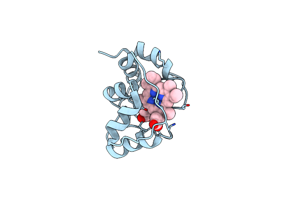
X-Ray Structure Of Ferric Cytochrome C-550 From Paracoccus Versutus
Organism: Paracoccus versutus
Method: X-RAY DIFFRACTION Resolution:1.90 Å Release Date: 2005-05-11 Classification: ELECTRON TRANSFER Ligands: HEC |
Organism: Paracoccus versutus
Method: X-RAY DIFFRACTION
Release Date: 2005-05-11
Ligands: HEC
 |
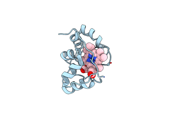
X-Ray Structure Of The M100K Variant Of Ferric Cyt C-550 From Paracoccus Versutus Determined At 100 K.
Organism: Paracoccus versutus
Method: X-RAY DIFFRACTION Resolution:1.55 Å Release Date: 2005-05-11 Classification: ELECTRON TRANSFER Ligands: HEC |
Organism: Paracoccus versutus
Method: X-RAY DIFFRACTION
Release Date: 2005-05-11
Ligands: HEC
 |
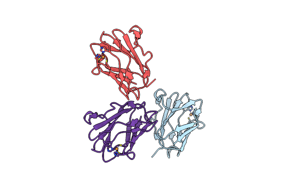
X-Ray Structure Of The M100K Variant Of Ferric Cyt C-550 From Paracoccus Versutus Determined At 295 K.
Organism: Paracoccus versutus
Method: X-RAY DIFFRACTION Resolution:1.95 Å Release Date: 2005-05-11 Classification: ELECTRON TRANSFER Ligands: HEC |
Organism: Paracoccus versutus
Method: X-RAY DIFFRACTION
Release Date: 2005-05-11
Ligands: HEC
 |
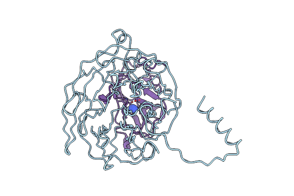
Crystal Structure Of Amicyanin From Paracoccus Versutus (Thiobacillus Versutus)
Organism: Paracoccus versutus
Method: X-RAY DIFFRACTION Resolution:2.15 Å Release Date: 2001-04-11 Classification: ELECTRON TRANSPORT Ligands: CU |
Organism: Paracoccus versutus
Method: X-RAY DIFFRACTION
Release Date: 2001-04-11
Ligands: CU
 |
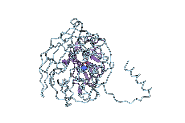
The Active Site Structure Of Methylamine Dehydrogenase: Hydrazines Identify C6 As The Reactive Site Of The Tryptophan Derived Quinone Cofactor
Organism: Paracoccus versutus
Method: X-RAY DIFFRACTION Resolution:2.80 Å Release Date: 1994-01-31 Classification: OXIDOREDUCTASE(CHNH2(D)-DEAMINATING) Ligands: HDZ |
Organism: Paracoccus versutus
Method: X-RAY DIFFRACTION
Release Date: 1994-01-31
Ligands: HDZ
 |
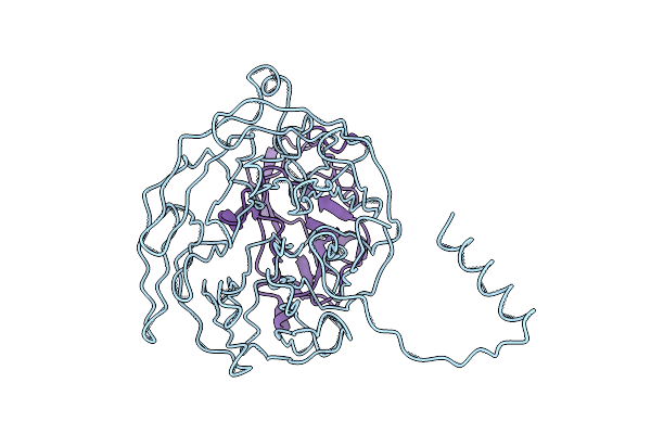
The Active Site Structure Of Methylamine Dehydrogenase: Hydrazines Identify C6 As The Reactive Site Of The Tryptophan Derived Quinone Cofactor
Organism: Paracoccus versutus
Method: X-RAY DIFFRACTION Resolution:2.60 Å Release Date: 1994-01-31 Classification: OXIDOREDUCTASE(CHNH2(D)-DEAMINATING) Ligands: HDZ |
Organism: Paracoccus versutus
Method: X-RAY DIFFRACTION
Release Date: 1994-01-31
Ligands: HDZ
 |
The Active Site Structure Of Methylamine Dehydrogenase: Hydrazines Identify C6 As The Reactive Site Of The Tryptophan Derived Quinone Cofactor
Organism: Paracoccus versutus
Method: X-RAY DIFFRACTION Resolution:2.25 Å Release Date: 1994-01-31 Classification: OXIDOREDUCTASE(CHNH2(D)-DEAMINATING) |
Organism: Paracoccus versutus
Method: X-RAY DIFFRACTION
Release Date: 1994-01-31