

Search Count: 53
 |
Crystal Structure Of Hsp72 In Complex With Ligand 11 At 1.43 Mgy X-Ray Dose
Organism: Homo sapiens
Method: X-RAY DIFFRACTION Resolution:2.15 Å Release Date: 2024-12-11 Classification: CHAPERONE Ligands: PO4, DMS, EDO, PEG, 1PS, A1ACZ |
Organism: Homo sapiens
Method: X-RAY DIFFRACTION
Release Date: 2024-12-11
Ligands: PO4, DMS, EDO, PEG, 1PS, A1ACZ
 |
Crystal Structure Of Hsp72 In Complex With Ligand 11 At 2.86 Mgy X-Ray Dose
Organism: Homo sapiens
Method: X-RAY DIFFRACTION Resolution:2.15 Å Release Date: 2024-12-11 Classification: CHAPERONE Ligands: PO4, DMS, EDO, PEG, 1PS, A1ACZ |
Organism: Homo sapiens
Method: X-RAY DIFFRACTION
Release Date: 2024-12-11
Ligands: PO4, DMS, EDO, PEG, 1PS, A1ACZ
 |
Crystal Structure Of Hsp72 In Complex With Ligand 11 At 4.29 Mgy X-Ray Dose
Organism: Homo sapiens
Method: X-RAY DIFFRACTION Resolution:2.15 Å Release Date: 2024-12-11 Classification: CHAPERONE Ligands: PO4, DMS, EDO, PEG, 1PS, A1ACZ |
Organism: Homo sapiens
Method: X-RAY DIFFRACTION
Release Date: 2024-12-11
Ligands: PO4, DMS, EDO, PEG, 1PS, A1ACZ
 |
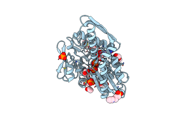
Crystal Structure Of Hsp72 In Complex With Ligand 11 At 5.72 Mgy X-Ray Dose
Organism: Homo sapiens
Method: X-RAY DIFFRACTION Resolution:2.15 Å Release Date: 2024-12-11 Classification: CHAPERONE Ligands: PO4, DMS, EDO, PEG, 1PS, A1ACZ |
Organism: Homo sapiens
Method: X-RAY DIFFRACTION
Release Date: 2024-12-11
Ligands: PO4, DMS, EDO, PEG, 1PS, A1ACZ
 |
Crystal Structure Of Hsp72 In Complex With Ligand 11 At 7.15 Mgy X-Ray Dose
Organism: Homo sapiens
Method: X-RAY DIFFRACTION Resolution:2.15 Å Release Date: 2024-12-11 Classification: CHAPERONE Ligands: PO4, DMS, EDO, PEG, 1PS, A1ACZ |
Organism: Homo sapiens
Method: X-RAY DIFFRACTION
Release Date: 2024-12-11
Ligands: PO4, DMS, EDO, PEG, 1PS, A1ACZ
 |
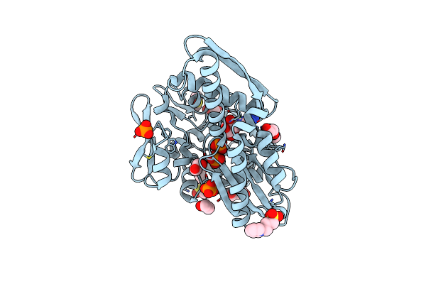
Crystal Structure Of Hsp72 In Complex With Ligand 11 At 8.58 Mgy X-Ray Dose
Organism: Homo sapiens
Method: X-RAY DIFFRACTION Resolution:2.15 Å Release Date: 2024-12-11 Classification: CHAPERONE Ligands: PO4, DMS, EDO, PEG, 1PS, A1ACZ |
Organism: Homo sapiens
Method: X-RAY DIFFRACTION
Release Date: 2024-12-11
Ligands: PO4, DMS, EDO, PEG, 1PS, A1ACZ
 |
Crystal Structure Of Hsp72 In Complex With Ligand 11 At 10.01 Mgy X-Ray Dose
Organism: Homo sapiens
Method: X-RAY DIFFRACTION Resolution:2.15 Å Release Date: 2024-12-11 Classification: CHAPERONE Ligands: PO4, DMS, EDO, PEG, 1PS, A1ACZ |
Organism: Homo sapiens
Method: X-RAY DIFFRACTION
Release Date: 2024-12-11
Ligands: PO4, DMS, EDO, PEG, 1PS, A1ACZ
 |
Crystal Structure Of Hsp72 In Complex With Ligand 11 At 11.44 Mgy X-Ray Dose
Organism: Homo sapiens
Method: X-RAY DIFFRACTION Resolution:2.15 Å Release Date: 2024-12-11 Classification: CHAPERONE Ligands: PO4, DMS, EDO, PEG, 1PS, A1ACZ |
Organism: Homo sapiens
Method: X-RAY DIFFRACTION
Release Date: 2024-12-11
Ligands: PO4, DMS, EDO, PEG, 1PS, A1ACZ
 |
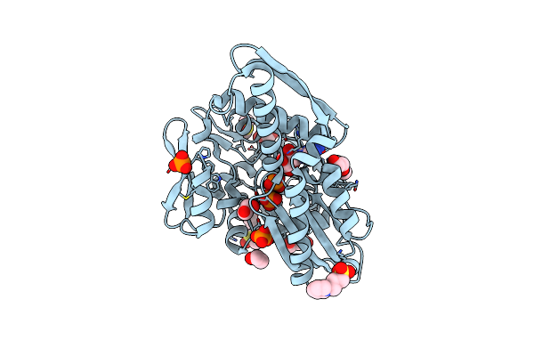
Crystal Structure Of Hsp72 In Complex With Ligand 11 At 12.87 Mgy X-Ray Dose
Organism: Homo sapiens
Method: X-RAY DIFFRACTION Resolution:2.15 Å Release Date: 2024-12-11 Classification: CHAPERONE Ligands: PO4, DMS, EDO, PEG, 1PS, A1ACZ |
Organism: Homo sapiens
Method: X-RAY DIFFRACTION
Release Date: 2024-12-11
Ligands: PO4, DMS, EDO, PEG, 1PS, A1ACZ
 |
Crystal Structure Of Hsp72 In Complex With Ligand 11 At 14.30 Mgy X-Ray Dose
Organism: Homo sapiens
Method: X-RAY DIFFRACTION Resolution:2.15 Å Release Date: 2024-12-11 Classification: CHAPERONE Ligands: PO4, DMS, EDO, PEG, 1PS, A1ACZ |
Organism: Homo sapiens
Method: X-RAY DIFFRACTION
Release Date: 2024-12-11
Ligands: PO4, DMS, EDO, PEG, 1PS, A1ACZ
 |
Crystal Structure Of Hsp72 In Complex With Ligand 11 At 15.73 Mgy X-Ray Dose
Organism: Homo sapiens
Method: X-RAY DIFFRACTION Resolution:2.15 Å Release Date: 2024-12-11 Classification: CHAPERONE Ligands: PO4, DMS, EDO, PEG, 1PS, A1ACZ |
Organism: Homo sapiens
Method: X-RAY DIFFRACTION
Release Date: 2024-12-11
Ligands: PO4, DMS, EDO, PEG, 1PS, A1ACZ
 |
Crystal Structure Of Hsp72 In Complex With Ligand 11 At 17.16 Mgy X-Ray Dose
Organism: Homo sapiens
Method: X-RAY DIFFRACTION Resolution:2.15 Å Release Date: 2024-12-11 Classification: CHAPERONE Ligands: PO4, DMS, EDO, PEG, 1PS, A1ACZ |
Organism: Homo sapiens
Method: X-RAY DIFFRACTION
Release Date: 2024-12-11
Ligands: PO4, DMS, EDO, PEG, 1PS, A1ACZ
 |
Crystal Structure Of Hsp72 In Complex With Ligand 11 At 18.59 Mgy X-Ray Dose
Organism: Homo sapiens
Method: X-RAY DIFFRACTION Resolution:2.15 Å Release Date: 2024-12-11 Classification: CHAPERONE Ligands: PO4, DMS, EDO, PEG, 1PS, A1ACZ |
Organism: Homo sapiens
Method: X-RAY DIFFRACTION
Release Date: 2024-12-11
Ligands: PO4, DMS, EDO, PEG, 1PS, A1ACZ
 |
Crystal Structure Of Hsp72 In Complex With Ligand 11 At 20.02 Mgy X-Ray Dose
Organism: Homo sapiens
Method: X-RAY DIFFRACTION Resolution:2.15 Å Release Date: 2024-12-11 Classification: CHAPERONE Ligands: PO4, DMS, EDO, PEG, 1PS, A1ACZ |
Organism: Homo sapiens
Method: X-RAY DIFFRACTION
Release Date: 2024-12-11
Ligands: PO4, DMS, EDO, PEG, 1PS, A1ACZ
 |
Crystal Structure Of Hsp72 In Complex With Ligand 11 At 21.45 Mgy X-Ray Dose
Organism: Homo sapiens
Method: X-RAY DIFFRACTION Resolution:2.15 Å Release Date: 2024-12-11 Classification: CHAPERONE Ligands: PO4, DMS, EDO, PEG, 1PS, A1ACZ |
Organism: Homo sapiens
Method: X-RAY DIFFRACTION
Release Date: 2024-12-11
Ligands: PO4, DMS, EDO, PEG, 1PS, A1ACZ
 |
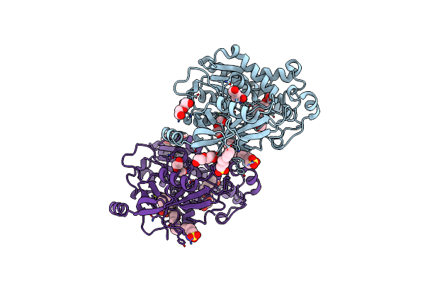
Crystal Structure Of Tick Tyrosylprotein Sulfotransferase Reveals The Activation Mechanism Of Tick Anticoagulant Protein Madanin
Organism: Ixodes, Longicornis species complex
Method: X-RAY DIFFRACTION Resolution:1.55 Å Release Date: 2024-04-10 Classification: TRANSFERASE Ligands: A3P, 1PS, TRS, MG |
Organism: Ixodes, Longicornis species complex
Method: X-RAY DIFFRACTION
Release Date: 2024-04-10
Ligands: A3P, 1PS, TRS, MG
 |
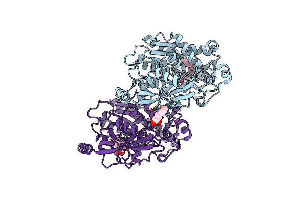
Structure Of The Thermolabile Hemolysin From Vibrio Alginolyticus (Apo Form)
Organism: Vibrio alginolyticus
Method: X-RAY DIFFRACTION Resolution:1.81 Å Release Date: 2023-08-16 Classification: HYDROLASE Ligands: 1PE, 1PS, PEG, MG |
Organism: Vibrio alginolyticus
Method: X-RAY DIFFRACTION
Release Date: 2023-08-16
Ligands: 1PE, 1PS, PEG, MG
 |
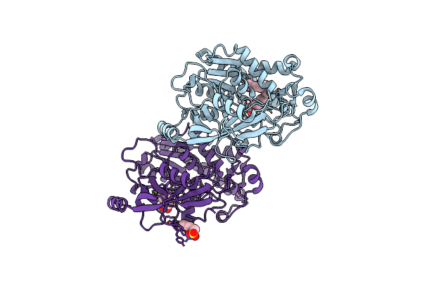
Structure Of The Thermolabile Hemolysin From Vibrio Alginolyticus (In Complex With Arachidonic Acid)
Organism: Vibrio alginolyticus
Method: X-RAY DIFFRACTION Resolution:1.98 Å Release Date: 2023-08-16 Classification: HYDROLASE Ligands: ACD, 1PS, MG |
Organism: Vibrio alginolyticus
Method: X-RAY DIFFRACTION
Release Date: 2023-08-16
Ligands: ACD, 1PS, MG
 |
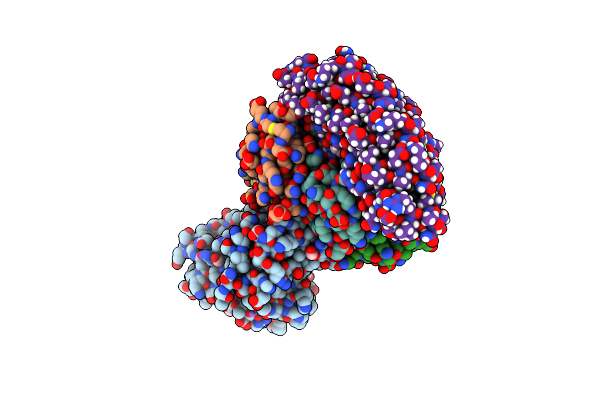
Structure Of The Thermolabile Hemolysin From Vibrio Alginolyticus (In Complex With Docosahexaenoic Acid)
Organism: Vibrio alginolyticus
Method: X-RAY DIFFRACTION Resolution:2.01 Å Release Date: 2023-08-16 Classification: HYDROLASE Ligands: HXA, 1PS, MG |
Organism: Vibrio alginolyticus
Method: X-RAY DIFFRACTION
Release Date: 2023-08-16
Ligands: HXA, 1PS, MG
 |
Stx2A And Darpin Complex
Organism: Synthetic construct, Escherichia phage 933w
Method: ELECTRON MICROSCOPY Release Date: 2023-04-12 Classification: ANTITOXIN Ligands: 1PS, FMT, NA, EDO |
Organism: Synthetic construct, Escherichia phage 933w
Method: ELECTRON MICROSCOPY
Release Date: 2023-04-12
Ligands: 1PS, FMT, NA, EDO