
Search Count: 4,231
 |
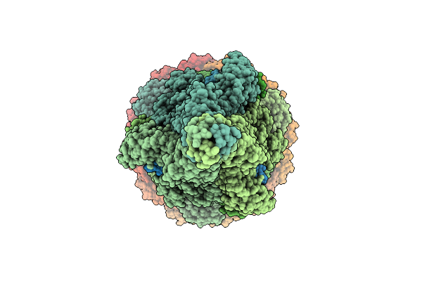
Organism: Francisella tularensis subsp. novicida (strain u112)
Method: ELECTRON MICROSCOPY Resolution:3.40 Å Release Date: 2026-06-10 Classification: TOXIN |
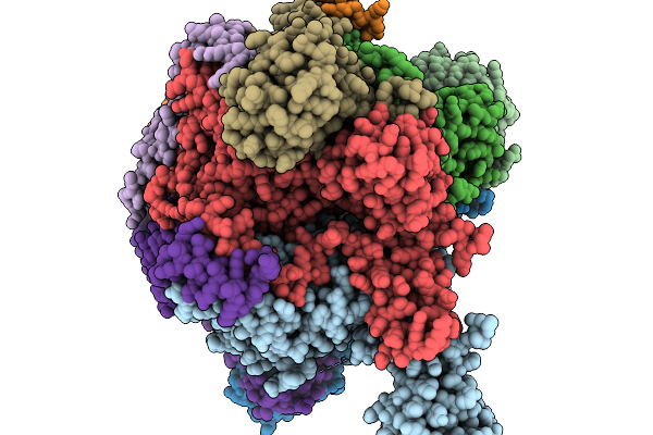 |
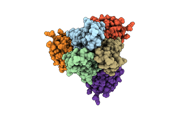
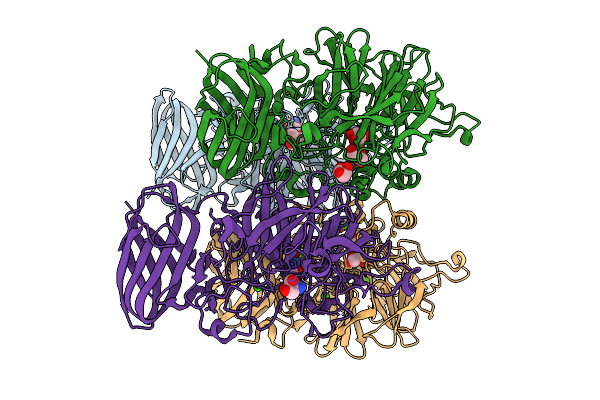
Ecoli Dnab Helicase And Phage Lambda Loader P With Adp-Mg In A 6:5 Stoichiometry Ratio
Organism: Escherichia coli, Escherichia phage lambda
Method: ELECTRON MICROSCOPY Release Date: 2026-03-04 Classification: DNA BINDING PROTEIN Ligands: ADP, MG |
 |
Organism: Streptomyces phage phi-c31, Escherichia coli
Method: ELECTRON MICROSCOPY Release Date: 2026-03-04 Classification: DNA BINDING PROTEIN Ligands: ZN |
 |
Organism: Francisella tularensis subsp. novicida u112, Synthetic construct
Method: ELECTRON MICROSCOPY Resolution:3.26 Å Release Date: 2026-02-11 Classification: DNA BINDING PROTEIN |
 |
Organism: Francisella tularensis subsp. novicida u112, Synthetic construct
Method: ELECTRON MICROSCOPY Resolution:4.00 Å Release Date: 2026-02-11 Classification: DNA BINDING PROTEIN |
 |
Organism: Francisella tularensis subsp. novicida u112, Synthetic construct
Method: ELECTRON MICROSCOPY Resolution:3.68 Å Release Date: 2026-02-11 Classification: DNA BINDING PROTEIN |
 |
Structure Of S. Typhimurium 14028 Gifsy-1 Prophage Heps Bound To Bacteriophage Lambda J Tail Tip
Organism: Salmonella enterica subsp. enterica serovar typhimurium str. atcc 14028, Escherichia phage lambda
Method: X-RAY DIFFRACTION Resolution:1.86 Å Release Date: 2026-01-28 Classification: VIRAL PROTEIN |
 |
Organism: Escherichia phage lambda, Escherichia coli o157:h7 str. edl933
Method: ELECTRON MICROSCOPY Release Date: 2026-01-28 Classification: VIRAL PROTEIN |
 |
Open State Of A8 Gpj 713 Central Tail Fiber With Ompc G17 From E. Coli Edl933
Organism: Escherichia phage lambda, Escherichia coli o157:h7 str. edl933
Method: ELECTRON MICROSCOPY Release Date: 2026-01-28 Classification: VIRAL PROTEIN |
 |
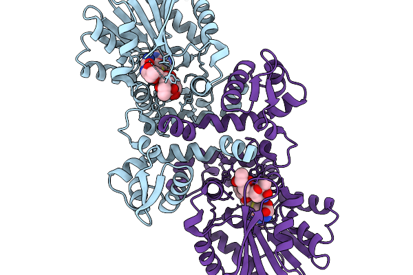
Comparative Analysis Of Functions And Catalytic Mechanisms Of Methyltransferases Involved In Anthracycline Biosynthesis
Organism: Streptomyces coeruleorubidus
Method: X-RAY DIFFRACTION Resolution:1.56 Å Release Date: 2026-01-07 Classification: TRANSFERASE Ligands: SAH, A1L3V |
 |
Comparative Analysis Of Functions And Catalytic Mechanisms Of Methyltransferases Involved In Anthracycline Biosynthesis
Organism: Streptomyces coeruleorubidus
Method: X-RAY DIFFRACTION Resolution:1.70 Å Release Date: 2026-01-07 Classification: TRANSFERASE Ligands: A1EI6, SAH |
 |
Organism: Parastagonospora nodorum sn15
Method: X-RAY DIFFRACTION Resolution:2.09 Å Release Date: 2025-12-10 Classification: OXIDOREDUCTASE Ligands: FAD, GOL |
 |
Cryoem Structure Of H7 Hemagglutinin In Complex With A Human Neutralizing Antibody 6Y13
Organism: Influenza a virus (a/duck/chiba/25-51-14/2013(h7n1)), Homo sapiens
Method: ELECTRON MICROSCOPY Release Date: 2025-12-03 Classification: VIRAL PROTEIN Ligands: NAG |
 |
Organism: Escherichia phage lambda
Method: ELECTRON MICROSCOPY Release Date: 2025-11-26 Classification: VIRAL PROTEIN |
 |
Organism: Escherichia phage lambda
Method: ELECTRON MICROSCOPY Release Date: 2025-11-05 Classification: VIRAL PROTEIN Ligands: SF4 |
 |
The Bifunctional Arabinofuranosidase/Xylosidase From Metagenome Of Pseudacanthotermes Militaris.
Organism: Pseudacanthotermes militaris
Method: X-RAY DIFFRACTION Resolution:2.05 Å Release Date: 2025-10-08 Classification: HYDROLASE Ligands: CA, TRS, GOL |
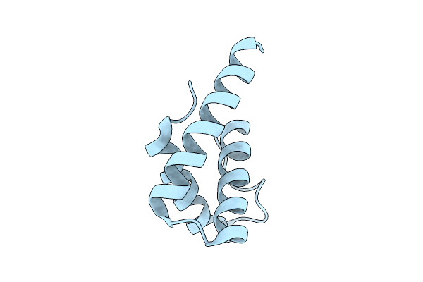 |
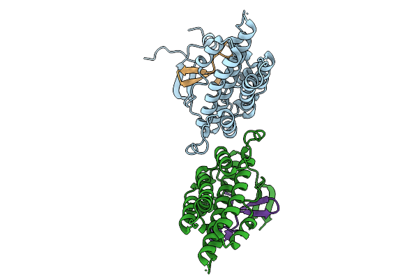
Distinct Quaternary States, Intermediates, And Autoinhibition During Loading Of The Dnab-Replicative Helicase By The Phage Lambda P Helicase Loader
Organism: Escherichia phage lambda
Method: X-RAY DIFFRACTION Resolution:1.86 Å Release Date: 2025-07-16 Classification: REPLICATION |
 |
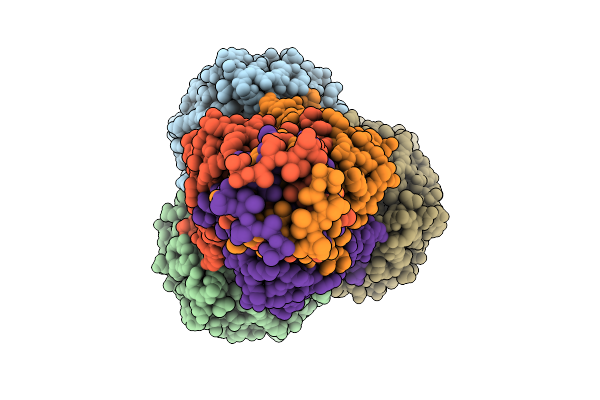
Ecoli Dnab Helicase And Phage Lambda Loader P With Adp-Mg In A 6:5 Stoichiometry Ratio.
Organism: Escherichia coli, Escherichia phage lambda
Method: ELECTRON MICROSCOPY Release Date: 2025-07-16 Classification: DNA BINDING PROTEIN Ligands: ADP, MG |
 |
Ecoli Dnab Helicase And Phage Lambda Loader P With Adp-Mg In A 6:6 Stoichiometry Ratio.
Organism: Escherichia coli, Escherichia phage lambda
Method: ELECTRON MICROSCOPY Release Date: 2025-07-16 Classification: DNA BINDING PROTEIN Ligands: ADP, MG |
 |
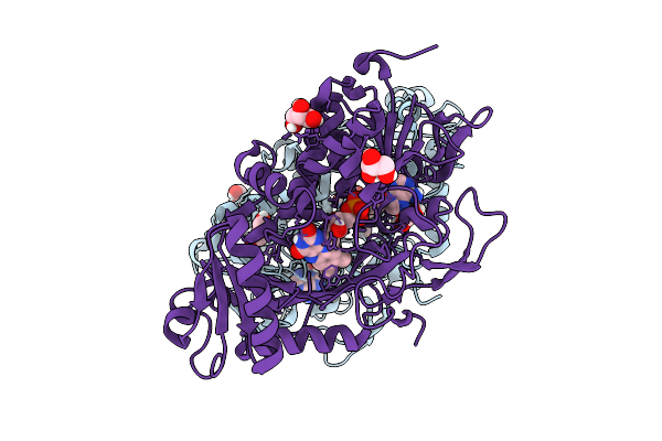
Crystal Structure Of An Inverse Charged Cutinase Mutant From Saccharopolyspora Flava (611)
Organism: Saccharopolyspora flava
Method: X-RAY DIFFRACTION Resolution:1.17 Å Release Date: 2025-04-16 Classification: HYDROLASE |