

Search Count: 3
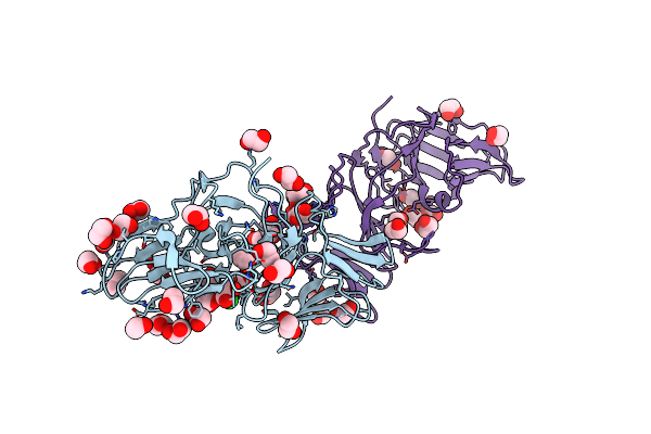 |
The Crystal Structure Of Pyocin L1 At 2.09 Angstroms
Organism: Pseudomonas aeruginosa
Method: X-RAY DIFFRACTION Resolution:2.09 Å Release Date: 2014-02-19 Classification: SUGAR BINDING PROTEIN Ligands: EDO, CL |
Organism: Pseudomonas aeruginosa
Method: X-RAY DIFFRACTION
Release Date: 2014-02-19
Ligands: EDO, CL
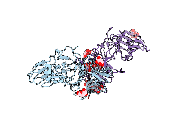 |
The Crystal Structure Of Pyocin L1 Bound To D-Mannose At 2.55 Angstroms
Organism: Pseudomonas aeruginosa
Method: X-RAY DIFFRACTION Resolution:2.55 Å Release Date: 2014-02-19 Classification: SUGAR BINDING PROTEIN Ligands: BMA |
Organism: Pseudomonas aeruginosa
Method: X-RAY DIFFRACTION
Release Date: 2014-02-19
Ligands: BMA
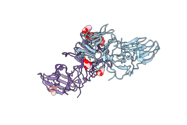 |
The Crystal Structure Of Pyocin L1 Bound To D-Rhamnose At 2.37 Angstroms
Organism: Pseudomonas aeruginosa
Method: X-RAY DIFFRACTION Resolution:2.37 Å Release Date: 2014-02-19 Classification: SUGAR BINDING PROTEIN Ligands: XXR |
Organism: Pseudomonas aeruginosa
Method: X-RAY DIFFRACTION
Release Date: 2014-02-19
Ligands: XXR